. 2013;8(1):e49783.
doi: 10.1371/journal.pone.0049783. Epub 2013 Jan 2.

The P body protein Dcp1a is hyper-phosphorylated during mitosis

Affiliations

The P body protein Dcp1a is hyper-phosphorylated during mitosis

Adva Aizer et al. PLoS One. 2013.

Abstract

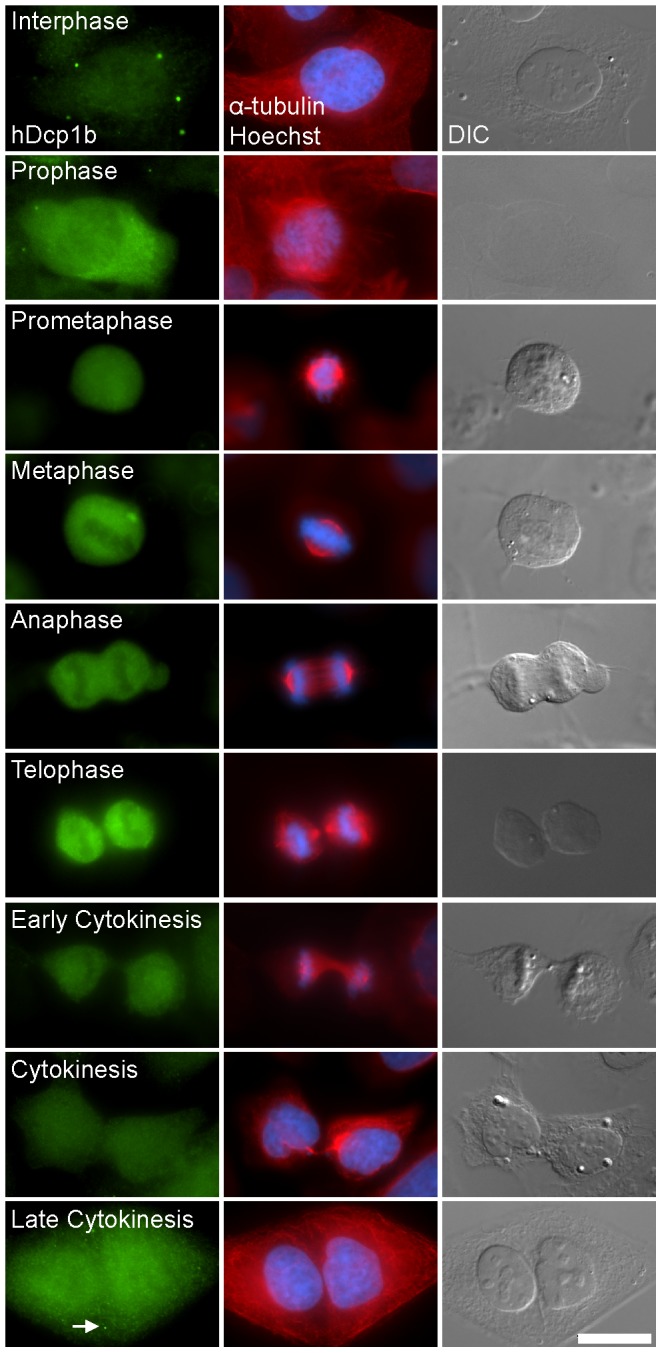
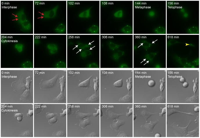
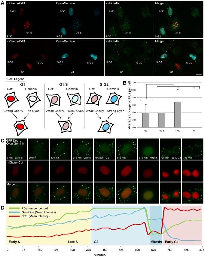
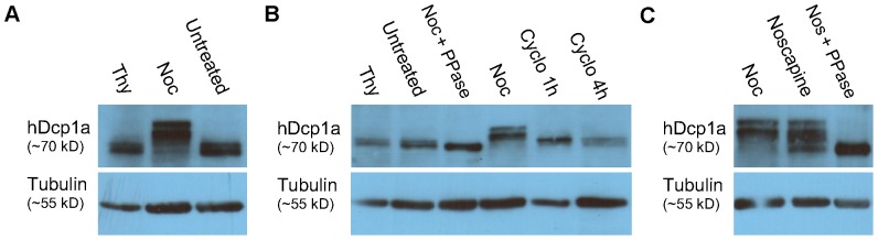
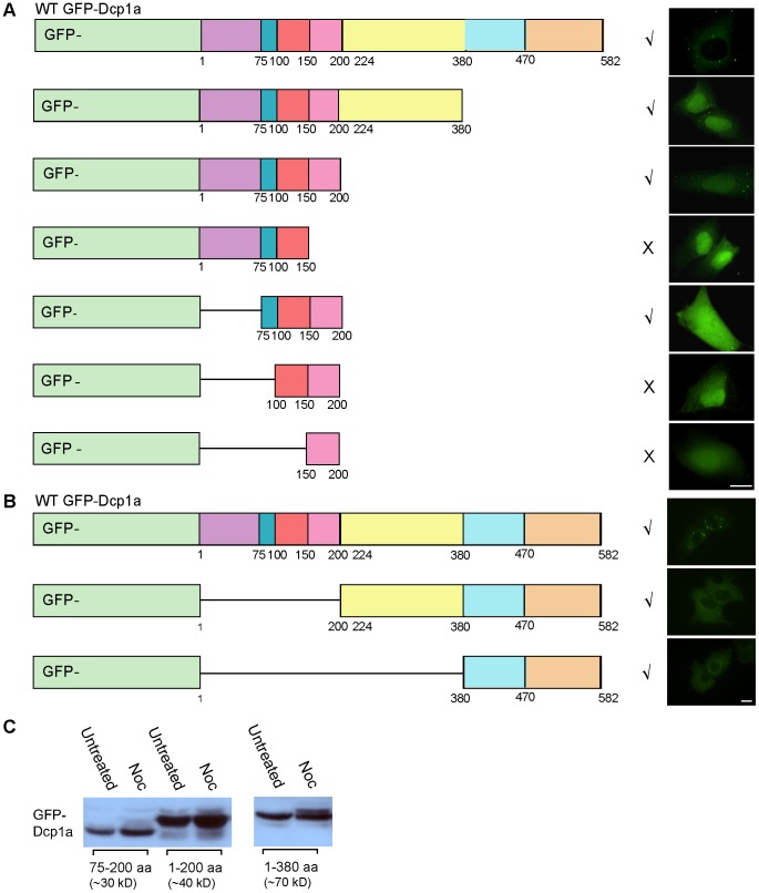
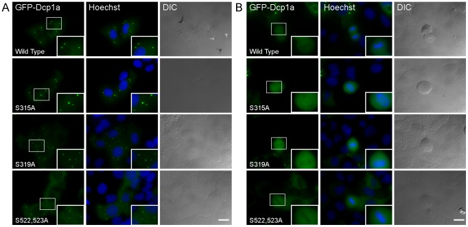
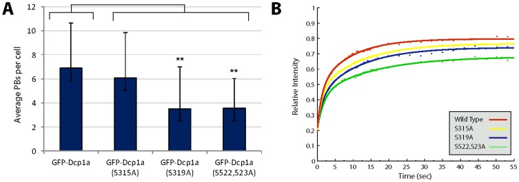
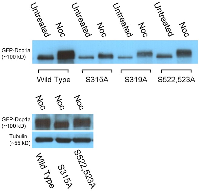
Processing bodies (PBs) are non-membranous cytoplasmic structures found in all eukaryotes. Many of their components such as the Dcp1 and Dcp2 proteins are highly conserved. Using live-cell imaging we found that PB structures disassembled as cells prepared for cell division, and then began to reassemble during the late stages of cytokinesis. During the cell cycle and as cells passed through S phase, PB numbers increased. However, there was no memory of PB numbers between mother and daughter cells. Examination of hDcp1a and hDcp1b proteins by electrophoresis in mitotic cell extracts showed a pronounced slower migrating band, which was caused by hyper-phosphorylation of the protein. We found that hDcp1a is a phospho-protein during interphase that becomes hyper-phosphorylated in mitotic cells. Using truncations of hDcp1a we localized the region important for hyper-phosphorylation to the center of the protein. Mutational analysis demonstrated the importance of serine 315 in the hyper-phosphorylation process, while other serine residues tested had a minor affect. Live-cell imaging demonstrated that serine mutations in other regions of the protein affected the dynamics of hDcp1a association with the PB structure. Our work demonstrates the control of PB dynamics during the cell cycle via phosphorylation.


Conflict of interest statement

Competing Interests: The authors have declared that no competing interests exist.

Figures

Figure 1
Figure 1. PB assembly and disassembly during the cell cycle.
Immunofluoresence staining of endogenous hDcp1b (green), α-tubulin (red), DNA (Hoechst, blue) and DIC images show that PB structures disassemble during cell division. (Bar 20 µm).
Figure 2
Figure 2. PB assembly and disassembly during cell division in living cells.
Cells stably expressing GFP-Dcp1a were simultaneously imaged in GFP and DIC showing the assembly and disassembly of PBs from a movie acquired for 14 hours. Cells were imaged every 6 min. Red arrows: PBs in the cell before mitosis. White arrows: PBs in daughter cells after mitosis. Yellow arrow head: PB in a retraction fiber.
Figure 3
Figure 3. PB numbers increase as cells reach S/G2 phase of the cell cycle.
(A) The Fucci markers mCherry-Cdt1 (red) and AmCyan1-Geminin (cyan) were expressed in U2OS cells and then cells were stained with an anti-Hedls (green) antibody to mark PBs. (Bar 20 µm). It was possible to detect the cell cycle phase using the intensity combination of the red and cyan markers in the cell, as explained in scheme below. (B) The number of PBs in each cell was counted and assigned a cell cycle phase according to the Fucci colors. The plot designates the average PB number in each phase (G1 n = 40, G1/S n = 15, S/G2 n = 40, M n = 10). Error bars represent STDEV and a T-Test was performed. (C) U2OS cells stably expressing GFP-Dcp1a were co-transfected with AmCyan1-Geminin and mCherry-Cdt1 and imaged for 15 hours. Frames show the cytoplasmic GFP-Dcp1a signal together with nuclear AmCyan1-Geminin staining that looks green due to the filter used. The plot represents the relative intensity analysis of all markers as quantified throughout the movie. Red – mean Cdt1 intensity, cyan – mean Geminin intensity, green – number of PBs.
Figure 4
Figure 4. Dcp1a is hyper-phosphorylated during cell division.
Western blot analysis of (A) endogenous hDcp1a protein in U2OS cell extracts during interphase (untreated), metaphase (nocodazole block, Noc) and at G1/S (thymidine block, Thy), showed the appearance of slower migrating Dcp1a bands in metaphase cells. (B) Treatment of U2OS protein extracts from metaphase cells with a phosphatase (Noc+PPase) caused a reduction in the molecular weight of hDcp1a, compared to untreated, G1/S blocked (Thy), and metaphase blocked cells (Noc). This demonstrated that Dcp1a is hyper-phosphorylated during mitosis. Treatment with cycloheximide (Cyclo) for 1 or 4 hrs did not change the mobility of hDcp1a indicating that hyper-phosporylation is cell cycle dependent. (C) Shift in mobility due to hyper-phosphorylation in mitotic cells is seen using two different cell cycle blockers, nocodazole (Noc) and noscapine. Similarly, phosphatase treatment (Nos+PPase) caused a reduction in the molecular weight of Dcp1a from noscapine treated cells. Tubulin was used as a loading control.
Figure 5
Figure 5. Phosphorylation of Dcp1a occurs in 200–380 region of the protein.
GFP constructs containing different fragments of the hDcp1a protein were transfected into U2OS cells and their assembly into PBs was monitored. The symbol √ indicates accumulation in PBs and the symbol×indicates no accumulation. (A) C-terminal truncations showing that region 1–200 is important for assembly into PBs. (B) N-terminal truncations. (Bar 20 µm). (C) Change in SDS-PAGE mobility in extracts from mitotic cells was detected for the 1–380 aa GFP-Dcp1a truncated protein, but not in the 1–200 and 75–200 aa forms. The blots were reacted with anti-GFP. Tubulin was used as a loading control.
Figure 6
Figure 6. Serine mutated Dcp1a proteins assembled into PBs.
(A) Serine to alanine mutated GFP-Dcp1a proteins assembled in PBs in U2OS cells, and (B) disassembled during mitosis. Enlarged insets are boxed. DNA was counterstained with Hoechst. (Bar 20 µm).
Figure 7
Figure 7. Serine 522 and 523 mutations affect Dcp1a association/dissociation dynamics in PBs.
(A) Plot showing the average number of PBs in cells expressing the different serine mutated forms of Dcp1a. A statistically significant reduction in PB numbers was seen in cells expressing the S522,523A and S319A mutations (n = 40). Error bars represent STDEV and a T-Test was performed. (B) PBs containing the different serine mutated proteins were photobleached and the kinetics of recovery were analyzed. The curves represent an average of 20 PBs in 10 cells. The recovery curves were statistically different as seen by Mann-Whitney (non parametric test). The recovery curves were fit by Matlab and the t1/2 recovery times and fixed fractions were calculated and showed a reduction in Dcp1a association with the PB structure in cells expressing the S522,423A mutant.
Figure 8
Figure 8. Serine 315 is important for the hyper-phosphorylation of Dcp1a during cell division.
Top - The S319A and S522,523A mutated GFP-Dcp1a proteins showed prominent hyper-phosphorylation patterns compared to the S315A protein. Bottom - blot comparing the mobility shifts of the mutated proteins from mitotic cell extracts showing that S315A is the least affected.

References

    1. Teixeira D, Sheth U, Valencia-Sanchez MA, Brengues M, Parker R (2005) Processing bodies require RNA for assembly and contain nontranslating mRNAs. RNA 11: 371–382. - PMC - PubMed
    1. Eulalio A, Behm-Ansmant I, Schweizer D, Izaurralde E (2007) P-body formation is a consequence, not the cause, of RNA-mediated gene silencing. Mol Cell Biol 27: 3970–3981. - PMC - PubMed
    1. Cougot N, Babajko S, Seraphin B (2004) Cytoplasmic foci are sites of mRNA decay in human cells. J Cell Biol 165: 31–40. - PMC - PubMed
    1. Ingelfinger D, Arndt-Jovin DJ, Luhrmann R, Achsel T (2002) The human LSm1-7 proteins colocalize with the mRNA-degrading enzymes Dcp1/2 and Xrnl in distinct cytoplasmic foci. RNA 8: 1489–1501. - PMC - PubMed
    1. Sheth U, Parker R (2003) Decapping and decay of messenger RNA occur in cytoplasmic processing bodies. Science 300: 805–808. - PMC - PubMed

Publication types

MeSH terms