. 2013;8(1):e53017.
doi: 10.1371/journal.pone.0053017. Epub 2013 Jan 2.

Proteomic analysis of lipid droplets from Caco-2/TC7 enterocytes identifies novel modulators of lipid secretion

Affiliations

Proteomic analysis of lipid droplets from Caco-2/TC7 enterocytes identifies novel modulators of lipid secretion

Frauke Beilstein et al. PLoS One. 2013.

Abstract

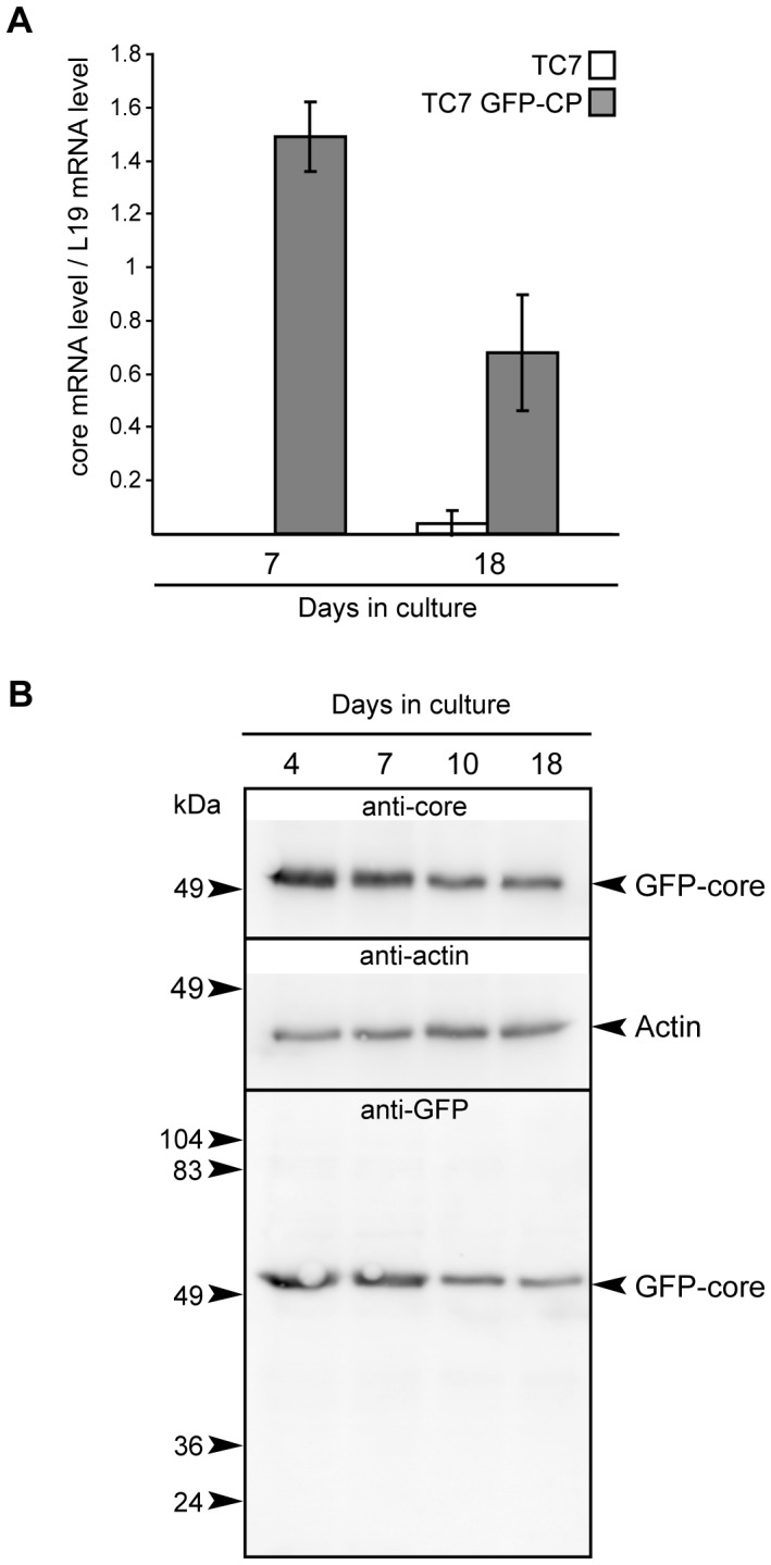
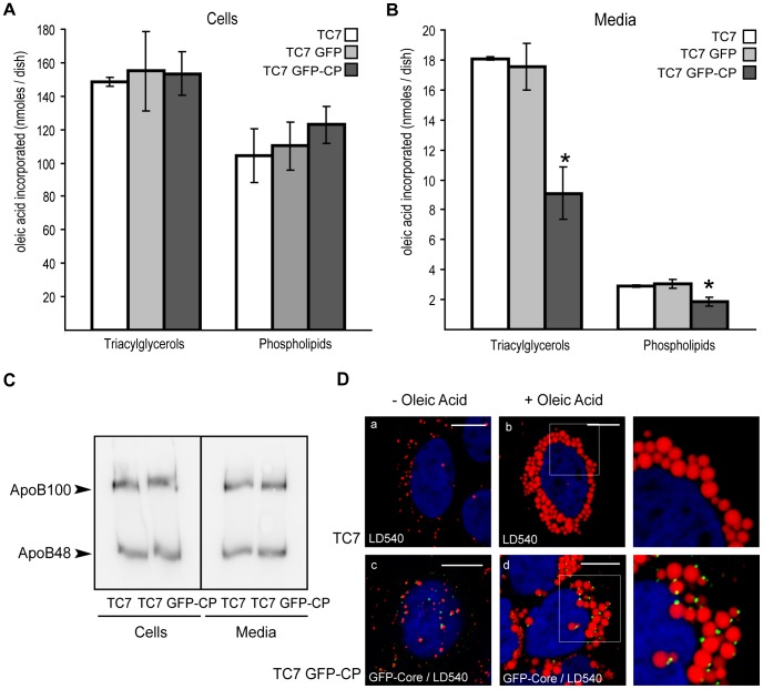
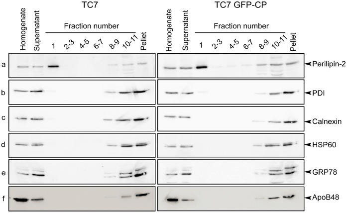
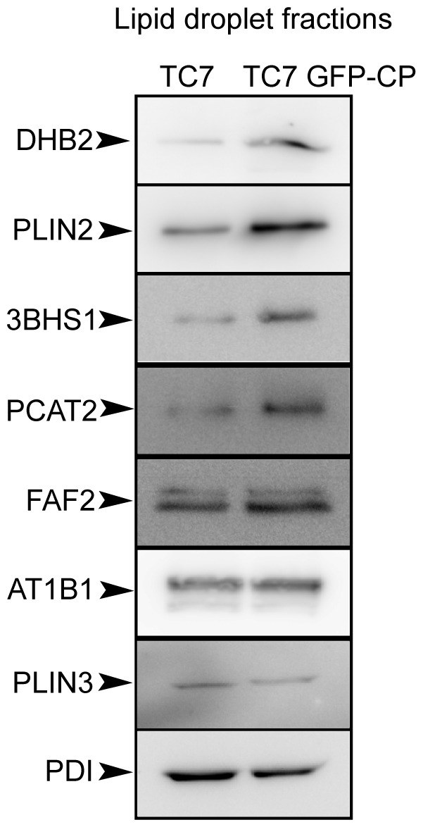
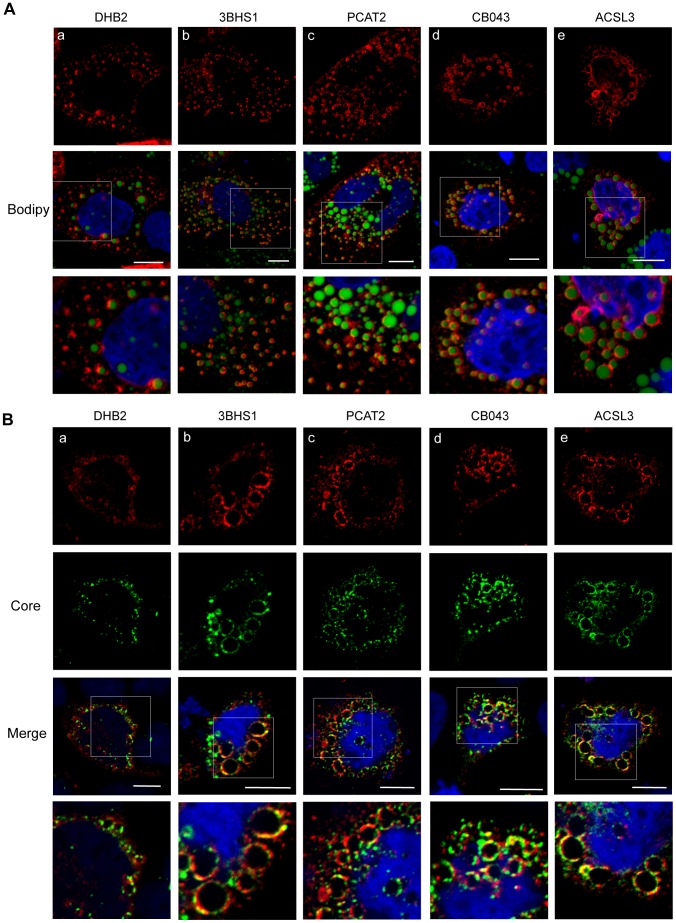
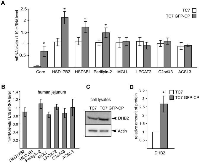
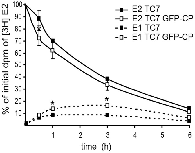
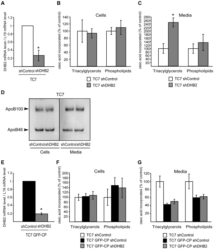
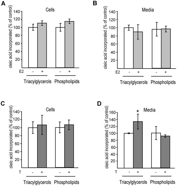
In enterocytes, the dynamic accumulation and depletion of triacylglycerol (TAG) in lipid droplets (LD) during fat absorption suggests that cytosolic LD-associated TAG contribute to TAG-rich lipoprotein (TRL) production. To get insight into the mechanisms controlling the storage/secretion balance of TAG, we used as a tool hepatitis C virus core protein, which localizes onto LDs, and thus may modify their protein coat and decrease TRL secretion. We compared the proteome of LD fractions isolated from Caco-2/TC7 enterocytes expressing or not hepatitis C virus core protein by a differential proteomic approach (isobaric tag for relative and absolute quantitation (iTRAQ) labeling coupled with liquid chromatography and tandem mass spectrometry). We identified 42 proteins, 21 being involved in lipid metabolism. Perilipin-2/ADRP, which is suggested to stabilize long term-stored TAG, was enriched in LD fractions isolated from Caco-2/TC7 expressing core protein while perilipin-3/TIP47, which is involved in LD synthesis from newly synthesized TAG, was decreased. Endoplasmic reticulum-associated proteins were strongly decreased, suggesting reduced interactions between LD and endoplasmic reticulum, where TRL assembly occurs. For the first time, we show that 17β-hydroxysteroid dehydrogenase 2 (DHB2), which catalyzes the conversion of 17-keto to 17 β-hydroxysteroids and which was the most highly enriched protein in core expressing cells, is localized to LD and interferes with TAG secretion, probably through its capacity to inactivate testosterone. Overall, we identified potential new players of lipid droplet dynamics, which may be involved in the balance between lipid storage and secretion, and may be altered in enterocytes in pathological conditions such as insulin resistance, type II diabetes and obesity.


Conflict of interest statement

Competing Interests: The authors have declared that no competing interests exist.

Figures

Figure 1
Figure 1. HCV core protein expression in Caco-2/TC7 GFP-CP cells as a function of time in culture.
Caco-2/TC7 cells expressing HCV core protein-GFP (TC7 GFP-CP) or not (TC7) were grown on filters for indicated days: confluence is reached on day 7 then cells differentiate i.e. TRL secretion increases gradually with time in culture. Cells were analyzed for expression of HCV core transcripts by quantitative RT-PCR (A) and for HCV core protein and GFP content by western blot (B) using antibodies against HCV core protein or against GFP. Blots were probed for actin as protein loading control.
Figure 2
Figure 2. HCV core protein leads to decreased lipid secretion in Caco-2/TC7 enterocytes and localizes on lipid droplets.
Caco-2/TC7 cells expressing HCV core protein-GFP (TC7 GFP-CP) and control cells [Caco-2/TC7 cells (TC7) and Caco-2/TC7 cells expressing the empty vector (TC7 GFP)] were cultured on filters for 17 days for differentiation then supplied with lipid micelles for 24 h. Lipid micelles were supplemented (A, B) or not (C) with [1-14C]oleic acid. Lipids extracted from cells (A) and basolateral media (B) were fractionated by thin layer chromatography and the radioactivity in the spots was counted. Results for triacylglycerols and phospholipids are shown as means ± SD, expressed as nmoles of oleic acid incorporated per dish, obtained in three independent experiments performed in triplicate (*, p<0.05 compared to control cells). (C) Cell lysates and basolateral media were analyzed for apoB by western blot. (D) Caco-2/TC7 (TC7) and Caco-2/TC7 GFP-CP (TC7 GFP-CP) cells were cultured for one day then incubated (+) or not (−) with 0.6 mM oleic acid/BSA for 24 h. Lipid droplets were visualized using the neutral lipid stain LD540 (red). The core-GFP fusion protein appears in green. Scale bars, 10 µm.
Figure 3
Figure 3. Protein analysis of sucrose gradient fractions prepared from Caco-2/TC7 and Caco-2/TC7 GFP-CP cells.
Caco-2/TC7 cells (TC7, left panel) and Caco-2/TC7 GFP-CP cells (TC7 GFP-CP, right panel) were cultured on filters for 17 days then supplied with lipid micelles for 24 h. Cell homogenates were centrifuged for 10 min at 1000×g and the supernatants were fractionated onto sucrose gradients. Top to bottom fractions (1 to 11) and pellets were analyzed by western blot using antibodies specific for (a) perilipin-2/ADRP, (b) protein disulfide isomerase (PDI), (c) calnexin, (d) heat shock protein 60 (HSP 60), (e) 78 kDa glucose-regulated protein (GRP78) and (f) apolipoprotein B48 (ApoB48). The same percentage of each fraction of the sucrose gradient was loaded on gels except for fraction 1 which was 2-fold loaded in order to evaluate the presence of organelle markers with greater sensitivity.
Figure 4
Figure 4. Western blot analysis of some proteins identified by the differential proteomic approach in the lipid droplet fractions isolated from Caco-2/TC7 (TC7) and Caco-2/TC7 GFP-CP (TC7 GFP-CP) cells.
The lipid droplet fractions (fraction 1) were prepared as described in figure 3, freeze-dried for concentration and analyzed by western blot using antibodies against 17β-hydroxysteroid dehydrogenase type 2 (DHB2), perilipin-2/ADRP (PLIN2), 3-beta-hydroxysteroid dehydrogenase (3BHS1), lysophosphatidylcholine acyltransferase 2 (PCAT2), FAS-associated factor 2/UBXD8 (FAF2), Na+/K+ATPase α1 (AT1B1), perilipin-3/TIP47 (PLIN3) and protein disulfide isomerase (PDI). The amount of material loaded per well was 10 times higher than in Fig.3, fraction 1.
Figure 5
Figure 5. Proteins identified by LC-MS/MS in the lipid droplet fractions isolated from Caco-2/TC7 cells localise to lipid droplets (A) and co-localise with HCV core protein around lipid droplets (B).
Caco-2/TC7 cells were transfected with plasmids encoding the proteins of interest fused to a myc-tag (A) or double transfected with plasmids expressing proteins of interest fused to a myc-tag and the core expressing plasmid pGFP-CP (B), and incubated with 0.6 mM oleic acid/BSA for 24 h to induce lipid droplet formation. The myc-tag was detected with mAb9E10 and Alexa Fluor 568–conjugated anti-mouse IgG (red) and the core protein by GFP fluorescence (green). (a) 17β-hydroxysteroid dehydrogenase type 2 (DHB2), (b) 3-beta-hydroxysteroid dehydrogenase (3BHS1), (c) lysophosphatidylcholine acyltransferase type 2 (PCAT2), (d) UPF0554 C2orf43 (CB043) and (e) long-chain-fatty-acid–CoA ligase 3 (ACSL3). Scale bars, 10 µm.
Figure 6
Figure 6. Gene and protein expression in Caco-2/TC7 cells (TC7), Caco-2/TC7 GFP-CP cells (TC7 GFP-CP) and human jejunum of some proteins identified by LC-MS/MS in lipid droplet fractions.
(A) Caco-2/TC7 and Caco-2/TC7 GFP-CP cells were cultured on filters for 17 days then supplied with lipid micelles for 24 h. mRNA levels were measured by quantitative RT-PCR for core (HCV core protein), HSD17B2 (17β-hydroxysteroid dehydrogenase type 2), HSD3B1 (3-beta-hydroxysteroid dehydrogenase), PLIN2 (perilipin-2), MGLL (monoacylglycerol lipase), LPCAT2 (lysophosphatidylcholine acyltransferase 2), C2orf43 (UPF0554 protein C2orf43) and ACSL3 (long-chain-fatty-acid–CoA ligase 3). (B) mRNA levels for the same genes were quantified in human jejunum mRNA samples. (C) Lysates of Caco-2/TC7 and Caco-2/TC7 GFP-CP cells were analyzed by western blot for 17β-hydroxysteroid dehydrogenase type 2 (DHB2) and actin. (D) The immunoblot shown in C was quantified and standardized to actin used as the loading control. Results shown are the means ± SD from three independent experiments performed in triplicate, except for human jejunum (one sample measured in triplicate). *, p<0.05 compared to control cells.Caco-2 cell line derives from a human epithelial colorectal adenocarcinoma and TC7 is a clone of Caco-2 cells . Although these cells differentiate such that their phenotype resembles absorptive enterocytes of the small intestine, it still remains a cell line with a cancerous origin and thus proteins might be differently expressed in normal cells from human intestine. Therefore, to assess the physiological relevance of these results, we performed similar experiments on mRNA isolated from human small intestine (Fig. 6B). All above-mentioned genes were expressed in human small intestine except HSD3B1. Therefore, since 3BHS1 protein was also not significantly up-regulated in LD isolated from Caco-2/TC7 GFP-CP as compared to Caco-2/TC7 cells, it was not studied further.
Figure 7
Figure 7. DHB2 activity in Caco-2/TC7 (TC7) and Caco-2/TC7 GFP-CP (TC7 GFP-CP) cells.
Cells were cultured on filters for 17 days then incubated with [3H] estradiol (E2) for the indicated times. Steroids were extracted from the basolateral media and separated by TLC. The radioactivity in the spots corresponding to estradiol (E2) and estrone (E1) was quantified by scintillation counting. Results are expressed as percentage of dpm of [3H]estradiol present in medium at time 0. Data are mean ± SD from triplicate determinations of three independent experiments. *, p<0.05 compared to control cells.
Figure 8
Figure 8. Depletion of DHB2 leads to increased lipid secretion by Caco-2/TC7 cells.
Caco-2/TC7 cells (A−D) and Caco-2/TC7 GFP-CP cells (E−G) cultured on filters were transduced with lentiviral vectors expressing shRNA directed against the luciferase gene (shControl) or HSD17B2 (shDHB2). After 17 days in culture, cells were incubated with lipid micelles for 24 h. Lipid micelles were supplemented (B, C, F, G) or not (A, D, E) with [1-14C]oleic acid. The efficiency of silencing in Caco-2/TC7 cells (A) and Caco-2/TC7 GFP-CP cells (E) was analyzed by quantitative RT-PCR. shControl values were set at 1. Lipids extracted from cells (B and F) and basolateral media (C and G) were fractionated by thin layer chromatography and the radioactivity in triacylglycerols and phospholipids was counted. Results are expressed as a percentage of control cells (TC7 shControl). Data are means ± SD of at least four independent experiments. *, p<0.05 compared to control cells.
Figure 9
Figure 9. Effect of estradiol (E2) and testosterone (T) on lipid synthesis and secretion by Caco-2/TC7 cells.
Caco-2/TC7 cells were cultured on filters for 17 days then incubated with lipid micelles supplemented with [1-14C]oleic acid, in the presence or not of estradiol (A, B) or testosterone (C, D) for 24 h. Lipids extracted from cells (A and C) and basolateral media (B and D) were fractionated by thin layer chromatography and the radioactivity in triacylglycerols and phospholipids was counted. Results are expressed as percentage of control cells. Data are means ± SD of four independent experiments. *, p<0.05 compared to control cells.

References

    1. Walther TC, Farese RV Jr (2008) The life of lipid droplets. Biochim Biophys Acta 1791: 459–466. - PMC - PubMed
    1. Fujimoto T, Parton RG (2011) Not just fat: the structure and function of the lipid droplet. Cold Spring Harb Perspect Biol 3. - PMC - PubMed
    1. Murphy DJ, Vance J (1999) Mechanisms of lipid-body formation. Trends Biochem Sci 24: 109–115. - PubMed
    1. Ohsaki Y, Cheng J, Suzuki M, Shinohara Y, Fujita A, et al. (2009) Biogenesis of cytoplasmic lipid droplets: From the lipid ester globule in the membrane to the visible structure. Biochim Biophys Acta 1791: 399–407. - PubMed
    1. Lehner R, Lian J, Quiroga AD (2012) Lumenal lipid metabolism: implications for lipoprotein assembly. Arterioscler Thromb Vasc Biol 32: 1087–1093. - PubMed

Publication types