. 2013;8(1):e53575.
doi: 10.1371/journal.pone.0053575. Epub 2013 Jan 3.

RNA-Seq profiling of spinal cord motor neurons from a presymptomatic SOD1 ALS mouse

Affiliations

RNA-Seq profiling of spinal cord motor neurons from a presymptomatic SOD1 ALS mouse

Urmi Bandyopadhyay et al. PLoS One. 2013.

Abstract

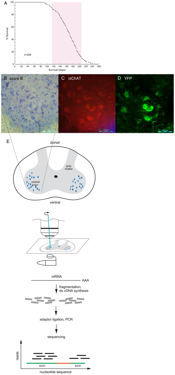
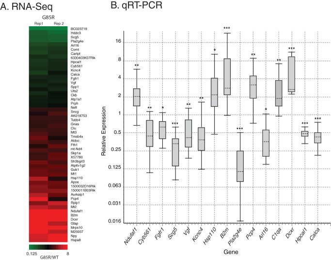
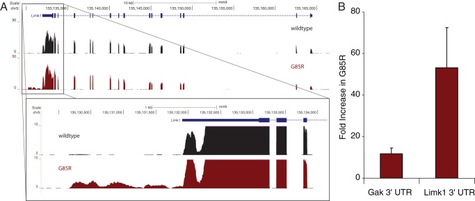
Mechanisms involved with degeneration of motor neurons in amyotrophic lateral sclerosis (ALS; Lou Gehrig's Disease) are poorly understood, but genetically inherited forms, comprising ~10% of the cases, are potentially informative. Recent observations that several inherited forms of ALS involve the RNA binding proteins TDP43 and FUS raise the question as to whether RNA metabolism is generally disturbed in ALS. Here we conduct whole transcriptome profiling of motor neurons from a mouse strain, transgenic for a mutant human SOD1 (G85R SOD1-YFP), that develops symptoms of ALS and paralyzes at 5-6 months of age. Motor neuron cell bodies were laser microdissected from spinal cords at 3 months of age, a time when animals were presymptomatic but showed aggregation of the mutant protein in many lower motor neuron cell bodies and manifested extensive neuromuscular junction morphologic disturbance in their lower extremities. We observed only a small number of transcripts with altered expression levels or splicing in the G85R transgenic compared to age-matched animals of a wild-type SOD1 transgenic strain. Our results indicate that a major disturbance of polyadenylated RNA metabolism does not occur in motor neurons of mutant SOD1 mice, suggesting that the toxicity of the mutant protein lies at the level of translational or post-translational effects.


Conflict of interest statement

Competing Interests: The authors have declared that no competing interests exist.

Figures

Figure 1
Figure 1. RNA-Seq of laser capture microdissected spinal cord motor neurons from wtSOD1-YFP and G85R SOD1-YFP transgenic mice.
A. Survival curve of G85R SOD1-YFP mice with copy number greater than 200. 80% of the mice were paralyzed (and euthanized) between ∼115 days and 205 days (red bar). N = 226. For the present study of motor neuron RNA, presymptomatic mice at ∼90 days of age were used. B.-D. Spinal cord from a 3 month old G85R SOD1-YFP mouse. Frozen section of right ventral horn region is shown, stained with Azure B dye, panel B (see Methods); incubated with anti-ChAT antibodies, panel C; or directly examined for YFP fluorescence, panel D. The large blue-stained cell bodies in panel B are motor neurons as indicated by anti-ChAT staining in panel C. Note that the same cells have YFP fluorescence in panel D. E. Large Azure B-stained cell bodies were laser captured directly into a guanidine thiocyanate solution (see Methods) and subsequent steps carried out as diagrammed (see Methods).
Figure 2
Figure 2. Differential expression in G85R motor neurons.
A. Heatmap of selected genes from Table S2 significantly differentially expressed (raw p-value <0.005) between wild-type and G85R SOD1-YFP mice. For each gene listed, the ratio of RPKM values (G85R/WT) for individual pairs of biological replicates (Rep1, Rep2) is plotted according to the color code below. B. Validation of differentially expressed genes in G85R by qRT-PCR. Shown are box plots representing relative expression values of each gene in G85R versus wild-type motor neurons. Upper whisker represents top 25% of values, box represents the middle 50% of values, and lower whisker represents bottom 25% of values. Median value is indicated by horizontal dashed line. Statistical significance calculated by REST 2009 is indicated by *  =  p<0.05, **  =  p<0.005, ***  =  p<0.0005. RNAs from at least three different mouse pairs were compared for each gene. Note that the Hsp110 and B2m validations used one exon-junction-spanning and one non-spanning primer set; minus reverse transcriptase controls for these samples were negative for DNA contamination. The expression changes in the left nine genes were validated with 0.19 ng of total RNA, while the remaining six were validated with 1.5 ng of total RNA.
Figure 3
Figure 3. Differential expression of novel TARs in G85R motor neurons.
A. RNA-Seq signal plots in reads per million mapped reads at the Limk1 gene from wild-type (black) and G85R (red) motor neurons. The canonical Limk1 gene map is shown at the top. The enlarged region shows a clear 3′-UTR extension in G85R motor neuron RNA. Similar data were observed for Gak (not shown). B. Validation of novel 3′-UTRs of Gak and Limk1 by qRT-PCR. Values are relative expression for each 3′-UTR in G85R versus wild-type motor neuron RNA.

References

    1. Rosen DR, Siddique T, Patterson D, Figlewicz DA, Sapp P, et al. (1993) Mutations in Cu/Zn superoxide dismutase gene are associated with familial amyotrophic lateral sclerosis. Nature 362: 59–62. - PubMed
    1. Hart PJ (2006) Pathogenic superoxide dismutase structure, folding, aggregation and turnover. Curr Op Chem Biol 10: 131–138. - PubMed
    1. Deng H-X, Chen W, Hong ST, Boycott KM, Gorrie GH, et al. (2011) Mutations in UBQLN2 cause dominant X-linked juvenile and adult-onset ALS and ALS/dementia. Nature 477: 211–215. - PMC - PubMed
    1. Johnson JO, Mandrioli J, Benatar M, Abramzon Y, Van Deerlin VM, et al. (2010) Exome sequencing reveals VCP mutations as a cause of familial ALS. Neuron 68: 857–864. - PMC - PubMed
    1. Maruyama H, Morino H, Ito H, Izumi Y, Kato H, et al. (2010) Mutations of optineurin in amyotrophic lateral sclerosis. Nature 465: 223–226. - PubMed

Publication types

Substances