. 2012 Dec;45(4):241-58.
doi: 10.5115/acb.2012.45.4.241. Epub 2012 Dec 14.

Nuclear expression of PG-21, SRC-1, and pCREB in regions of the lumbosacral spinal cord involved in pelvic innervation in young adult and aged rats

Affiliations

Nuclear expression of PG-21, SRC-1, and pCREB in regions of the lumbosacral spinal cord involved in pelvic innervation in young adult and aged rats

Richard N Ranson et al. Anat Cell Biol. 2012 Dec.

Abstract

In rats, ageing results in dysfunctional patterns of micturition and diminished sexual reflexes that may reflect degenerative changes within spinal circuitry. In both sexes the dorsal lateral nucleus and the spinal nucleus of the bulbospongiosus, which lie in the L5-S1 spinal segments, contain motor neurons that innervate perineal muscles, and the external anal and urethral sphincters. Neurons in the sacral parasympathetic nucleus of these segments provide autonomic control of the bladder, cervix and penis and other lower urinary tract structures. Interneurons in the dorsal gray commissure and dorsal horn have also been implicated in lower urinary tract function. This study investigates the cellular localisation of PG-21 androgen receptors, steroid receptor co-activator one (SRC-1) and the phosphorylated form of c-AMP response element binding protein (pCREB) within these spinal nuclei. These are components of signalling pathways that mediate cellular responses to steroid hormones and neurotrophins. Nuclear expression of PG-21 androgen receptors, SRC-1 and pCREB in young and aged rats was quantified using immunohistochemistry. There was a reduction in the number of spinal neurons expressing these molecules in the aged males while in aged females, SRC-1 and pCREB expression was largely unchanged. This suggests that the observed age-related changes may be linked to declining testosterone levels. Acute testosterone therapy restored expression of PG-21 androgen receptor in aged and orchidectomised male rats, however levels of re-expression varied within different nuclei suggesting a more prolonged period of hormone replacement may be required for full restoration.

Keywords: Androgens; Hormone replacement therapy; Pelvic function; Perineal motor neuron.


Figures

Fig. 1
Fig. 1
Diagram showing the areas of the spinal cord (rectangles) analysed in the fifth and sixth lumbar (L5, L6) and first sacral (S1) segments. The dimensions of the rectangles are 120×120 µm (1), 180×180 µm (2), and 240×240 µm (3). DGC, dorsal grey commissure; DH, dorsal horn; DLN, dorsolateral nucleus; RDLN, retrodorsolateral nucleus; SNB, spinal nucleus of the bulbospongiosus; SPN, sacral parasympathetic nucleus, VMN, ventromedial nucleus.
Fig. 2
Fig. 2
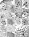
PG-21 (androgen receptor) immunoreactivity within lumbosacral spinal cord. (A) Low power photomicrograph of PG-21 immunostaining in a section from a young male rat. Staining (black punctate dots) is seen predominantly within the nuclei of pudendal motor neurons of the dorsolateral nucleus (DLN) and spinal nucleus of the bulbospongiosus (SNB), and within the neurons of the dorsal grey commissure (DGC). Moderate numbers of cells in the dorsal horn (DH) and sacral parasympathetic nucleus (SPN) are immunoreactive. By contrast labelled motor neurons within the retrodorsolateral nucleus (RDLN) are sparser. Dashed lines indicate the approximate boundaries of the RDLN and SPN. (B-E) show labelled motor neurons in the SNB. More cells are labelled in intact young male rats (B) or those orchidectomised with testosterone replacement (C) than in aged intact males (D). Testosterone replacement is also effective in orchidectomised aged rats (E) although expression does not match that seen for young animals. (F-I) show motor neurons within the DLN. Young male rats show robust nuclear immunoreactivity for PG-21 which is completely abolished by orchidectomy (G) and only partially restored by testosterone propionate treatment (H). The number of PG-21 immunoreactive motor neurons within the DLN of aged male rats (I) is significantly smaller than in young rats (F). A similar situation prevails in the DGC where numbers of PG-21 immunoreactive cells were significantly higher in young animals (J) compared to orchidectomised animals +TP treatment (K) and aged animals (L). CC, spinal central canal. In young males (M), labelled nuclei in the DH appeared largely confined to laminae I-III. (N) Immunoreactivity of nuclei within the SPN of an aged male rat. The larger nuclei (large arrowheads) probably belong to preganglionic neurons whilst the smaller nuclei (small arrowheads) may belong to glial cells or interneurons. Scale bars=200 µm (A), 50 µm (B-N).
Fig. 3
Fig. 3
Graphs showing the number of cells with nuclei immunoreactive for PG-21 in segments L5-S1. Data is presented for young and aged (3 and 24 months) intact male rats and for orchidectomised animals (orchid) with and without testosterone propionate replacement (+TP). For the sacral parasympathetic nucleus (SPN) a distinction was made between immunoreactive cells with a diameter of 20-25 µm which probably represent preganglionic neurons (SPNp), and those that were smaller and likely to be interneurons or glial cells (SPNi). DGC, dorsal grey commissure; DH, dorsal horn; DLN, dorsolateral nucleus; SNB, spinal nucleus of the bulbospongiosus.
Fig. 4
Fig. 4
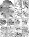
Immunolocalisation of steroid receptor co-activator one (SRC-1) in lumbosacral spinal cord. (A) Section from L6 of a young intact male rat showing neurons with labelled nuclei distributed widely throughout the spinal grey matter. DH, dorsal horn. (B-E) Large labelled nuclei in the spinal nucleus of the bulbospongiosus (SNB) probably belong to motor neurons (e.g., black arrowhead in C) while smaller nuclei belong to glial cells or interneurons (white arrowhead). SRC-1 immunolabelling is robust in intact young male rats (B) and appears unaffected by orchidectomy (C), regardless of testosterone replacement (D), but the number of labelled nuclei is reduced by ageing (E). A similar relationship is seen in the dorsolateral nucleus (DLN) of young (F) vs. aged (G) intact male rats. There are no differences in numbers of labelled nuclei in DLN motor neurons between young (H) and aged (I) female rats. In (F-I), the larger cells (black arrowhead in G) are likely to be motor neurons whilst the smaller cells (white arrowhead in G) are probably glial cells or interneurons. The retrodorsolateral nucleus (RDLN) in male rats also appears susceptible to the effects of ageing since more motor neurons with labelled nuclei are present in young (J) than in aged animals (K). Black arrowheads indicate likely motor neurons whilst white arrowheads point to nuclei of interneurons or glia. A small group of L6 motor neurons with SRC-1-labelled are present in ventromedial nucleus (VMN) (L, young female rat). SRC-1 was abundant in laminae I-III of the dorsal horn (M, aged male rat). Many labelled nuclei were seen in the dorsal grey commissure (DGC) (N, aged male rat). The cell marked by the arrowhead is seen at higher magnification in the inset. (O) Labelling in the sacral parasympathetic nucleus (SPN). The highlighted area is shown at higher magnification in (P) in which putative preganglionic neurons (black arrowheads) can be distinguished from smaller cells (white arrowheads) that may represent interneurons or glial cells. (N-P) are from an aged male rat. Scale bars=200 µm (A), 50 µm (B-M), 100 µm (N, O), 25 µm (P).
Fig. 5
Fig. 5
Graphs showing the numbers of steroid receptor co-activator one immunoreactive cells within lumbosacral spinal nuclei. Counts were performed in spinal sections derived from gonadally intact males and females aged 3 and 24 months and male rats that were orchidectomised (orchid) with or without testosterone replacement (+TP). Values are presented as mean±SEM. DGC, dorsal grey commissure; DH, dorsal horn; DLN, dorsolateral nucleus; i, interneurons orglial cells; p, preganglionic neurons; RDLN, retrodorsolateral nucleus; SNB, spinal nucleus of the bulbospongiosus; SPN, sacral parasympathetic nucleus; VMN, ventromedial nucleus.
Fig. 6
Fig. 6
Phosphorylated form of c-AMP response element binding protein (pCREB) localisation within lumbosacral spinal cord. Cells expressing immunoreactivity for pCREB are extensively located throughout the grey matter and are also observed to a lesser extent within the white matter (A, from a young female rat). VMN, ventral medial nucleus. (B-E) illustrate labelled cells within the region of the spinal nucleus of the bulbospongiosus (SNB) which are relatively few in females. (B) Probable motor neurons (black arrowhead) and interneurons or glial cells (white arrowhead) in an intact young male rat. Nuclear expression of pCREB appears similar to this in orchidectomised rats regardless of whether they did (D) or did not (C) receive testosterone injections. However the numbers of pCREB expressing motor neurons within the SNB appears reduced in aged (E) in comparison to young male rats (B). (F-I) show the immunolocalisation of pCREB within the dorsolateral nucleus. pCREB expressing motor neurons in young intact males (F) are present in greater numbers then in aged intact males (G) or either young (H) or aged (I) females, however there is no age-related decline in the females. (J) Nuclear immunoreactivity for pCREB in the retrodorsolateral nucleus (RDLN) of an aged male rat is seen both in putative motor neurons (black arrowhead) and other cells (glia or interneurons, white arrowhead). A similar situation prevails in the sacral parasympathetic nucleus (SPN) (K, young intact male). In the dorsal horn (DH), (L, M) cell nuclei are immunoreactive for pCREB are particularly abundant in laminae I-III (L, young intact male) and their numbers increase significantly following orchidectomy (M, young male). pCREB containing cells are also densely packed within the dorsal grey commissure (DGC) (N, O) and exhibit an age related decline in number (N, young intact male; O, aged intact male). Scale bars=200 µm (A), 50 µm (B-J), 100 µm (K-O).
Fig. 7
Fig. 7
Graphs showing numbers of phosphorylated form of c-AMP response element binding protein immunoreactive cells in lumbosacral spinal nuclei. For each region data is provided for male and female rats, that are gonadally intact, and at two ages. Further data is provided for males that have been orchidectomised (orchid) with or without testosterone replacement (+TP). All counts represent mean±SEM. DGC, dorsal grey commissure; DH, dorsal horn; DLN, dorsolateral nucleus; i, interneurons orglial cells; p, preganglionic neurons; RDLN, retrodorsolateral nucleus; SNB, spinal nucleus of the bulbospongiosus; SPN, sacral parasympathetic nucleus; VMN, ventromedial nucleus.

References

    1. Dering MA, Santer RM, Watson AH. Age-related changes in the morphology of preganglionic neurons projecting to the rat hypogastric ganglion. J Neurocytol. 1996;25:555–563. - PubMed
    1. Hancock MB, Peveto CA. Preganglionic neurons in the sacral spinal cord of the rat: an HRP study. Neurosci Lett. 1979;11:1–5. - PubMed
    1. Nadelhaft I, Booth AM. The location and morphology of preganglionic neurons and the distribution of visceral afferents from the rat pelvic nerve: a horseradish peroxidase study. J Comp Neurol. 1984;226:238–245. - PubMed
    1. Ranson RN, Santer RM, Watson AH. The relationship between serotonin, dopamine beta hydroxylase and GABA immunoreactive inputs and spinal preganglionic neurones projecting to the major pelvic ganglion of Wistar rats. Neuroscience. 2006;141:1935–1949. - PubMed
    1. Santer RM, Dering MA, Ranson RN, Waboso HN, Watson AH. Differential susceptibility to ageing of rat preganglionic neurones projecting to the major pelvic ganglion and of their afferent inputs. Auton Neurosci. 2002;96:73–81. - PubMed

LinkOut - more resources