

Assessment of the relationship between the maxillary molars and adjacent structures using cone beam computed tomography
- PMID: 23301207
- PMCID: PMC3534175
- DOI: 10.5624/isd.2012.42.4.219
Assessment of the relationship between the maxillary molars and adjacent structures using cone beam computed tomography
Abstract
Purpose: This study investigated the relationship between the roots of the maxillary molars and the maxillary sinus using cone beam computed tomography (CBCT), and measured the distances between the roots of the maxillary molars and the sinus floor as well as the thickness of the bone between the root and the alveolar cortical plate.
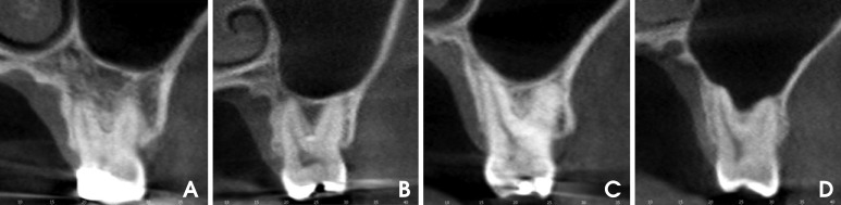
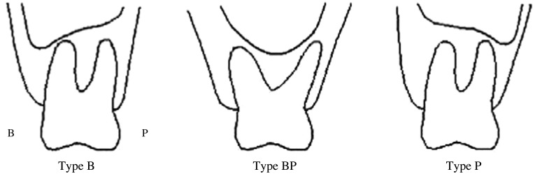
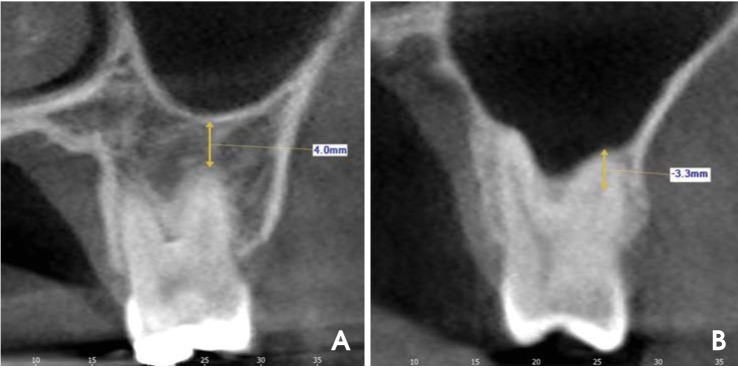
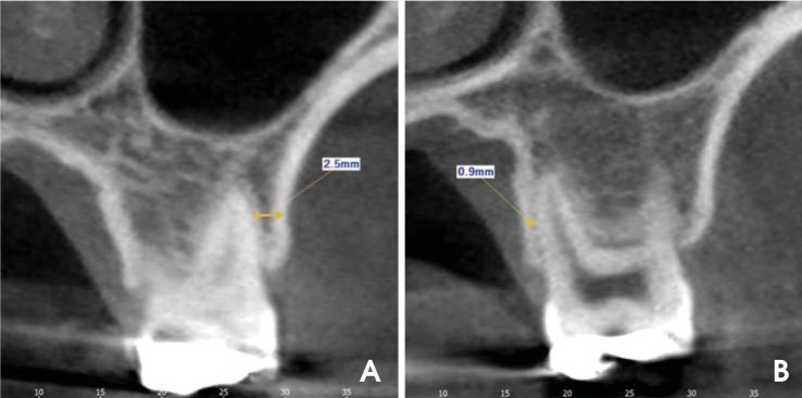
Materials and methods: The study sample consisted of 83 patients with normally erupted bilateral maxillary first and second molars. A total of 332 maxillary molars were examined using CBCT images. The vertical relationship of each root with the maxillary sinus was classified into four types on CBCT cross-sectional images. The distance between the sinus floor and root and the bone thickness between the root and alveolar cortical plate were measured.
Results: In the buccal roots of the maxillary molars, a root protruding into the sinus occurred most frequently. A root projecting laterally along the sinus cavity was most common in the palatal roots of the maxillary first molars. The mesiobuccal roots of the maxillary second molar were closest to the sinus. The mesiobuccal roots of the first molars were closest to the cortical plate.
Conclusion: The relationship between the roots of the maxillary molars and the sinus differed between the buccal and palatal roots. A root protruding into the sinus occurred more frequent in the buccal roots of the maxillary molars. The mesiobuccal root of the maxillary second molar was closest to the maxillary sinus floor and farthest from the alveolar cortical plate.
Keywords: Bone and Bones; Cone-Beam Computed Tomography; Maxillary Sinus; Molar.
Figures
References
-
- Watzek G, Bernhart T, Ulm C. Complications of sinus perforations and their management in endodontics. Dent Clin North Am. 1997;41:563–583. - PubMed
-
- Hauman CH, Chandler NP, Tong DC. Endodontic implications of the maxillary sinus: a review. Int Endod J. 2002;35:127–141. - PubMed
-
- Fuhrmann R, Bücker A, Diedrich P. Radiological assessment of artificial bone defects in the floor of the maxillary sinus. Dentomaxillofac Radiol. 1997;26:112–116. - PubMed
-
- Engström H, Chamberlain D, Kiger R, Egelberg J. Radiographic evaluation of the effect of initial periodontal therapy on thickness of the maxillary sinus mucosa. J Periodontol. 1988;59:604–608. - PubMed
