

Measurements of simulated periodontal bone defects in inverted digital image and film-based radiograph: an in vitro study
- PMID: 23301211
- PMCID: PMC3534179
- DOI: 10.5624/isd.2012.42.4.243
Measurements of simulated periodontal bone defects in inverted digital image and film-based radiograph: an in vitro study
Abstract
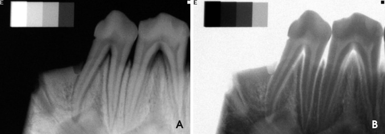
Purpose: This study was performed to compare the inverted digital images and film-based images of dry pig mandibles to measure the periodontal bone defect depth.
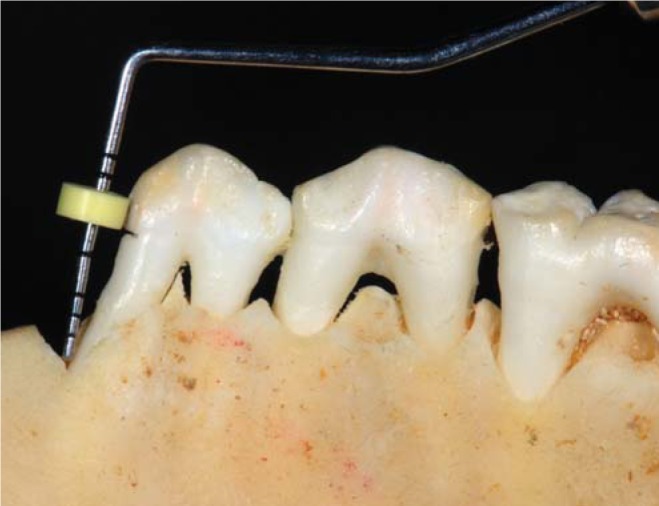
Materials and methods: Forty 2-wall bone defects were made in the proximal region of the premolar in the dry pig mandibles. The digital and conventional radiographs were taken using a Schick sensor and Kodak F-speed intraoral film. Image manipulation (inversion) was performed using Adobe Photoshop 7.0 software. Four trained examiners made all of the radiographic measurements in millimeters a total of three times from the cementoenamel junction to the most apical extension of the bone loss with both types of images: inverted digital and film. The measurements were also made in dry mandibles using a periodontal probe and digital caliper. The Student's t-test was used to compare the depth measurements obtained from the two types of images and direct visual measurement in the dry mandibles. A significance level of 0.05 for a 95% confidence interval was used for each comparison.
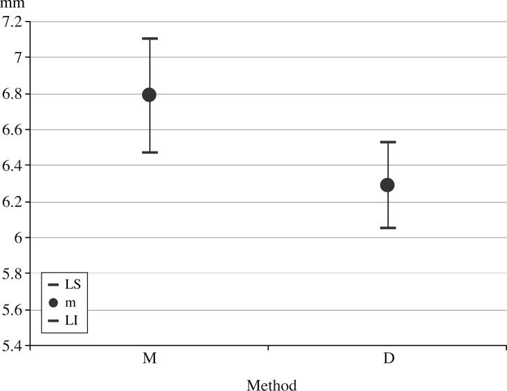
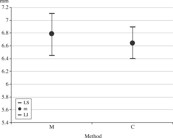
Results: There was a significant difference between depth measurements in the inverted digital images and direct visual measurements (p>|t|=0.0039), with means of 6.29 mm (IC(95%):6.04-6.54) and 6.79 mm (IC(95%):6.45-7.11), respectively. There was a non-significant difference between the film-based radiographs and direct visual measurements (p>|t|=0.4950), with means of 6.64mm(IC(95%):6.40-6.89) and 6.79mm(IC(95%):6.45-7.11), respectively.
Conclusion: The periodontal bone defect measurements in the inverted digital images were inferior to film-based radiographs, underestimating the amount of bone loss.
Keywords: Alveolar Bone Loss; Digital Radiography, Dental; Radiographic Image Enhancement.
Figures
Similar articles
-
Comparison of simulated periodontal bone defect depth measured in digital radiographs in dedicated and non-dedicated software systems.Dentomaxillofac Radiol. 2006 Nov;35(6):422-5. doi: 10.1259/dmfr/61300663. Dentomaxillofac Radiol. 2006. PMID: 17082333
-
Comparison between inverted and unprocessed digitized radiographic imaging in periodontal bone loss measurements.J Appl Oral Sci. 2007 Dec;15(6):492-4. doi: 10.1590/s1678-77572007000600007. J Appl Oral Sci. 2007. PMID: 19089186 Free PMC article.
-
Comparison of two imaging modalities: F-speed film and digital images for detection of osseous defects in patients with interdental vertical bone defects.Dentomaxillofac Radiol. 2007 Dec;36(8):500-5. doi: 10.1259/dmfr/29704550. Dentomaxillofac Radiol. 2007. PMID: 18033948
-
Evaluation of simulated periodontal defects via various radiographic methods.Clin Oral Investig. 2015 Nov;19(8):2053-8. doi: 10.1007/s00784-015-1421-8. Epub 2015 Feb 14. Clin Oral Investig. 2015. PMID: 25677242
-
Computer-aided image manipulation of intraoral radiographs to enhance diagnosis in dental practice: a review.Int Dent J. 1993 Apr;43(2):99-108. Int Dent J. 1993. PMID: 8320010 Review.
Cited by
-
Treatment of periodontal intrabony defects using autologous periodontal ligament stem cells: a randomized clinical trial.Stem Cell Res Ther. 2016 Feb 19;7:33. doi: 10.1186/s13287-016-0288-1. Stem Cell Res Ther. 2016. PMID: 26895633 Free PMC article. Clinical Trial.
-
Comparison of changes in dental and bone radiographic densities in the presence of different soft-tissue simulators using pixel intensity and digital subtraction analyses.Dentomaxillofac Radiol. 2013;42(9):20130235. doi: 10.1259/dmfr.20130235. Epub 2013 Sep 4. Dentomaxillofac Radiol. 2013. PMID: 24005061 Free PMC article.
-
Effect of digital noise reduction on the accuracy of endodontic file length determination.Imaging Sci Dent. 2013 Sep;43(3):185-90. doi: 10.5624/isd.2013.43.3.185. Epub 2013 Sep 23. Imaging Sci Dent. 2013. PMID: 24083212 Free PMC article.
-
Detection of Simulated Periapical Lesion in Intraoral Digital Radiography with Different Brightness and Contrast.Eur Endod J. 2019 Nov 22;4(3):133-138. doi: 10.14744/eej.2019.46036. eCollection 2019. Eur Endod J. 2019. PMID: 32161900 Free PMC article.
-
Diagnostic Accuracy of Inverted and Unprocessed Digitized Periapical Radiographs for Detection of Peri-Implant Defects.J Dent (Tehran). 2015 Aug;12(8):571-6. J Dent (Tehran). 2015. PMID: 27123016 Free PMC article.
References
-
- Scaf G, Sakakura CE, Kalil PF, Dearo de Morais JA, Loffredo LC, Wenzel A. Comparison of simulated periodontal bone defect depth measured in digital radiographs in dedicated and non-dedicated software systems. Dentomaxillofac Radiol. 2006;35:422–425. - PubMed
-
- de Molon RS, Sakakura CE, Morais-Camillo JA, de Almeida Junior PC, de Castro Monteiro Loffredo L, Scaf G. Comparison between embossed digital imaging and unprocessed film-based radiography in detecting periodontal bone defects: an in vitro study. Oral Radiol. 2012;28:95–100.
-
- Borg E, Gröndahl K, Gröndahl HG. Marginal bone level buccal to mandibular molars in digital radiographs from charge-coupled device and storage phosphor systems. An in vitro study. J Clin Periodontol. 1997;24:306–312. - PubMed
