. 2013 May 1;30(9):702-15.
doi: 10.1089/neu.2012.2630. Epub 2013 May 7.

Behavioral and histopathological alterations resulting from mild fluid percussion injury

Affiliations

Behavioral and histopathological alterations resulting from mild fluid percussion injury

Michael J Hylin et al. J Neurotrauma. .

Abstract

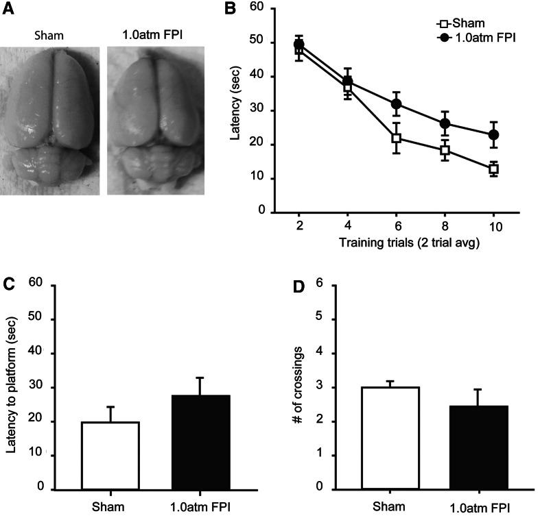
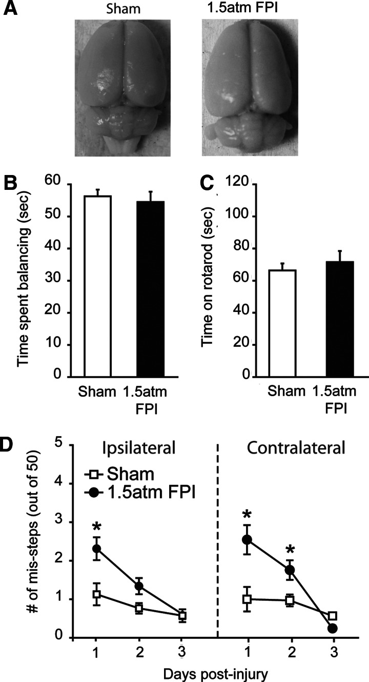
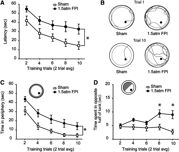
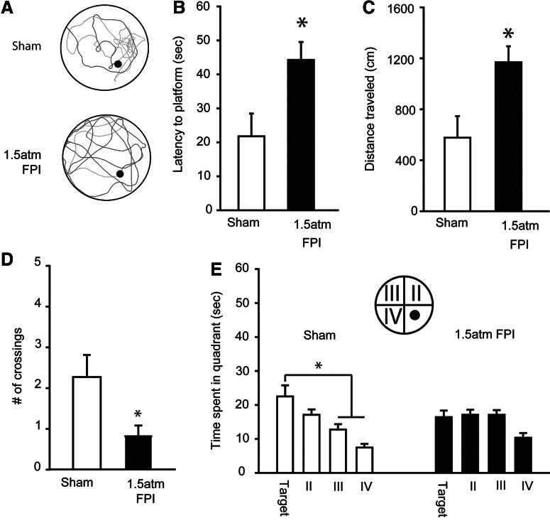
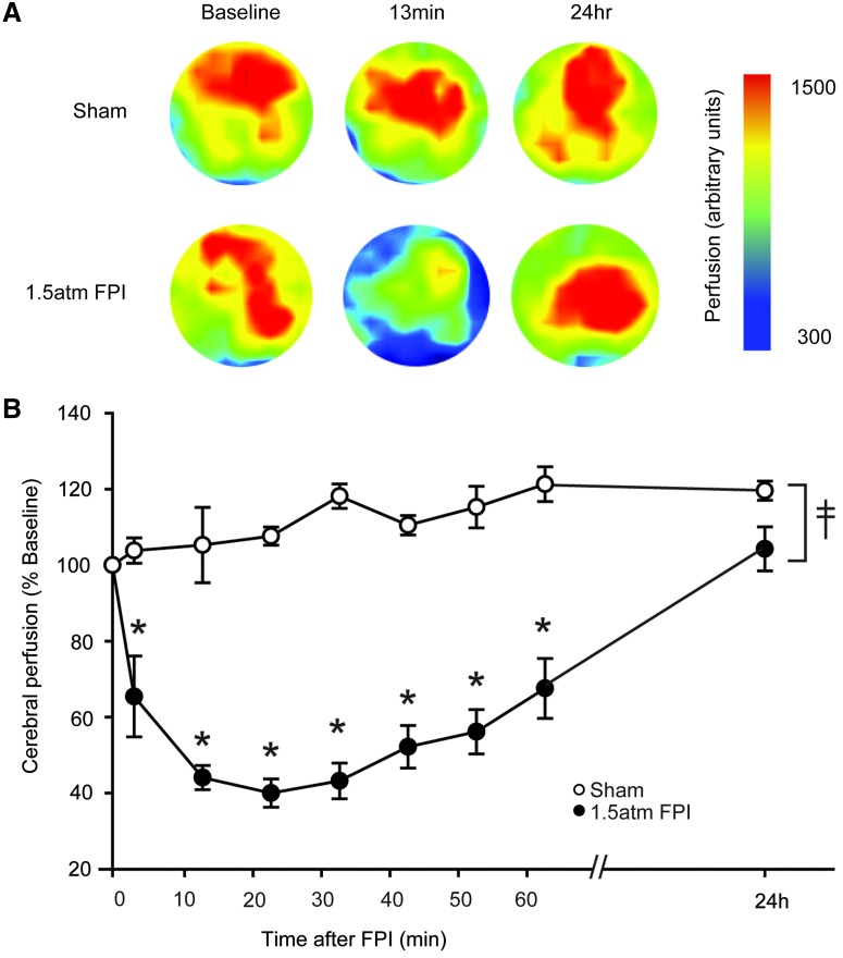
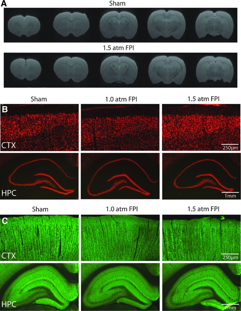
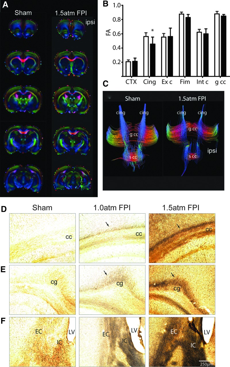
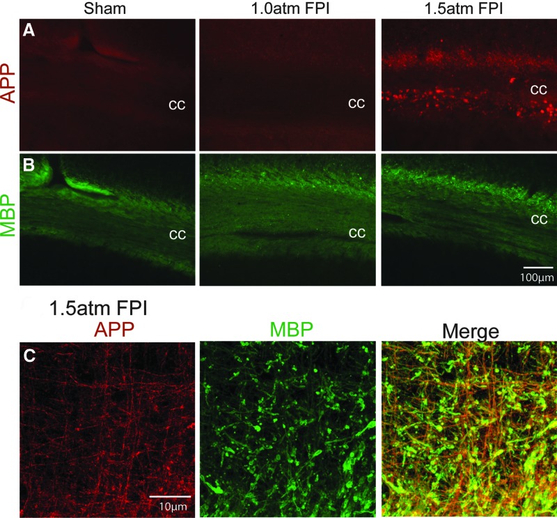
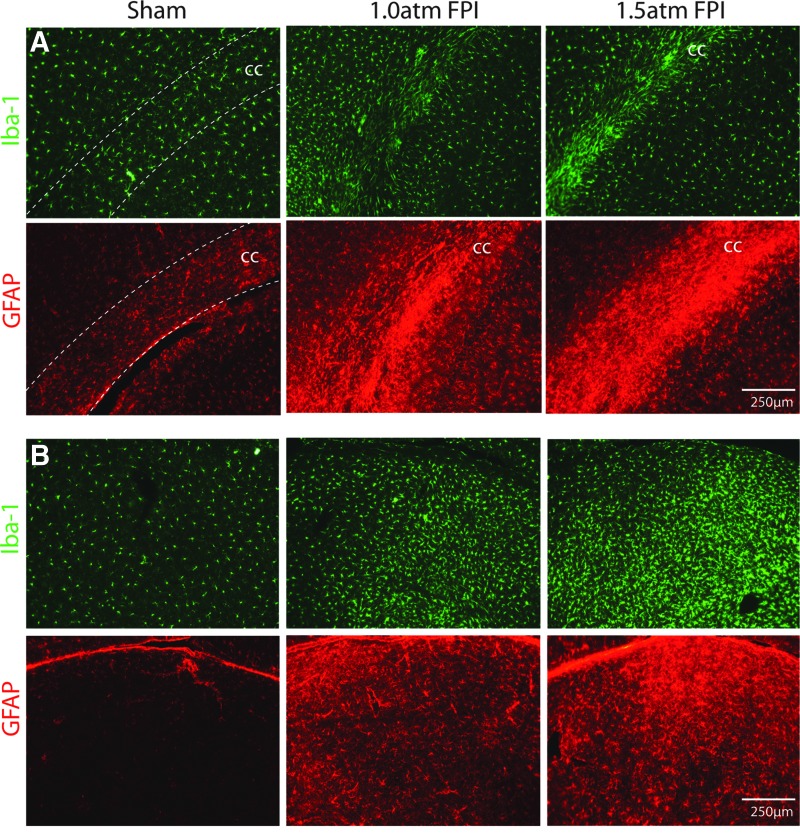
The majority of people who sustain a traumatic brain injury (TBI) have an injury that can be classified as mild (often referred to as concussion). Although head CT scans for most subjects who have sustained a mild TBI (mTBI) are negative, these persons may still suffer from neurocognitive and neurobehavioral deficits. In order to expedite pre-clinical research and develop therapies, there is a need for well-characterized animal models of mTBI that reflect the neurological, neurocognitive, and pathological changes seen in human patients. In the present study, we examined the motor, cognitive, and histopathological changes resulting from 1.0 and 1.5 atmosphere (atm) overpressure fluid percussion injury (FPI). Both 1.0 and 1.5 atm FPI injury caused transient suppression of acute neurological functions, but did not result in visible brain contusion. Animals injured with 1.0 atm FPI did not show significant motor, vestibulomotor, or learning and memory deficits. In contrast, 1.5 atm injury caused transient motor disturbances, and resulted in a significant impairment of spatial learning and short-term memory. In addition, 1.5 atm FPI caused a marked reduction in cerebral perfusion at the site of injury that lasted for several hours. Consistent with previous studies, 1.5 atm FPI did not cause visible neuronal loss in the hippocampus or in the neocortex. However, a robust inflammatory response (as indicated by enhanced GFAP and Iba1 immunoreactivity) in the corpus callosum and the thalamus was observed. Examination of fractional anisotropy color maps after diffusion tensor imaging (DTI) revealed a significant decrease of FA values in the cingulum, an area found to have increased silver impregnation, suggesting axonal injury. Increased silver impregnation was also observed in the corpus callosum, and internal and external capsules. These findings are consistent with the deficits and pathologies associated with mild TBI in humans, and support the use of mild FPI as a model to evaluate putative therapeutic options.


Figures

FIG. 1.
FIG. 1.
FPI of 1.0 atm did not cause learning or memory dysfunction when tested 5 days post-injury. (A) Representative pictures of brains from a sham and a 1.0 atm FPI injured animal. (B) Training curves in the abbreviated Morris water maze task for sham-operated controls (n=17) and 1.0 atm FPI (n=20) animals. During memory testing 30 min after training, 1.0 atm FPI rats were not significantly different than controls in their (C) latency to first platform crossing or in (D) the number of platform crossings during the 60 sec probe trial. Data are presented as the mean±SEM.
FIG. 2.
FIG. 2.
FPI of 1.5 atm caused transient motor coordination deficits. (A) Representative pictures of brains from a sham and a 1.5 atm FPI injured animal. When tested on day 1 post-injury, 1.5 atm FPI animals (n=11) were not significantly different than shams (n=11) in their ability to perform the (B) beam balance, or (C) rotarod tasks. (D) In contrast, injured animals displayed transient ipsilateral and contralateral paw placement deficits that recovered by day 3 post-injury. Data are presented as the mean±SEM; *p<0.05.
FIG. 3.
FIG. 3.
FPI of 1.5 atm caused significant spatial learning deficits. (A) When trained in the abbreviated version of the Morris water maze task on day 5 post-injury, 1.5 atm FPI rats were significantly impaired in their ability to learn the location of the hidden platform compared to sham-operated controls. (B) Representative traces of the swimming paths taken by a sham and a 1.5 atm FPI rat at the beginning and end of training. Poor performance in the water maze task was in part due to injured animals spending more time searching in (C) the perimeter and (D) opposite half of the tank for a means of escape. Data are presented as the mean±SEM; *p=0.05.
FIG. 4.
FIG. 4.
FPI of 1.5 atm caused significant short-term memory deficits. (A) Representative traces of swimming paths taken by a sham and a 1.5 atm FPI rat in a probe trial given 30 min after training. Injured rats performed poorly in the short-term memory tasks as evidence by (B) longer latencies to first platform crossing, (C) longer path lengths to first platform crossing, (D) fewer number of platform crossings during the 60 sec probe trial, and (E) a lack of preference for searching in the quadrant in which the platform was located. Data are presented as the mean±SEM; *p<0.05.
FIG. 5.
FIG. 5.
FPI of 1.5 atm caused transient disturbances in cerebral perfusion. Cerebral perfusion at the site of injury was monitored using a Perimed PIM3 Laser Doppler Blood Perfusion Imager. (A) Representative colorized images indicating cerebral perfusion over time from a sham and a 1.5 atm FPI rat. (B) Injury (n=6) caused a significant decrease in cerebral perfusion, as indicated by lower perfusion values than detected in shams (n=4), throughout the 1 h contiguous monitoring period. Cerebral perfusion returned to baseline 24 h after injury. Color image is available online at www.liebertpub.com/neu
FIG. 6.
FIG. 6.
FPI of 1.5 atm did not cause overt cell loss or dendritic damage. (A) Representative RARE images in the coronal plane from a sham and a 1.5 atm FPI rat. Representative photomicrographs of tissue sections collected 15 days post-injury from a sham, a 1.0 atm FPI, and a 1.5 atm FPI rat showing (B) NeuN and (C) microtubule-associated protein 2 (MAP-2) immunoreactivity from the ipsilateral cortex (CTX) and hippocampus (HPC). Color image is available online at www.liebertpub.com/neu
FIG. 7.
FIG. 7.
Mild FPI increased markers of axonal damage. (A) Representative colorized FA maps in the coronal plane from a sham and a 1.5 atm FPI rat. (B) Summary data showing the quantification of FA values from defined ROIs corresponding to the cortex (CTX) immediately beneath the injury site, the cingulum (Cing), the external capsule (Ext c), the fimbria (Fim), the internal capsule (Int c), and the genu of the corpus callosum (g cc) from sham and 1.5 atm rats (n=6/group). (C) Representative images of tract tracings from a sham and a 1.5 atm rat, demonstrating the fibers passing through the area of the cingulum. Representative photomicrographs of silver-stained tissue sections collected 15 days post-injury from a sham, a 1.0 atm FPI, and a 1.5 atm FPI rat. 1.0 atm FPI, and to a greater degree 1.5 atm FPI, caused enhanced silver impregnation in fibers of (D) the cc, (E) the cingulum (cg), and (F) the internal (IC) and external (EC) capsule. LV, lateral ventricle. Color image is available online at www.liebertpub.com/neu
FIG. 8.
FIG. 8.
Mild FPI enhanced APP immunoreactivity. (A) 1.5 atm FPI, but not 1.0 atm FPI, increased amyloid precursor protein (APP) immunoreactivity in the ipsilateral corpus callosum (cc), but did not appear to (B) reduce myelin basic protein (MBP) immunostaining. (C) Representative confocal images showing that the enhanced APP immunoreactivity (red) observed in 1.5 atm FPI rats co-localizes with MBP (green)-positive myelinated fibers. Areas of co-localization appear yellow in the merged image. Color image is available online at www.liebertpub.com/neu
FIG. 9.
FIG. 9.
FPI of 1.5 atm increased Iba-1 and GFAP immunoreactivity. Representative photomicrographs of tissue sections taken 15 days after sham surgery, 1.0 atm FPI or 1.5 atm FPI are shown. (A) Images of the ipsilateral corpus callosum (cc) showing that 1.5 atm FPI increased Iba-1 (a marker of microglia) and GFAP (a marker of astrocytes) immunoreactivity, suggesting ongoing microglial and astrocytic activation, respectively. (B) Similar increases in Iba-1 and GFAP immunoreactivity were observed in the thalamus, suggesting a prolonged period of neuroinflammation. Color image is available online at www.liebertpub.com/neu

References

    1. Alexander MP. Mild traumatic brain injury: Pathophysiology, natural history, and clinical management. Neurology. 1995;45:1253–1260. - PubMed
    1. Binder LM. A review of mild head trauma. Part II: Clinical implications. J Clin Exp Neuropsychol. 1997;19:432–457. - PubMed
    1. d'Hemecourt P. Subacute symptoms of sports-related concussion: Outpatient management and return to play. Clin Sports Med. 2011;30:63–72. - PubMed
    1. Ruff RM. Camenzuli L. Mueller J. Miserable minority: Emotional risk factors that influence the outcome of a mild traumatic brain injury. Brain Inj. 1996;10:551–565. - PubMed
    1. Mac Donald CL. Johnson AM. Cooper D, et al. Detection of blast-related traumatic brain injury in U.S. military personnel. N Engl J Med. 2011;364:2091–2100. - PMC - PubMed

Publication types