. 2013 Mar 15;73(6):1892-9.
doi: 10.1158/0008-5472.CAN-12-2609. Epub 2013 Jan 9.

Focused ultrasound delivers targeted immune cells to metastatic brain tumors

Affiliations

Focused ultrasound delivers targeted immune cells to metastatic brain tumors

Ryan Alkins et al. Cancer Res. .

Abstract

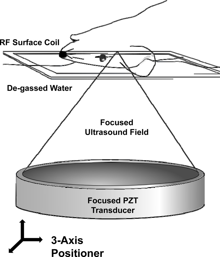
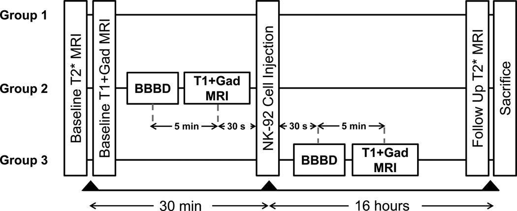
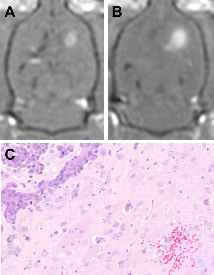
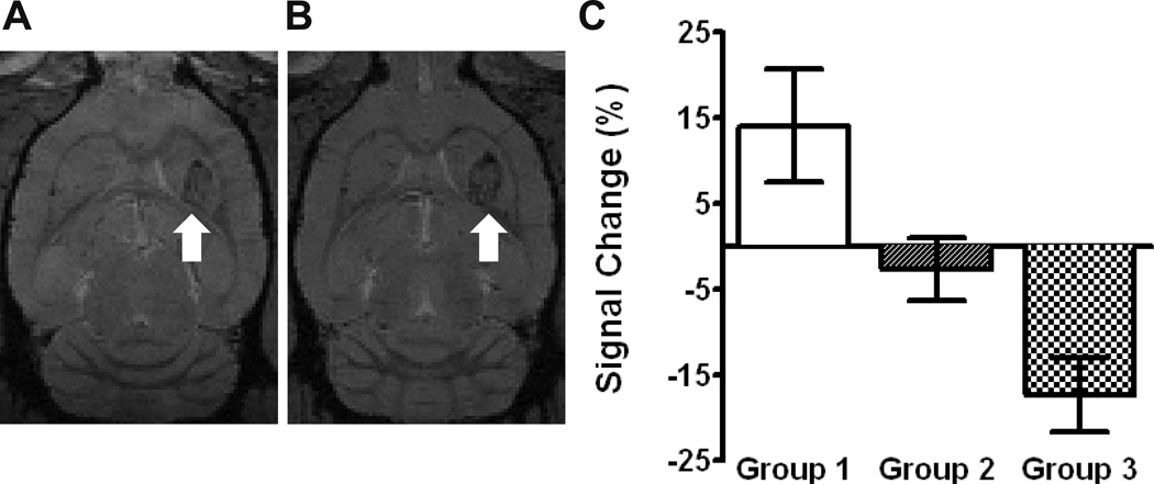
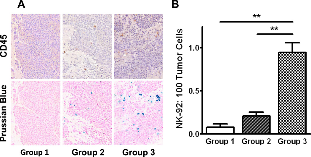
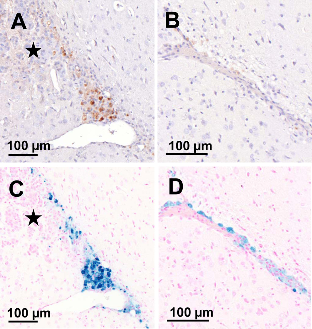
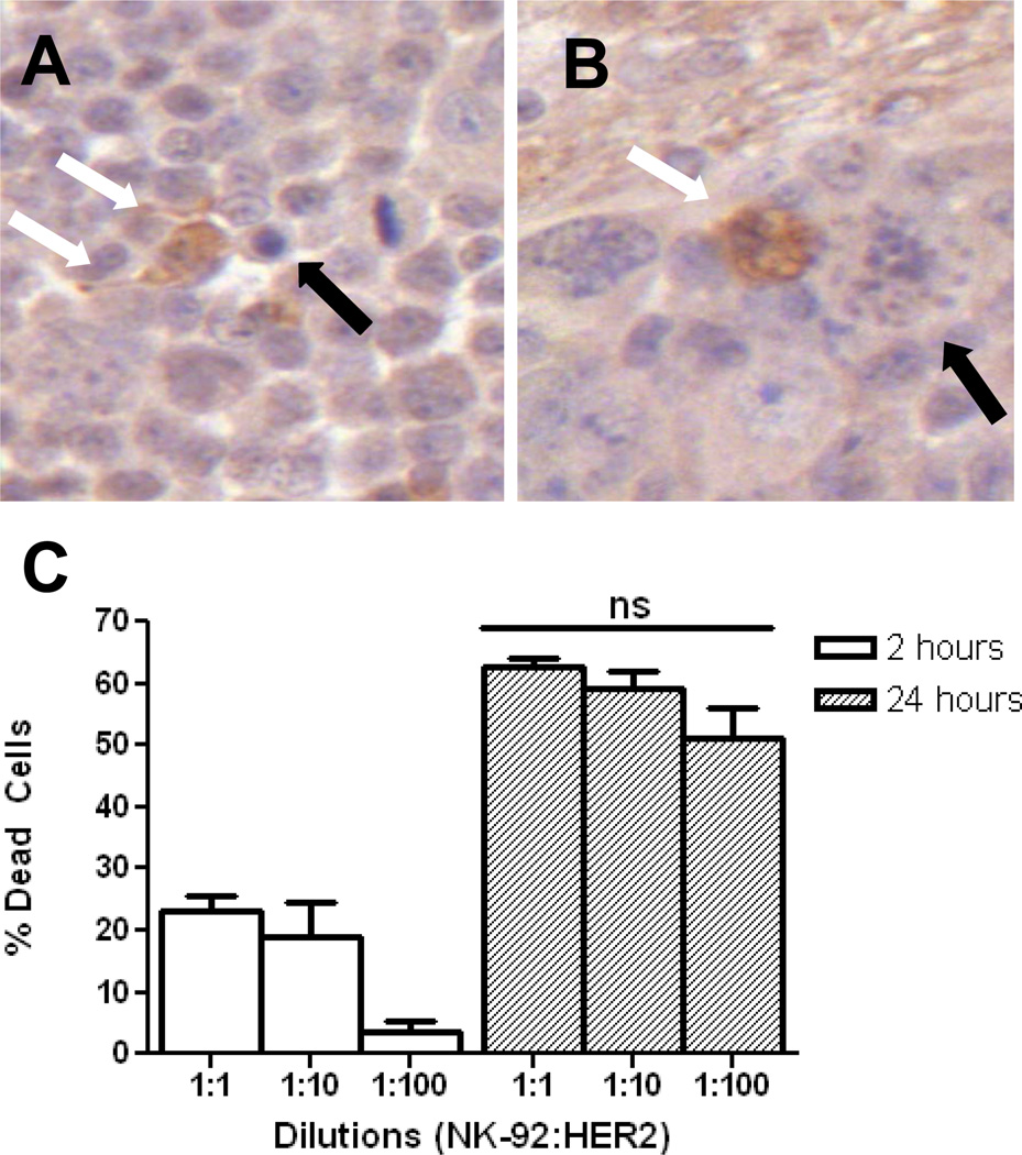
Natural killer (NK) cells are cytotoxic lymphocytes involved in innate immunity. NK-92, a human NK cell line, may be targeted to tumor-associated antigens in solid malignancies where it exhibits antitumor efficacy, but its clinical utility for treating brain tumors is limited by an inability to cross the blood-brain barrier (BBB). We investigated the potential for focused ultrasound (FUS) to deliver targeted NK-92 cells to the brain using a model of metastatic breast cancer. HER2-expressing human breast tumor cells were implanted into the brain of nude rats. The NK-92-scFv(FRP5)-zeta cell line expressing a chimeric HER2 antigen receptor was transfected with superparamagnetic iron oxide nanoparticles before intravenous injection, before and following BBB disruption using focused ultrasound (551.5 kHz focused transducer, 0.33 MPa average peak rarefaction pressure) in the presence of a microbubble contrast agent. Baseline and posttreatment 1.5T and 7T MR imaging was done, and histology used to identify NK-92 cells post-mortem. Contrast-enhanced MRI showed reproducible and consistent BBB disruption. 7T MR images obtained at 16 hours posttreatment revealed a significant reduction in signal indicating the presence of iron-loaded NK-92 cells at the tumor site. The average ratio of NK-92 to tumor cells was 1:100 when NK cells were present in the vasculature at the time of sonication, versus 2:1,000 and 1:1,000 when delivered after sonication and without BBB disruption, respectively. Our results offer a preclinical proof-of-concept that FUS can improve the targeting of immune cell therapy of brain metastases.


Conflict of interest statement

The authors disclose no potential conflicts of interest.

Figures

Figure 1
Figure 1
The experimental MRI-guided FUS setup. The anesthetized animal was positioned supine over an MRI RF surface coil and coupled to the piezo-ceramic focused transducer through a bath of de-gassed water. The transducer was repositioned with the aid of a computer-controlled three-axis positioning system, allowing the ultrasound focus to reach any point within the brain. The entire setup fits in the bore of the 1.5T MR scanner, allowing for coordinate co-registration, targeting and confirmation of BBBD.
Figure 2
Figure 2
Experimental timeline for the three groups. All groups initially underwent baseline imaging to assess the tumour size and location. In group 1 (control) this was followed by injection of SPIO nanoparticle labeled HER2-specific NK-92 cells. Follow-up imaging was performed at 16 hours following cell injection and immediately prior to sacrifice. Group 2 underwent BBBD, followed 5 minutes later by T1-weighted MRI with contrast to assess the change in contrast extravasation. Cells were then injected approximately 30 s after the completion of the imaging. In group 3, cells were injected via the tail vein and BBBD initiated 30 s after the injection. T1-weighted MRI was performed 5 minutes post-BBBD again to assess the change in contrast extravasation. Both groups 2 and 3 were imaged at 16 hours following the cell injection and immediately euthanized.
Figure 3
Figure 3
The results of blood-brain barrier disruption with FUS. Representative T1-weighted MR images with Omniscan contrast of the tumor before and after BBBD, A and B respectively. The average enhancement of untreated tumors was 17 ± 8 % but increased to 34 ± 10 % following exposure to ultrasound and microbubbles (mean ± SD, p<0.05). A small region of erythrocyte extravasation was seen in one of the treatment animals, C. There was no further evidence of tissue injury.
Figure 4
Figure 4
HER2-specific NK-92 cell detection with 7T MRI. A baseline axial T2*-weighted MR image from group 3 is shown with the tumor identified in the left frontal striatum (white arrow), A. The corresponding post-treatment image demonstrates a signal reduction at the tumor site (white arrow), B. C shows the average signal intensity change (mean ± SEM) at the tumor site following treatment. A negative change suggests the accumulation of iron-labelled effector cells. There was a statistically significant difference between group 3 and group 1.
Figure 5
Figure 5
Histological quantification of HER2-specific NK-92 cells accumulating at the tumor site. Effector cells were co-localized with CD45 IHC (upper panel) and Prussian blue histochemistry (lower panel) in the three experimental groups, A. HER2-specific NK-92 cells reaching the tumor were quantitatively assessed (mean ± SEM), B. When NK-92 cells were injected prior to BBBD, the number reaching the tumor was significantly higher than if they were injected following or without BBBD (group 3 vs groups 1 and 2: 0.95 ± 0.23 vs 0.09 ± 0.11, 0.21 ± 0.15, p < 0.01). There was no statistical difference between groups 1 and 2. These results are in agreement with the iron-sensitive MR imaging in Figure 4.
Figure 6
Figure 6
FUS causes the translocation of HER2-specific NK-92 cells from the vasculature into the brain and tumor when they are present in the circulation at the time of BBBD. A, CD45 IHC depicting a vessel from which a large number of cells have extravasated and appear to track to the tumor (indicated by the star). B, the corresponding Prussian blue stained section is shown, colocalizing the HER2-specific NK-92 cells. C, a normal capillary adjacent the tumor but within the sonicated region, shows HER2-specific NK-92 cells forced to the adluminal surface of the vessel. FUS results in HER2-specific NK-92 cells circumventing both the BBB and BTB. D, the corresponding Prussian blue section. These cell distributions were seen exclusively in group 3 animals.
Figure 7
Figure 7
HER2-specific NK-92 cells accumulate at the tumor and have preserved function. A, IHC for granzyme B highlights an NK-92 cell releasing granzyme B into the surrounding extra-cellular space (white arrows). An adjacent apoptotic tumor cell can be seen (black arrow). B, granzyme B-containing NK-92 cell (white arrow) causing apoptosis in a tumor cell (black arrow). At 24 hrs, a 1:100 ratio of effector:tumor cells is statistically no different (p > 0.05) in causing tumor cell lysis than higher starting ratios, C. This is the ratio of effector-to-tumor cells that was achieved in vivo in group 3.

Similar articles

Cited by

References

    1. Lee Y-T. Breast carcinoma: Pattern of metastasis at autopsy. J Surg Oncol. 1983;23:175–180. - PubMed
    1. Lin NU, Bellon JR, Winer EP. CNS Metastases in Breast Cancer. J Clin Oncol. 2004;22:3608–3617. - PubMed
    1. Abbott NJ, Rönnbäck L, Hansson E. Astrocyte-endothelial interactions at the blood-brain barrier. Nat Rev Neurosci. 2006;7:41–53. - PubMed
    1. Lockman PR, Mittapalli RK, Taskar KS, Rudraraju V, Gril B, Bohn KA, et al. Heterogeneous blood–tumor barrier permeability determines drug efficacy in experimental brain metastases of breast cancer. Clin Cancer Res. 2010;16:5664–5678. - PMC - PubMed
    1. Linn SC, Giaccone G, van Diest PJ, Blokhuis WM, van Der Valk P, van Kalken CK, et al. Prognostic relevance of p-glycoprotein expression in breast cancer. Ann Oncol. 1995;6:679–685. - PubMed

Publication types