. 2013 Jan 22;110(4):1446-51.
doi: 10.1073/pnas.1214608110. Epub 2013 Jan 9.

Cardiomyocyte proliferation contributes to heart growth in young humans

Affiliations

Cardiomyocyte proliferation contributes to heart growth in young humans

Mariya Mollova et al. Proc Natl Acad Sci U S A. .

Abstract

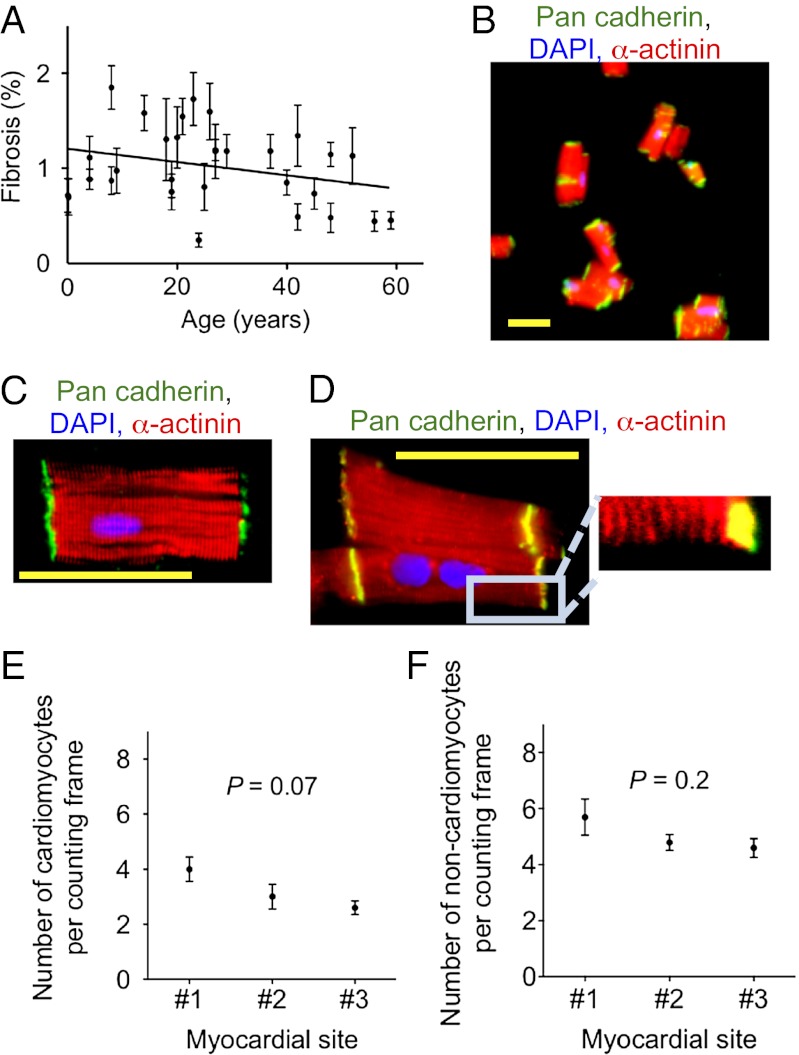
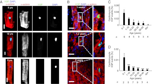
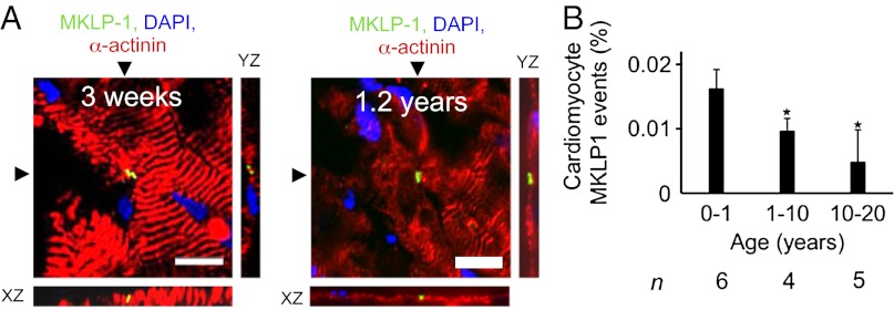
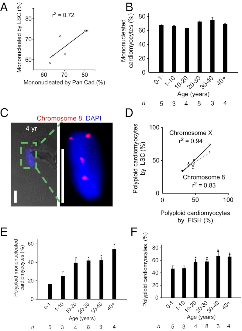
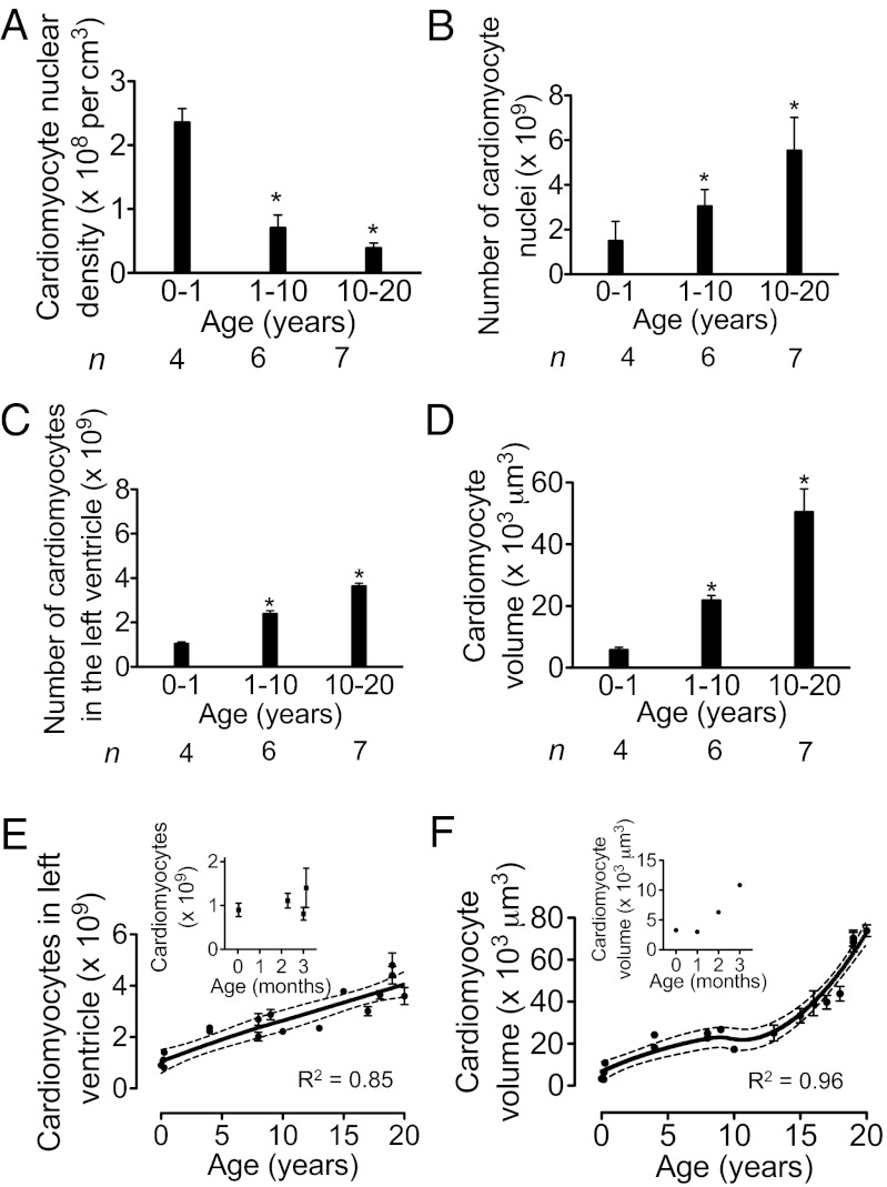
The human heart is believed to grow by enlargement but not proliferation of cardiomyocytes (heart muscle cells) during postnatal development. However, recent studies have shown that cardiomyocyte proliferation is a mechanism of cardiac growth and regeneration in animals. Combined with evidence for cardiomyocyte turnover in adult humans, this suggests that cardiomyocyte proliferation may play an unrecognized role during the period of developmental heart growth between birth and adolescence. We tested this hypothesis by examining the cellular growth mechanisms of the left ventricle on a set of healthy hearts from humans aged 0-59 y (n = 36). The percentages of cardiomyocytes in mitosis and cytokinesis were highest in infants, decreasing to low levels by 20 y. Although cardiomyocyte mitosis was detectable throughout life, cardiomyocyte cytokinesis was not evident after 20 y. Between the first year and 20 y of life, the number of cardiomyocytes in the left ventricle increased 3.4-fold, which was consistent with our predictions based on measured cardiomyocyte cell cycle activity. Our findings show that cardiomyocyte proliferation contributes to developmental heart growth in young humans. This suggests that children and adolescents may be able to regenerate myocardium, that abnormal cardiomyocyte proliferation may be involved in myocardial diseases that affect this population, and that these diseases might be treatable through stimulation of cardiomyocyte proliferation.


Conflict of interest statement

The authors declare no conflict of interest.

Figures

Fig. 1.
Fig. 1.
Sampling and isolation methods yield representative probes of healthy human hearts. (A) Analysis of AFOG-stained tissue sections reveals no increased fibrosis; data fitted with linear regression, slope: –0.007 ± 0.002, P = 0.007. (B) Isolation from a 9-y-old donor heart using the fixation-digestion method yields intact cardiomyocytes. (C) Staining of desmosomes with an antibody against pan-cadherin shows intact cardiomyocytes. (D) α-actinin shows intact sarcomeres. (E) Optical dissector method quantifying cardiomyocyte and (F) noncardiomyocyte nuclei on three random myocardial sections from different sites of the same LV shows no significant differences in the nuclear density in different compartments of the same LV. Statistical significance was tested with ANOVA. The results are mean ± SD. Scale bar: 50 μm (B–D).
Fig. 2.
Fig. 2.
Human hearts show evidence for cardiomyocyte cycling after birth. (A) Isolated cardiomyocytes in M-phase were visualized by immunofluorescent microscopy using antibodies against phosphorylated histone H3 (H3P, green) and α-actinin (red). H3P-negative cardiomyocyte is shown as a control (Lower). (B) H3P-positive cardiomyocytes on tissue sections. (C and D) Quantification of isolated cardiomyocytes by LSC (C) and on tissue sections (D) shows that the percentage of cycling cardiomyocytes decreases with age. Columns represent mean values of the number of hearts investigated per age group (n) that are indicated under each column. One-way ANOVA revealed a significant difference between 0–1 y and the other age groups. *P < 0.05, ***P < 0.001. Scale bar: 25 µm.
Fig. 3.
Fig. 3.
Humans show evidence for cardiomyocyte cytokinesis in the first two decades of life. (A) Cardiomyocyte cytokinesis was detected with an antibody against MKLP-1; XY and XZ reconstruction planes are indicated with arrowheads. (B) Frequency of MKLP-1–positive cardiomyocyte-specific events declines over first two decades of life. Scale bar: 25 µm. *P < 0.05 compared with 0–1 y (ANOVA); n, number of hearts analyzed. Cardiomyocyte-specific MKLP-1 activity was not found in samples >20 y of age (n = 9). For 3D reconstructions of the photomicrographs in A, see Movies S3, S4, S5, and S6.
Fig. 4.
Fig. 4.
Human cardiomyocytes show increased formation of polyploid nuclei between birth and 20 y. (A) Validation of LSC method for quantification of mononucleated cardiomyocytes. (B) In humans, the percentage of mononucleated cardiomyocytes remains unchanged after birth. (C) Tetraploid cardiomyocyte nucleus (DAPI, blue) with FISH probe against chromosome 8. Scale bar: 25 μm. (D) Comparison of quantification of nuclear DNA content using FISH and LSC shows significant correlation. (E) The percentage of polyploid mononucleated cardiomyocytes increases with age. (F) Percentage of cardiomyocytes with DNA content >2N determined by LSC increases with age. Statistical difference between 0–1 y and other age groups was tested with one-way ANOVA: *P < 0.05. n, number of hearts analyzed.
Fig. 5.
Fig. 5.
Human cardiomyocytes proliferate and enlarge after birth. (A) Cardiomyocyte nuclear density, determined by the optical dissector method, decreases with age. (B) Number of cardiomyocyte nuclei per LV increases with age. (C) Number of cardiomyocytes per LV, calculated from number of cardiomyocyte nuclei (B) and percentages of mono-, bi-, and multinucleated cardiomyocytes. (D) Mean volume of cardiomyocytes increases with age. (E and F) The number (E) and mean volume (F) of cardiomyocytes from individual LVs were graphed over age and modeled using locally weighted scatter plot smoothing (LOWESS). Blow-up graphs of results from the first 3.5 mo of life are shown. Dotted lines indicate 95% confidence intervals. R2 values are indicated. *P < 0.05. n, number of hearts analyzed.

References

    1. Roger VL, et al. American Heart Association Statistics Committee and Stroke Statistics Subcommittee Heart disease and stroke statistics—2012 update: A report from the American Heart Association. Circulation. 2012;125(1):e2–e220. - PMC - PubMed
    1. Linzbach AJ. Heart failure from the point of view of quantitative anatomy. Am J Cardiol. 1960;5:370–382. - PubMed
    1. Laflamme MA, Murry CE. Heart regeneration. Nature. 2011;473(7347):326–335. - PMC - PubMed
    1. Vela D, Buja LM. Quest for the cardiovascular holy grail: Mammalian myocardial regeneration. Cardiovasc Pathol. 2008;17(1):1–5. - PubMed
    1. Bolli R, et al. Cardiac stem cells in patients with ischaemic cardiomyopathy (SCIPIO): Initial results of a randomised phase 1 trial. Lancet. 2011;378(9806):1847–1857. - PMC - PubMed

Publication types

MeSH terms