. 2013 Jan 22;110(4):1440-5.
doi: 10.1073/pnas.1211179110. Epub 2013 Jan 9.

LRP-6 is a coreceptor for multiple fibrogenic signaling pathways in pericytes and myofibroblasts that are inhibited by DKK-1

Affiliations

LRP-6 is a coreceptor for multiple fibrogenic signaling pathways in pericytes and myofibroblasts that are inhibited by DKK-1

Shuyu Ren et al. Proc Natl Acad Sci U S A. .

Erratum in

Abstract

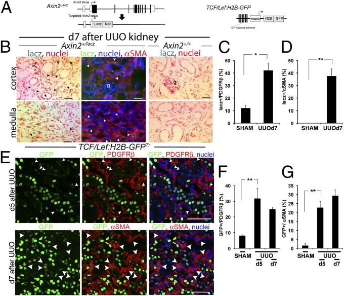
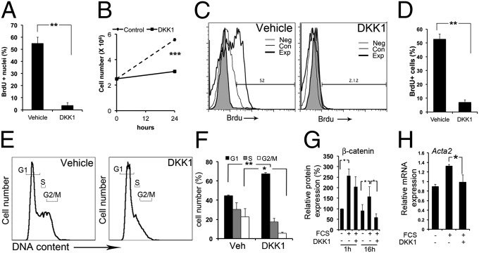
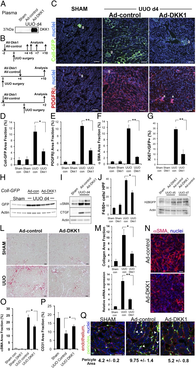
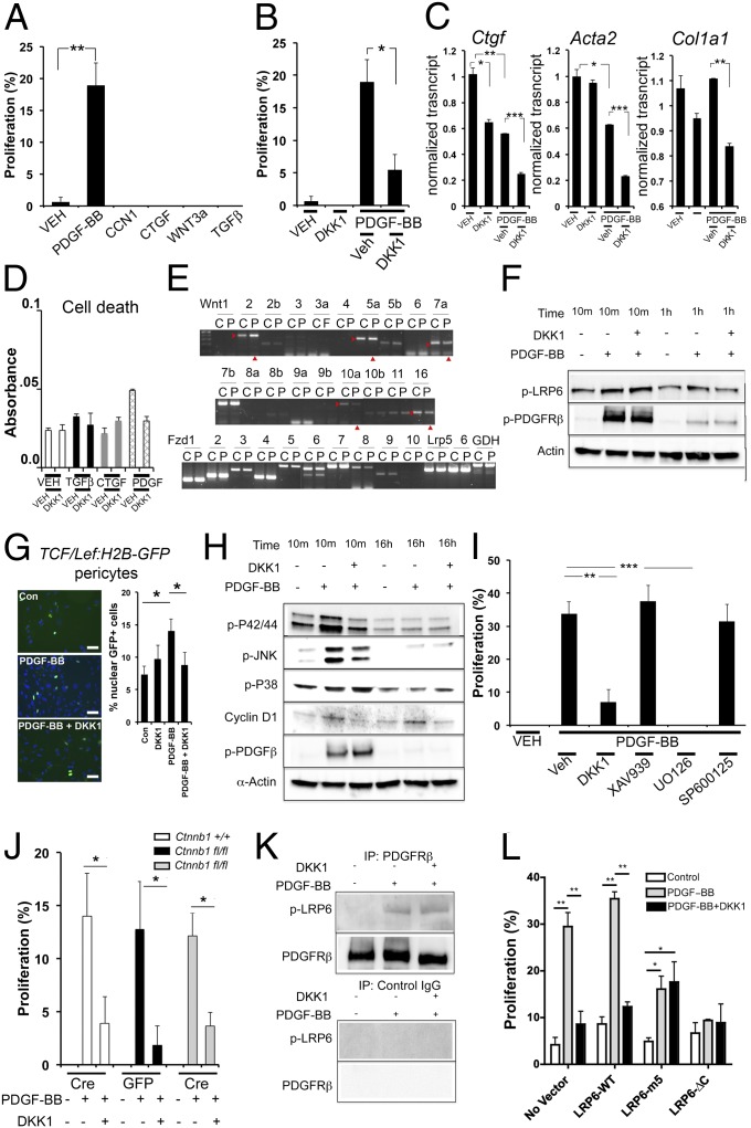
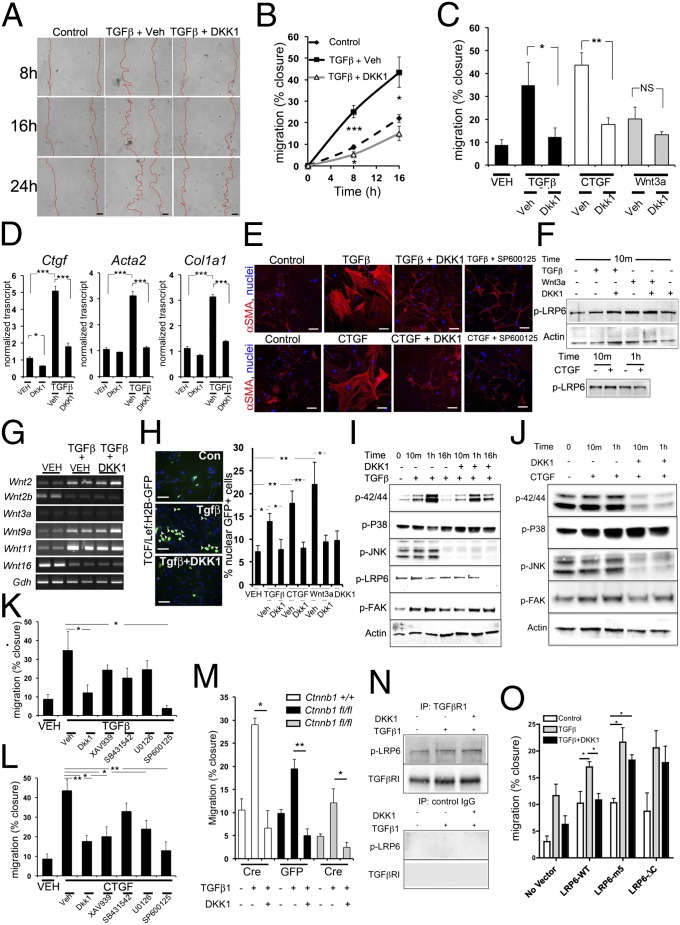
Fibrosis of vital organs is a major public health problem with limited therapeutic options. Mesenchymal cells including microvascular mural cells (pericytes) are major progenitors of scar-forming myofibroblasts in kidney and other organs. Here we show pericytes in healthy kidneys have active WNT/β-catenin signaling responses that are markedly up-regulated following kidney injury. Dickkopf-related protein 1 (DKK-1), a ligand for the WNT coreceptors low-density lipoprotein receptor-related proteins 5 and 6 (LRP-5 and LRP-6) and an inhibitor of WNT/β-catenin signaling, effectively inhibits pericyte activation, detachment, and transition to myofibroblasts in vivo in response to kidney injury, resulting in attenuated fibrogenesis, capillary rarefaction, and inflammation. DKK-1 blocks activation and proliferation of established myofibroblasts in vitro and blocks pericyte proliferation to PDGF, pericyte migration, gene activation, and cytoskeletal reorganization to TGF-β or connective tissue growth factor. These effects are largely independent of inhibition of downstream β-catenin signaling. DKK-1 acts predominantly by inhibiting PDGF-, TGF-β-, and connective tissue growth factor-activated MAPK and JNK signaling cascades, acting via LRP-6 with associated WNT ligand. Biochemically, LRP-6 interacts closely with PDGF receptor β and TGF-β receptor 1 at the cell membrane, suggesting that it may have roles in pathways other than WNT/β-catenin. In summary, DKK-1 blocks many of the changes in pericytes required for myofibroblast transition and attenuates established myofibroblast proliferation/activation by mechanisms dependent on LRP-6 and WNT ligands but not the downstream β-catenin pathway.


Conflict of interest statement

Conflict of interest statement: S.R. and J.S.D have submitted patent applications for the use of DKK-1 in fibrosis.

Figures

Fig. 1.
Fig. 1.
WNT/β-catenin signaling is activated during kidney injury in the pericyte/myofibroblast cell compartment. (A) Schema showing the Axin2LacZ allele and the TCF/Lef:H2B-GFP transgene, which report WNT/β-catenin signaling. (B–D) WNT responses identified by blue stain or green fluorescence in cells (B) and quantified in graphs (C and D) during kidney injury induced by UUO in Axin2+/lacz reporter mice are seen predominantly in myofibroblasts (arrowheads), but no stain is seen in Axin2+/+ kidneys. (E–G) WNT/β-catenin responses identified by nuclear GFP in confocal images (E) of kidneys from TCF/Lef:H2B-GFPTr reporter mice after UUO, highlighting PDGFRβ+ cells or αSMA cells, two markers for myofibroblasts in diseased kidney, and quantified (F and G) by PDGFRβ- or αSMA-staining cells expressing nuclear GFP. *P < 0.05, **P < 0.01. n = 4 per group. Error bars indicate SEM.
Fig. 2.
Fig. 2.
DKK-1 binds to myofibroblasts and blocks proliferation by G1/S cell-cycle arrest in vitro. (A) Graph showing BrdU nuclear incorporation in quiescent myofibroblasts stimulated for 3 h with 3% FCS and DKK-1 or vehicle. (B) Coulter-counted kidney quiescent myofibroblasts stimulated for 24 h with 3% FCS and DKK-1 or vehicle. (C and D) Flow cytometric plots (C) and graph (D) showing BrdU uptake in myofibroblasts stimulated for 3 h with 3% FCS and DKK-1 or vehicle. (E and F) Propidium iodide DNA content plots (E) and graph (F) showing quiescent myofibroblasts stimulated for 24 h with 3% FCS and DKK-1 or vehicle. (G) The effect of DKK-1 on cytoplasmic and nuclear β-catenin protein. Serum increases β-catenin, an effect not modulated by DKK-1 at 1 h, but DKK-1 markedly reduces β-catenin levels at later time points. (H) qPCR data showing myofibroblast expression of Acta2 after treatment with FCS or FCS + DKK-1. *P < 0.05, **P < 0.01, ***P < 0.001. n = 4 per group. Error bars indicate SEM.
Fig. 3.
Fig. 3.
DKK-1 blocks pericyte activation and transition to myofibroblasts and reverses myofibroblast activation in vivo, inhibiting fibrogenesis, capillary rarefaction, and inflammation. (A) Western blots of 5 µL of plasma from mice 5 d after i.v. injection of Ad-control or Ad-DKK-1 and from mice subjected to sham surgery and injected with control. (B) Experimental schemata for adenoviral administration, kidney injury, and analysis in the UUO model. (CM) Prevention studies. (C) Low-magnification confocal images of kidney cortex 4 d after sham operation or UUO in Coll-GFPTr mice that had received Ad-control or Ad-DKK-1 6 d previously, showing Coll-GFP cells or PDGFRβ cells. g, glomerulus; a, arteriole. (DF) Graphs showing quantification of Coll-GFP cells, PDGFRβ cells, and αSMA cells in kidney 4 d after UUO. (G) Proportion of Coll-GFP cells that express the proliferation marker Ki67. (H and I) Western blot of GFP (H) or αSMA/CTGF (I) in whole Coll-GFP mouse kidney 4 d after UUO. (J) Quantification of macrophage numbers in kidney sections detected by F4/80 staining. (K) Western blot quantifying canonical WNT signaling by detecting the H2B-GFP fusion protein after Ad-DKK-1 vs. Ad-control treatment of TCF/Lef:H2B-GFPTr reporter mice during UUO kidney injury. (L) Sirius red-stained kidneys 10 d after UUO. (M) Morphometry of Sirius red-stained collagen (Upper) or qPCR for Col1a1 transcripts (Lower) 10 d after UUO in mice treated with Ad-control vs. Ad-DKK-1. (NP) Reversal studies. Confocal Images (N) and morphometric quantification (O) of αSMA staining 10 d after UUO in mice treated with Ad-control or Ad-DKK-1 from day 4. (P) Quantification of capillary density 10 d after UUO. Note that rarefaction occurs in response to kidney disease, but DKK-1 partially reverses rarefaction. (Q) Pericyte detachment. Images and quantification of pericyte area in Coll-GFP mice 2 d after UUO in the presence of circulating DKK-1 or control. Note that injury to the kidney stimulated pericyte spreading and detachment from endothelium (arrowheads). *P < 0.05, **P < 0.01. n = 4–6 per group. Error bars indicate SEM.
Fig. 4.
Fig. 4.
DKK-1 inhibits PDGF-BB–mediated proliferation of pericytes in vitro by a noncanonical, LRP-6–dependent, P42/44 MAPK-dependent mechanism. (A) Graph of BrdU incorporation into quiescent kidney pericytes 6 h after stimulation with cytokines. (B) The effect of DKK-1 on PDGF-BB–stimulated proliferation. (C) qPCR of genes associated with cell activation in pericytes 48 h after stimulation. (D) Quantification of cell viability in pericytes stimulated with cytokines and DKK-1 for 24 h. (E) RT-PCR results showing the effect of PDGF-BB on WNT ligands and receptors in pericytes 12 h after stimulation. C, control; P, PDGF; arrowheads indicate regulated genes. (F) Western blot time course showing pPDGFRβ and pLRP-6 levels in pericytes. DKK-1 does not affect pLRP-6 at early time points but inhibits pLRP-6 at later time points. (G) Fluorescence images and data quantifying nuclear GFP+ (green) in TCF/Lef:H2B-GFPTr canonical WNT reporter pericytes 16 h after PDGF-BB or PDGF-BB + DKK-1. (H) Western blot time course of phosphorylated forms of P42/P44, JNK, and P38, PDGFRβ, and total cyclinD1 in pericytes activated by PDGF-BB or PDGF-BB + DKK-1. (I) Graph showing the effect of DKK-1 or the canonical WNT inhibitor XAV939, the P42/P44 inhibitor U0126, or the JNK inhibitor SP600125 on PDGF-BB–stimulated BrdU incorporation into quiescent pericytes. (J) Graph showing the effect of PDGF-BB on the proliferation of Ctnnb1fl/fl pericytes that underwent in vitro recombination by expressing Cre recombinase vs. Ctnnb1fl/fl pericytes that expressed control protein GFP. (K) Western blot of pericyte proteins immunoprecipitated by anti-PDGFRβ antibodies or control antibodies detecting pLRP-6 or PDGFRβ. (L) Graph showing the effect of expression of LRP-6 (wild type) or dominant-negative forms of LRP-6, LRP-6 with tyrosine-to-methionine mutations at the five tyrosine sites (5m), or LRP-6 lacking the cytoplasmic tail (ΔC) on 3T3 fibroblast proliferation in response to PDGF-BB and DKK-1. *P < 0.05, **P < 0.01, ***P < 0.01. n = 4–7 per group. All blots are representative of three experiments. (Scale bars, 25 µm.) Error bars indicate SEM.
Fig. 5.
Fig. 5.
DKK-1 blocks TGF-β– and CTGF-mediated migration of pericytes in vitro predominantly by a noncanonical, LRP-6–dependent, JNK-dependent mechanism. (A and B) Images (A) and time course graph (B) showing migration of kidney pericytes induced by TGF-β and blocked by DKK-1. (Scale bars, 50 µm.) (C) Graph of migration at 16 h by pericytes stimulated by TGF-β, CTGF, or WNT3a. All are blocked by DKK-1. (D) qPCR of genes associated with cell activation in pericytes. (E) Fluorescence images of αSMA showing the cytoskeleton of primary pericytes in control or stimulated conditions for 24 h. (Scale bars, 25 µm.) (F) Western blots showing phosphorylated LRP-6 levels in pericytes 10 min after activation with TGF-β or WNT3a in the presence of vehicle or DKK-1 (Upper), and after activation with CTGF (Lower) (G) Thirty-cycle RT-PCR showing the effect of TGF-β or TGF-β + DKK-1 on WNT ligand expression at 8 h. (H) Fluorescence images and data quantifying nuclear GFP+ (green) in TCF/Lef:H2B-GFPTr βcatenin reporter pericytes 16 h after stimulation with cytokines +/− DKK-1. (I) Western blot time course of phosphorylated forms of P42/P44, JNK, P38, LRP-6, and FAK in pericytes activated by TGF-β or TGF-β + DKK-1. (J) Western blots of phosphorylated forms of P42/P44, JNK, P38, and FAK in pericytes activated by CTGF or CTGF + DKK-1. (K and L) Graphs showing the effect of DKK-1, the canonical WNT inhibitor XAV939, the TGFβR1 kinase inhibitor SB431542, the P42/P44 inhibitor U0126, or the JNK inhibitor SP600125 on TGF-β–stimulated (K) or CTGF-stimulated (L) migration in quiescent pericytes. (M) Graph showing the effect of TGF-β on the migration of Ctnnb1fl/fl pericytes expressing Cre recombinase vs. Ctnnb1fl/fl pericytes expressing control protein GFP. (N) Western blots of pericyte proteins immunoprecipitated by anti-TGFβR1 antibodies or control antibodies detecting pLRP-6 or TGFβR1. (O) Graph showing the effect of expression of LRP-6 (wild type) or dominant-negative forms of LRP-6, 5m, or ΔC on 3T3 fibroblast migration in response to TGF-β and DKK-1. *P < 0.05, **P < 0.01, ***P < 0.01. Error bars indicate SEM. Experiments are from n = 4–7 per group. All blots are representative of three experiments.

References

    1. Lupher ML, Jr, Gallatin WM. Regulation of fibrosis by the immune system. Adv Immunol. 2006;89:245–288. - PubMed
    1. Humphreys BD, et al. Fate tracing reveals the pericyte and not epithelial origin of myofibroblasts in kidney fibrosis. Am J Pathol. 2010;176(1):85–97. - PMC - PubMed
    1. Lin SL, et al. Targeting endothelium-pericyte cross talk by inhibiting VEGF receptor signaling attenuates kidney microvascular rarefaction and fibrosis. Am J Pathol. 2011;178(2):911–923. - PMC - PubMed
    1. Keramati AR, et al. Wild-type LRP6 inhibits, whereas atherosclerosis-linked LRP6R611C increases PDGF-dependent vascular smooth muscle cell proliferation. Proc Natl Acad Sci USA. 2011;108(5):1914–1918. - PMC - PubMed
    1. Tamai K, et al. LDL-receptor-related proteins in Wnt signal transduction. Nature. 2000;407(6803):530–535. - PubMed

Publication types

MeSH terms