Review
. 2013 Jan 9;5(1):1.
doi: 10.1186/alzrt155. eCollection 2013.

Clinical, imaging, and pathological heterogeneity of the Alzheimer's disease syndrome

Affiliations
Review

Clinical, imaging, and pathological heterogeneity of the Alzheimer's disease syndrome

Benjamin Lam et al. Alzheimers Res Ther. .

Abstract

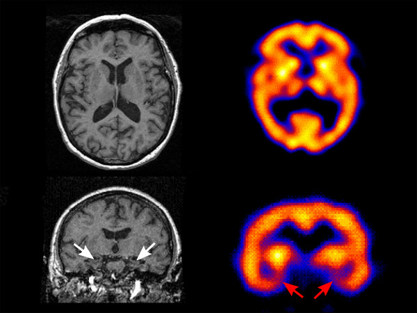
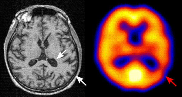
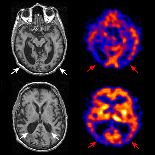
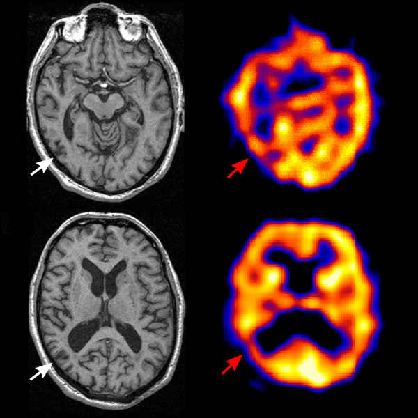
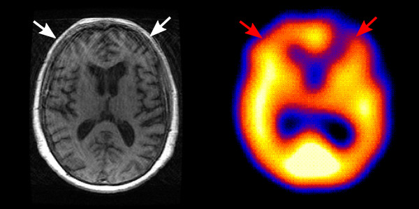
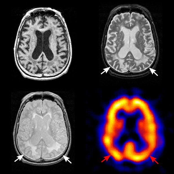
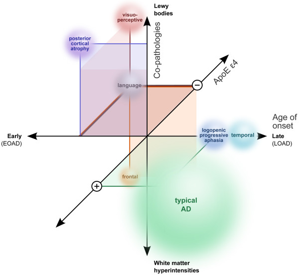
With increasing knowledge of clinical in vivo biomarkers and the pathological intricacies of Alzheimer's disease (AD), nosology is evolving. Harmonized consensus criteria that emphasize prototypic illness continue to develop to achieve diagnostic clarity for treatment decisions and clinical trials. However, it is clear that AD is clinically heterogeneous in presentation and progression, demonstrating variable topographic distributions of atrophy and hypometabolism/hypoperfusion. AD furthermore often keeps company with other conditions that may further nuance clinical expression, such as synucleinopathy exacerbating executive and visuospatial dysfunction and vascular pathologies (particularly small vessel disease that is increasingly ubiquitous with human aging) accentuating frontal-dysexecutive symptomatology. That some of these atypical clinical patterns recur may imply the existence of distinct AD variants. For example, focal temporal lobe dysfunction is associated with a pure amnestic syndrome, very slow decline, with atrophy and neurofibrillary tangles limited largely to the medial temporal region including the entorhinal cortex. Left parietal atrophy and/or hypometabolism/hypoperfusion are associated with language symptoms, younger age of onset, and faster rate of decline - a potential 'language variant' of AD. Conversely, the same pattern but predominantly affecting the right parietal lobe is associated with a similar syndrome but with visuospatial symptoms replacing impaired language function. Finally, the extremely rare frontal variant is associated with executive dysfunction out of keeping with degree of memory decline and may have prominent behavioural symptoms. Genotypic differences may underlie some of these subtypes; for example, absence of apolipoprotein E e4 is often associated with atypicality in younger onset AD. Understanding the mechanisms behind this variability merits further investigation, informed by recent advances in imaging techniques, biomarker assays, and quantitative pathological methods, in conjunction with standardized clinical, functional, neuropsychological and neurobehavioral evaluations. Such an understanding is needed to facilitate 'personalized AD medicine', and eventually allow for clinical trials targeting specific AD subtypes. Although the focus legitimately remains on prototypic illness, continuing efforts to develop disease-modifying therapies should not exclude the rarer AD subtypes and common comorbid presentations, as is currently often the case. Only by treating them as well can we address the full burden of this devastating dementia syndrome.


Figures

Figure 1
Figure 1
Temporal variant Alzheimer's disease. A 69 year old woman presenting with short-term memory loss and very slow progression with conversion to mild dementia about 5 years later. Her last neuropsychological testing after 11 years of follow-up revealed normal language and visuospatial function, with very mild executive dysfunction and significant impairment on memory items. Ten years into her course, imaging including coronal T1 MRI at the level of the hippocampus (bottom left) demonstrated marked hippocampal atrophy (white arrows) while single-photon emission computed tomography (SPECT) done at the same time (bottom right) showed mesiotemporal hypoperfusion (red arrows). Axial T1 MRI (top left) and SPECT (top right) at the level of the basal ganglia demonstrated no atrophy in the parietal and frontal association areas and no hypoperfusion in the same areas.
Figure 2
Figure 2
Left/language variant Alzheimer's disease. A 50 year old man presenting with short-term memory loss and significant aphasia, which progressed rapidly over the next few years. Axial T1 MRI at the level of the basal ganglia (left) demonstrated subtly greater left parietal atrophy and expansion of the lateral ventricle (white arrows). Single-photon emission computed tomography (SPECT) done at the same time (right) demonstrated left parietal hypoperfusion (red arrow). The patient died 8 years after symptom onset, and autopsy confirmed Alzheimer's disease (Braak V/VI).
Figure 3
Figure 3
Posterior cortical atrophy (PCA). A 67 year old man presenting with mild memory loss and striking visuospatial difficulties, later developing parkinsonism. Imaging 6 years into the disease course, including axial T1 MRI at the level of the midbrain (top left) and basal ganglia (bottom left), demonstrated temporal-occipital atrophy, posterior atrophy and ventricular expansion (white arrows), slightly greater on the right. Axial single-photon emission computed tomography (SPECT) imaging done at the same time (right) demonstrated bilateral posterior hypoperfusion (red arrows), greater on the right. The patient died 7 years into the course of disease, and autopsy revealed Alzheimer's disease (Braak IV/VI) with co-occurring Lewy bodies (Braak VI/VI).
Figure 4
Figure 4
Right/visuoperceptive variant Alzheimer's disease. A 64 year old man presenting with short-term memory loss and difficulty with visually guided tasks (reading clocks and music). Imaging done 3 years into the course of illness, including axial T1 MRI at the level of the midbrain (top left), demonstrated subtle right, greater than left, temporal and parietal atrophy with relative expansion of right lateral ventricle and increased right parietal sulcal markings (white arrows), less evident at the level of the basal ganglia (bottom left). At the same time, axial single-photon emission computed tomography (SPECT; right) demonstrated right hypoperfusion, particularly in the parietal region (red arrows). The patient died 5 years into the course of disease, and autopsy confirmed Alzheimer's disease (Braak V/VI).
Figure 5
Figure 5
Frontal variant Alzheimer's disease. A 57 year old woman presenting with mild short-term memory loss and inability to perform simple daily and occupational tasks. Prominent frontal symptoms were noted 2 years into the course of her illness, with prominent apathy, loss of empathy, and socially inappropriate behaviours. Axial T1 MRI at the level of the basal ganglia (left), done 1 year after symptom onset, demonstrated mild frontal atrophy with increased sulcal markings (white arrows). Axial single-photon emission computed tomography (SPECT) done within a few months at the same level (right) demonstrated frontal hypoperfusion, especially on the left (red arrows). The patient died 5 years after the start of her symptoms, and autopsy revealed Alzheimer's disease (Braak V/VI).
Figure 6
Figure 6
White matter hyperintensities in Alzheimer's disease. A 68 year old man presenting with short-term memory decline. Imaging done 7 years into the course of illness, including axial T1 MRI at the level of the centrum semiovale (upper left), demonstrated severe generalized atrophy. Axial T2 (top right) and proton density (bottom left) images at the same level showed marked white matter hyperintensities, especially posteriorly (white arrows). One year prior, axial single-photon emission computed tomography (SPECT) at the corresponding level (bottom right) demonstrated mild parietal hypoperfusion bilaterally (red arrows). The patient died 9 years later and autopsy confirmed Alzheimer's disease (Braak V/VI), with diffuse atherosclerosis throughout the white matter and basal ganglia, lacunar infarcts, remote microhaemorrhages and cortical microinfarcts. There had been no history of visual hallucinations, but parkinsonism developed very late into the disease course, and he was also found to have diffuse Lewy bodies within the brain stem and cingulum.
Figure 7
Figure 7
Proposed framework for Alzheimer's disease subtypes. Bubbles represent subpopulations of Alzheimer's disease (AD) by subtype, located along each of the three axes of heterogeneity: age of onset, apolipoprotein E status, and co-pathology. Exact size, positioning and degree of overlap for each subtype remain uncertain areas of active research.

References

    1. McKhann G, Drachman D, Folstein M, Katzman R, Price D, Stadlan EM. Clinical diagnosis of Alzheimer's disease: report of the NINCDS-ADRDA Work Group under the auspices of Department of Health and Human Services Task Force on Alzheimer's Disease. Neurology. 1984;34:939–944. doi: 10.1212/WNL.34.7.939. - DOI - PubMed
    1. Bartus RT, Dean RL, Beer B, Lippa AS. The cholinergic hypothesis of geriatric memory dysfunction. Science. 1982;217:408–414. doi: 10.1126/science.7046051. - DOI - PubMed
    1. Francis PT, Palmer AM, Snape M, Wilcock GK. The cholinergic hypothesis of Alzheimer's disease: a review of progress. J Neurol Neurosurg Psychiatry. 1999;66:137–147. doi: 10.1136/jnnp.66.2.137. - DOI - PMC - PubMed
    1. Braak H, Braak E. Neuropathological stageing of Alzheimer-related changes. Acta Neuropathol. 1991;82:239–259. doi: 10.1007/BF00308809. - DOI - PubMed
    1. McKhann GM, Knopman DS, Chertkow H, Hyman BT, Jack CR Jr, Kawas CH, Klunk WE, Koroshetz WJ, Manly JJ, Mayeux R, Mohs RC, Morris JC, Rossor MN, Scheltens P, Carrillo MC, Thies B, Weintraub S, Phelps CH. The diagnosis of dementia due to Alzheimer's disease: recommendations from the National Institute on Aging-Alzheimer's Association workgroups on diagnostic guidelines for Alzheimer's disease. Alzheimers Dement. 2011;7:263–269. doi: 10.1016/j.jalz.2011.03.005. - DOI - PMC - PubMed

Grants and funding

LinkOut - more resources