. 2013 Mar;87(6):3435-46.
doi: 10.1128/JVI.02906-12. Epub 2013 Jan 9.

Tetraspanin CD151 mediates papillomavirus type 16 endocytosis

Affiliations

Tetraspanin CD151 mediates papillomavirus type 16 endocytosis

Konstanze D Scheffer et al. J Virol. 2013 Mar.

Abstract

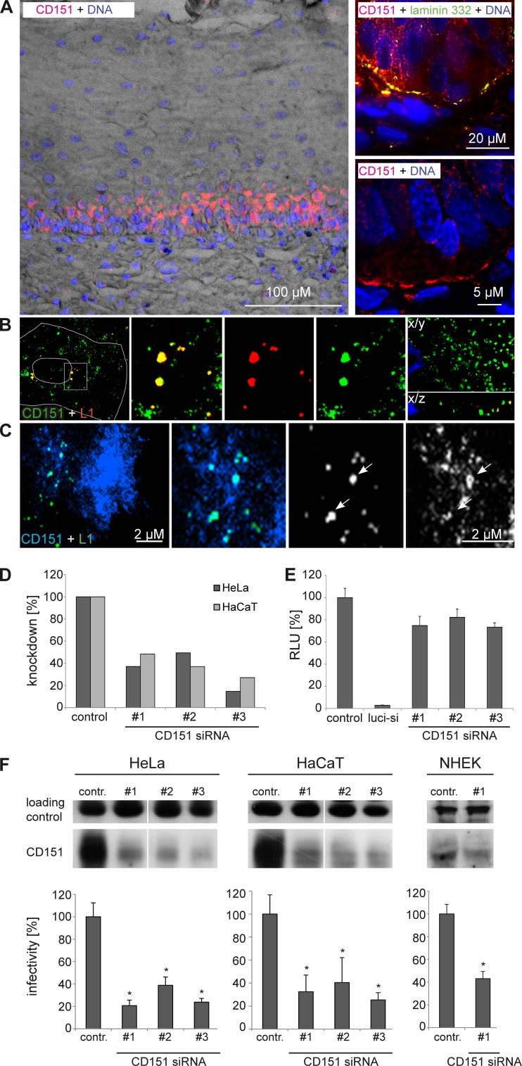
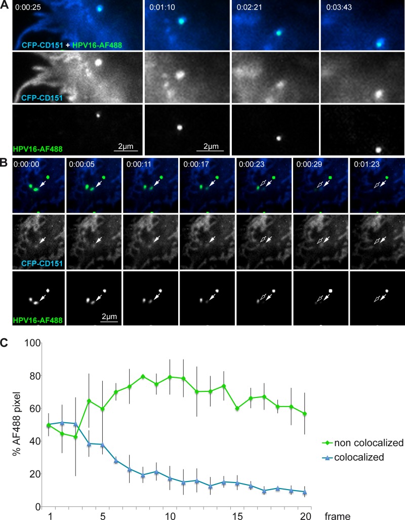
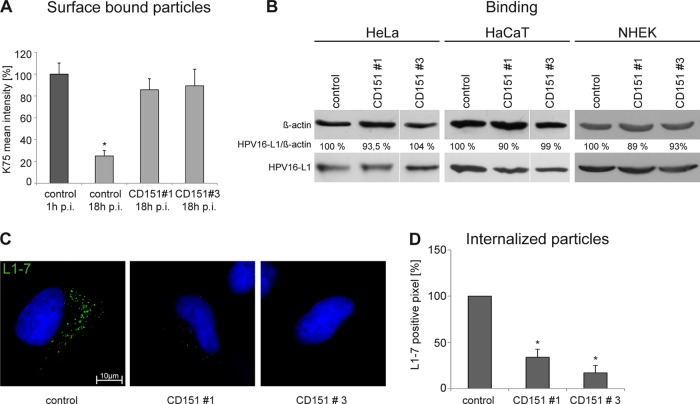
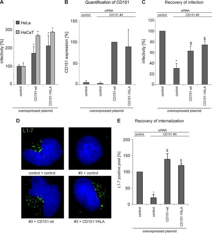
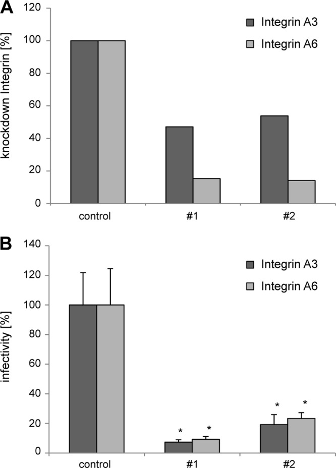
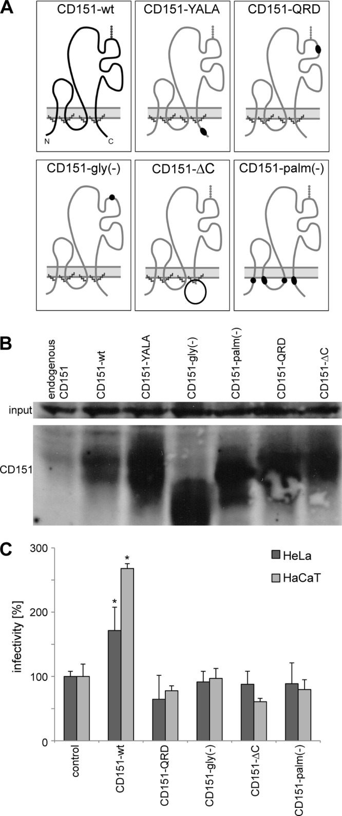
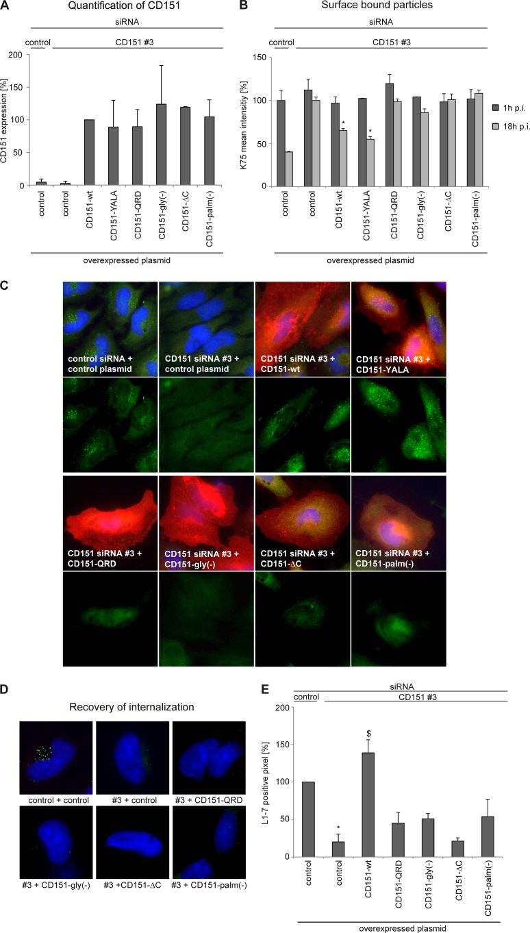
Human papillomavirus type 16 (HPV16) is the primary etiologic agent for cervical cancer. The infectious entry of HPV16 into cells occurs via a so-far poorly characterized clathrin- and caveolin-independent endocytic pathway, which involves tetraspanin proteins and actin. In this study, we investigated the specific role of the tetraspanin CD151 in the early steps of HPV16 infection. We show that surface-bound HPV16 moves together with CD151 within the plane of the membrane before they cointernalize into endosomes. Depletion of endogenous CD151 did not affect binding of viral particles to cells but resulted in reduction of HPV16 endocytosis. HPV16 uptake is dependent on the C-terminal cytoplasmic region of CD151 but does not require its tyrosine-based sorting motif. Reexpression of the wild-type CD151 but not mutants affecting integrin functions restored virus internalization in CD151-depleted cells. Accordingly, short interfering RNA (siRNA) gene knockdown experiments confirmed that CD151-associated integrins (i.e., α3β1 and α6β1/4) are involved in HPV16 infection. Furthermore, palmitoylation-deficient CD151 did not support HPV16 cell entry. These data show that complex formation of CD151 with laminin-binding integrins and integration of the complex into tetraspanin-enriched microdomains are critical for HPV16 endocytosis.


Figures

Fig 1
Fig 1
Tetraspanin CD151 is important for HPV16 infection in keratinocytes. (A) Distribution of CD151 in the cervical mucosa. Human cervical tissue sections are stained with anti-CD151 (red) showing a high expression level of basal cells (upper right panel; laminin 332 staining, indicating the basement membrane, is shown in green). Nuclei are shown in blue (A and B). (B) CD151 colocalizes with HPV16-L1 in primary keratinocytes. Primary keratinocytes (NHEK) were infected with HPV16 PsV for 4 h and analyzed by confocal and deconvolution fluorescence microscopy (right panel; the lower right shows the X/Z view of the z stack). (C) TIRF microscopy of HaCaT cells transfected with CFP-CD151 and infected with Alexa Fluor 488-labeled HPV16 pseudovirions for 1.5 h. Arrows depict colocalization of HPV16 and CD151. (D, E, and F) Knockdown of CD151 by CD151 siRNAs. Cells were transfected with CD151-specific siRNAs for 48 h. (D) The knockdown of the CD151 level on the cell surface was measured by flow cytometry in HeLa and HaCaT cells. (E) CD151-specific siRNAs have no significant influence on luciferase expression. HeLa cells were cotransfected with control, luciferase-specific, or CD151-specific siRNA and with luciferase-expressing plasmid to control the influence of siRNA transfection on luciferase expression. The luciferase-specific siRNA serves as a positive control. (F) Depletion of CD151 in HeLa, HaCaT, and NHEK cells correlates with reduced infection. SDS-PAGE (upper panels) was performed under nonreducing conditions and shows the amount of CD151. The loading control is an unspecific band produced by the CD151 polyclonal rabbit antibody that serves as a control for protein input. For infection assay (lower panels), cells were transfected with siRNA as indicated for 48 h and then infected for 24 h with HPV16 PsV. Infectivity was measured by luciferase activity and normalized by lactate dehydrogenase (LDH) measurements. The control siRNA infection rate was set to 100%. *, P < 0.05 compared to the control.
Fig 2
Fig 2
Lateral surface movement and cointernalization of HPV16-bound CD151. Shown are TIRF images of HeLa cells transfected with CFP-CD151, infected with Alexa Fluor 488 (AF488)-labeled HPV16 pseudovirions for 2.5 h, and analyzed by TIRF microscopy for 2 to 4 min. Time-lapse images show (A) lateral movement of HPV16-bound CD151 and (B) cointernalization of HPV16 and CD151. Arrows depict colocalization of HPV16 and CD151 during the process of internalization. (C) Quantitative analysis of HPV16 endocytosis. Cell surface areas with approximately 50% of the particles in colocalization with CFP-CD151 were selected, and the loss of fluorescent signal was determined over time. HPV16 Alexa Fluor 488 pixels noncolocalized (green) and colocalized with CFP-CD151 (blue) were analyzed by the Axiovision colocalization module with fixed thresholds for all images of two time-lapse sequences (20 frames every 5 s). The averages of the corresponding frames from two independent movies are shown with error bars representing the standard deviations of the pixel numbers.
Fig 3
Fig 3
CD151 is involved in HPV16 internalization but not in cell surface binding. (A) HeLa cells were treated with siRNAs for 48 h and subsequently incubated with HPV16 PsV for 1 or 18 h as indicated. The amount of surface-associated PsV was assessed by flow cytometry using anti-L1 PAb (K75). Mean fluorescence intensity for cells incubated with HPV16 PsV for 1 h was adjusted to 100%. Shown are compiled results of three independent experiments. (B) Primary binding of HPV16 to the cell surface is independent of CD151. HeLa, HaCaT, and NHEK cells were transfected with control or CD151-specific siRNAs for 48 h and subsequently incubated with HPV16 PsV for 1 h. Cell-bound PsV were detected in cell lysates by Western blotting using specific anti-L1 PAb. (C and D) HeLa cells were treated with siRNAs for 48 h and infected with HPV16 PsV for 7 h. Virus internalization was analyzed by immunofluorescence using monoclonal antibody (L1-7) recognizing an epitope that is located on the inner side of intact capsids and only accessible after internalization and capsid disassembly. Representative images are shown in panel C. (D) For quantification of virus internalization and disassembly, L1-7-positive pixels of at least 100 cells were analyzed using an ImageJ script. Shown are the results of three independent experiments normalized to control siRNA-treated and infected cells. *, P < 0.05 compared to the control.
Fig 4
Fig 4
Tyrosine-based sorting motif in CD151 is not required for HPV16 internalization and infection. (A) Overexpression of CD151-wt and CD151-YALA increases HPV16 infection. HeLa and HaCaT cells were transfected with control or CD151 plasmids as indicated for 24 h and then infected for 24 h with HPV16 PsV. Infectivity was measured by luciferase activity and normalized to LDH measurements. *, P < 0.05 compared to the control). (B to E) Reexpression of CD151-wt and -YALA in CD151-depleted cells recovers HPV16 infection and virus internalization in CD151-depleted cells. HeLa cells were transfected with control or CD151-specific siRNA (CD151#3) which targets the untranslated region of the mRNA. After 24 h, cells were transfected with a control plasmid or with a plasmid encoding CD151-wt or -YALA to recover CD151 expression. (B and C) Recovery of CD151 expression in CD151-depleted cells was controlled by flow cytometry and additionally monitored using a luciferase infectivity assay. (C) Infectivity was measured by luciferase activity and normalized by LDH measurements. The control siRNA infection rate was set to 100%. *, P < 0.05 compared to the control; $, P < 0.05 compared to CD151 knockdown. (D and E) Internalization and disassembly of HPV16 PsV. HeLa cells were treated with siRNAs for 48 h and infected with HPV16 PsV for 7 h. Virus internalization was analyzed by immunofluorescence using monoclonal antibody (L1-7) recognizing an epitope that is located on the inner side of intact capsids and only accessible after internalization and capsid disassembly. Representative images are shown in panel D. (E) For quantification, at least 100 CD151-overexpressing cells were analyzed using an ImageJ script. *, P < 0.05 compared to cells transfected with control siRNA and control plasmid; $, P < 0.05 compared to cells transfected with CD151#3 and control plasmid.
Fig 5
Fig 5
CD151-interacting integrins α3β1 and α6β1/4 are important for HPV16 infection. HeLa cells were transfected with siRNAs targeting α3β1- and α6β1/4-integrin subunits, and after 48 h cells were incubated with HPV16 PsV for 24 h. (A) The level of integrin knockdown was assessed by flow cytometry. Data are presented as percentages relative to the level expressed in cells transfected with an siRNA control. (B) Relative infectivity was measured by luciferase activity and normalized by LDH measurements. *, P < 0.05 compared to the control.
Fig 6
Fig 6
Integrin association and palmitoylation of CD151 are important for HPV16 infection. (A) Schematic diagram of CD151 mutants. (B) HeLa cells were transfected with control plasmid, CD151-wt, or CD151 mutants for 24 h. CD151 expression levels in cell lysates were analyzed by Western blotting. (C) Influence of CD151 mutants on HPV16 infection was analyzed after CD151 overexpression as described in the legend to Fig. 4A.
Fig 7
Fig 7
Recovery of endocytosis and disassembly using CD151 mutants. (A) HeLa cells were treated with siRNAs for 24 h and then transfected with plasmids as indicated. CD151 expression levels on the cell surface were analyzed by flow cytometry. (B) Primary binding of HPV16 to the cell surface is not affected by overexpression of CD151 mutants, but only CD151-wt and -YALA are able to reconstitute HPV16 endocytosis. HeLa cells were treated with CD151 siRNAs for 24 h, transfected with CD151 plasmids as indicated, and subsequently incubated with HPV16 PsV for 1 and 18 h. The amount of cell surface-bound virus particles was quantified by flow cytometry using anti-L1 pAb (K75). Mean of fluorescence intensity for cells treated with control siRNA and plasmid and incubated with HPV16 PsV for 1 h was adjusted to 100%. *, P < 0.05 compared to cells transfected with CD151#3 and control plasmid. (C) HeLa cells were treated with siRNAs and plasmids as described for panel B and infected with HPV16 PsV for 7 h. The major capsid protein L1 was detected with monoclonal anti-L1 antibody (L1-7; green) reacting with an L1 epitope that is accessible exclusively after viral entry, and CD151 was detected with monoclonal anti-CD151 antibody (red). The upper panels show the costaining of CD151 and HPV16-L1, and the lower panels show the staining of HPV16-L1 only. (D and E) Virus internalization was analyzed by immunofluorescence as described in the legend to Fig. 4D and E.

References

    1. Chen X, Garcea R, Goldberg I, Casini G, Harrison S. 2000. Structure of small virus-like particles assembled from the L1 protein of human papillomavirus 16. Mol. Cell 5:557–567 - PubMed
    1. Modis Y, Trus BL, Harrison SC. 2002. Atomic model of the papillomavirus capsid. EMBO J. 21:4754–4762 - PMC - PubMed
    1. Baker TS, Newcomb WW, Olson NH, Cowsert LM, Olson C, Brown JC. 1991. Structures of bovine and human papillomaviruses. Analysis by cryoelectron microscopy and three-dimensional image reconstruction. Biophys. J. 60:1445–1456 - PMC - PubMed
    1. Buck CB, Cheng N, Thompson CD, Lowy DR, Steven AC, Schiller JT, Trus BL. 2008. Arrangement of L2 within the papillomavirus capsid. J. Virol. 82:5190–5197 - PMC - PubMed
    1. Finnen RL, Erickson KD, Chen XS, Garcea RL. 2003. Interactions between papillomavirus L1 and L2 capsid proteins. J. Virol. 77:4818–4826 - PMC - PubMed

Publication types

LinkOut - more resources