. 2013 Mar;87(6):3471-83.
doi: 10.1128/JVI.01937-12. Epub 2013 Jan 9.

Role of interactions between Autographa californica multiple nucleopolyhedrovirus procathepsin and chitinase chitin-binding or active-site domains in viral cathepsin processing

Affiliations

Role of interactions between Autographa californica multiple nucleopolyhedrovirus procathepsin and chitinase chitin-binding or active-site domains in viral cathepsin processing

Jeffrey J Hodgson et al. J Virol. 2013 Mar.

Abstract

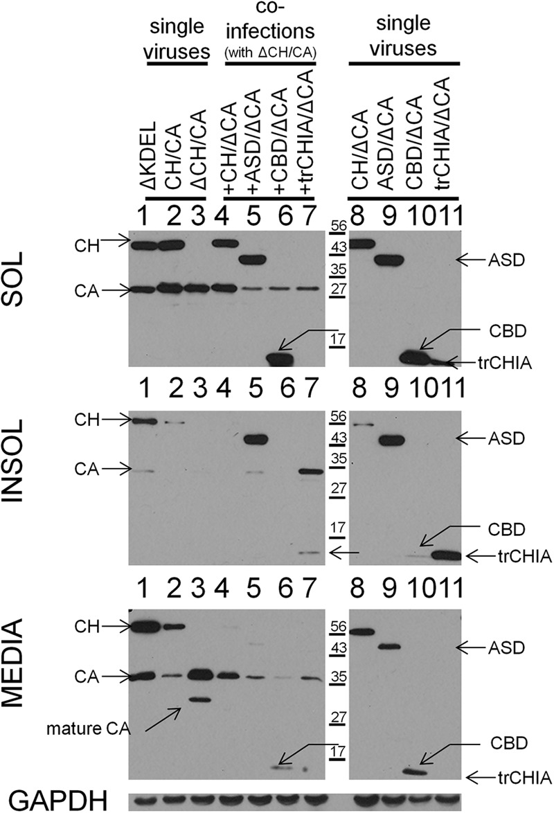
The binding of Autographa californica multiple nucleopolyhedrovirus chitinase (CHIA) to viral cathepsin protease progenitor (proV-CATH) governs cellular/endoplasmic reticulum (ER) coretention of CHIA and proV-CATH, thus coordinating simultaneous cellular release of both host tissue-degrading enzymes upon host cell death. CHIA is a proposed proV-CATH folding chaperone because insertional inactivation of chiA causes production of proV-CATH aggregates that are incompetent for proteolytic maturation into active V-CATH enzyme. We wanted to determine whether the N-terminal chitin-binding domain (CBD, 149 residues) and C-terminal CHIA active-site domain (ASD, 402 residues) of CHIA bind to proV-CATH independently of one another and whether either domain is dispensable for CHIA's putative proV-CATH folding chaperone activity. We demonstrate that N-terminally green fluorescent protein (GFP)-fused CHIA, ASD, and CBD each colocalize with proV-CATH-RFP in ER-like patterns and that both ASD and CBD independently associate with proV-CATH in vivo using bimolecular fluorescence complementation (BiFC) and in vitro using reciprocal nickel-histidine pulldown assays. Altogether, the data from colocalization, BiFC, and reciprocal copurification analyses suggest specific and independent interactions between proV-CATH and both domains of CHIA. These data also demonstrate that either CHIA domain is dispensable for normal proV-CATH processing. Furthermore, in contrast to prior evidence suggesting that a lack of chiA expression causes proV-CATH to become aggregated, insoluble, and unable to mature into V-CATH, a chiA deletion bacmid virus we engineered to express just v-cath produced soluble proV-CATH that was prematurely secreted from cells and proteolytically matured into active V-CATH enzyme.


Figures

Fig 1
Fig 1
Colocalization of proV-CATH-RFP (CA-RFP) with GFP-CH, GFP-CBD, or GFP-ASD in virus-infected cells. (a) Schematics showing the 31-amino-acid (aa) CHIA signal sequence (CHIA ER s.p.) and deduced (24) cleavage site, indicating which portions of CHIA comprise the CBD and ASD truncation constructs, and showing the position of GFP fusion to them. The CHIA residues 1 to 149 and 150 to 551 were used as the CBD and ASD portions, respectively. The GFP was fused into the SpeI sites inserted into chiA to produce the N-terminal fusions with full-length CHIA, CBD (aa 16 to 149), and ASD (aa 150 to 551) after the chiA 31 N-terminal amino acid coding sequence containing the CHIA signal peptide cleavage site at A17 (24). The adjacent CA-RFP construct was the same in all viruses and was the same as that used earlier (7). See Materials and Methods for details of the cloning steps. (b and c) Colocalization microscopy. (b) Virus construct schematics; (c) confocal microscopy results. The GFP-fused CH (or CH derivatives) and CA-RFP ORFs were expressed from the native intergenic chiA/v-cath promoters. Confocal microscopy images of CA-RFP coexpressed with GFP-CH, GFP-CBD, or GFP-ASD in individual live infected Hi5 cells, representative of the entire infected monolayers, were taken at 36 hpi.
Fig 2
Fig 2
Detection of proV-CATH interactions with CBD or ASD in vivo using bimolecular fluorescence complementation (BiFC). Virus constructs and BiFC results showing the infected monolayers and a single representative cell from each assay. For the BiFC-negative control, cells were coinfected at an MOI of 10 (for each virus) with CH-mRFPC/v-cath, which expressed the ER-targeted CH-mRFPC fusion protein, and unrelated MYC-mRFPN-KDEL/v-cath, which expressed the ER-targeted MYC-mRFPN-KDEL protein. The MYC-mRFPN-KDEL protein was targeted to the ER by fusion to the chiA signal peptide and was modified by addition of a C-terminal KDEL motif, so that it accumulates in the ER of infected cells. Both of the BiFC negative-control viruses carry unmodified v-cath. For the CBD and ASD BiFC assays, cells were infected (MOI, 10) with either the CBD-mRFPC/CA-mRFPN or the ASD-mRFPC/CA-mRFPN virus, respectively. In all three assays, cells were coinfected with the ER-GFP virus to identify the ER. Images of live virus-infected Hi5 cells were photographed at 48 hpi using confocal microscopy.
Fig 3
Fig 3
Immunoblots of soluble (SOL) and insoluble (INSOL) intracellular and extracellular (MEDIA) proteins extracted from Hi5 cells infected with viruses used for the truncation, colocalization (GFP/RFP), and interaction (BiFC and Ni/His) assays. Proteins from the soluble and insoluble cellular and extracellular medium fractions from each assay were immunoblotted to determine their solubility and distribution. Virus-infected cell lysates were simultaneously probed with anti-HA and anti-FLAG (a) or with anti-HA (for CA) and anti-GFP (for GFP-CH, GFP-CBD, and GFP-ASD) (b), or duplicate blots were probed with anti-FLAG (for CH-mRFPC CBD-mRFPC ASD-mRFPC) or anti-MYC (for MYC-mRFPN-KDEL and CA-MYC-mRFPN) antibodies (c). (c) Lanes CH, CBD, and ASD contain CH-mRFPC/CA-mRFPN, CBD-mRFPC/CA-mRFPN, and ASD-mRFPC/CA-mRFPN virus-infected protein samples (respectively); lane NEG contains lysate (or medium) from cells coinfected with the two BiFC negative-control viruses (CH-mRFPC/v-cath and MYC-mRFPN-KDEL/v-cath). Soluble cellular, insoluble cellular, and extracellular proteins from virus-infected cells were processed at 36 hpi and separated and immunoblotted as described in Materials and Methods.
Fig 4
Fig 4
Reciprocal Ni:6-His affinity copurification of proV-CATH with either CBD or ASD. (a and c) Viral constructs. Viruses coexpressed the indicated forms (6-His tagged or not) of v-cath with either cbd (a) or asd (c). “His” in the virus names indicates a 6-His-tagged isoform of the v-cath, cbd, or asd protein. Neither the CBD/CA nor ASD/CA control viruses contained any His-tagged proteins. Whether His-tagged or not, for immunodetection all cbd and asd proteins were FLAG tagged and all v-cath proteins were HA tagged. (b and d) Western blot analysis of purified proteins using anti-HA (to detect CA) and anti-FLAG (to detect CBD or ASD). Proteins from the input lysate (input) and those eluted from washed Ni-agarose beads (Ni/His) were analyzed. Lysate from 2 × 10⁷ or 6 × 10⁷ infected Sf21 cells (MOI, 10) at 40 hpi were used for the CBD-based or ASD-based purifications, respectively.
Fig 5
Fig 5
ProV-CATH solubility and distribution with and without chiA and/or N-glycosylation. Virus-infected serum-free medium-adapted Sf21 cells were collected at 40 hpi and fractionated into extracellular medium (MEDIA) and intracellular soluble (SOL) and insoluble (INSOL) components, as described in Materials and Methods. Duplicate gels were loaded with equal volumes of both the SOL and INSOL proteins, and the resulting blots were probed with either anti-FLAG (for CH) or anti-HA (for CA). The entire blot of extracellular medium samples is shown because a single gel was loaded, blotted, and probed with anti-FLAG and anti-HA simultaneously. Tunicamycin was supplemented to 10 μg ml−1 in growth medium from 0 hpi.
Fig 6
Fig 6
Distribution of proV-CATH in cell culture when coexpressed with CHIA, CBD, ASD, or trCHIA. Cell cultures were infected with control CH/CA and (ΔKDEL)CH/CA viruses or with the ΔCH/CA virus, which expressed only CA. Some cultures infected with ΔCH/CA were also coinfected with either CH/ΔCA (+CH, lane 4), CBD/ΔCA (+CBD, lane 6), ASD/ΔCA (+ASD, lane 5), or trCHIA/ΔCA (+trCHIA) in order to supply the various chitinase derivatives in trans during infection. Other cultures were infected with only the CH/ΔCA, CBD/ΔCA, ASD/ΔCA, or trCHIA/ΔCA virus to demonstrate their inherent distribution and to show that they each can differentially influence the distribution of proV-CATH (when CA was coexpressed in trans by the coinfecting virus) in infected cultures. All infected cultures were collected and fractionated into cellular soluble (SOL) and insoluble (INSOL) and extracellular (MEDIA) portions at 40 hpi as described in the Materials and Methods. Blots of the intracellular and extracellular proteins were each probed simultaneously with anti-FLAG (to detect CH, CBD, ASD, and trCHIA) and anti-HA (to detect CA) antibodies. The mature CA, detected only in the ΔCH/CA medium sample, is indicated. Blots of soluble proteins from each infected lysate sample were also probed with anti-GAPDH antibodies (bottom) to control for equivalency in protein loading and/or cell fractionation.
Fig 7
Fig 7
chiA-deficient bacmid V-CATH maturation and activity. (a) Induced maturation of V-CATH. Cell homogenate generated from chiA-deficient ΔCH/CATH40 virus-infected Sf21 cells was acidified to ∼pH 5.5 with 100 mM succinic acid and incubated at 37°C as described in Materials and Methods. Samples taken at the indicated times were supplemented with E64 protease inhibitor to halt V-CATH activation. Equal volumes of samples from each time point were analyzed by immunoblotting with anti-V-CATH antibody. (b) V-CATH protease assay. Cell homogenate (cells) or extracellular medium (media) from Sf21 cells infected with chiA/v-cath negative virus (ΔCC), chiA/v-cath repair virus (repair+CC), or chiA-deficient ΔCH/CATH40 virus (CATH40) was used to monitor V-CATH digestion of azocasein substrate in the presence or absence of cysteine protease inhibitor (E64, 30 μM) as described in Materials and Methods. Serum-containing (10%) and serum-free media were used for growing and infecting cells used for assaying cellular (cells) and extracellular (media) V-CATH, respectively. Activities are expressed as the mean A410 from the soluble dye released by azocasein proteolysis. The data are derived from duplicate assays of two replicate infections for the cellular samples or a single infection for the extracellular (serum-free) medium samples. Error bars denote standard errors.
Fig 8
Fig 8
Anti-CHIA immunoblots to detect CH and CHIA derivative proteins (CBD, trCHIA, and putative X-DZ1 CHIA remnant). Blots of soluble (SOL) and insoluble (INSOL) proteins (collected and fractionated at 40 hpi as described in Materials and Methods) were probed with an anti-CHIA antibody (anti-BmCHI-h). The CH, CBD, and trCHIA proteins (of each ΔCA virus) detected were the expected size relative to standards (indicated) and were distributed as in Fig. 6. The 25-kDa X-DZ1 peptide (putative CHIA remnant) was detected only in the insoluble fraction of X-DZ1 lysate. The anti-CHIA antibody detected similar nonspecific background bands for all samples, but none of them corresponded to the size of the putative X-DZ1 CHIA remnant protein.

References

    1. Hom LG, Ohkawa T, Trudeau D, Volkman LE. 2002. Autographa californica M nucleopolyhedrovirus ProV-CATH is activated during infected cell death. Virology 296:212–218 - PubMed
    1. Hawtin RE, Zarkowska T, Arnold K, Thomas CJ, Gooday GW, King LA, Kuzio JA, Possee RD. 1997. Liquefaction of Autographa californica nucleopolyhedrovirus-infected insects is dependent on the integrity of virus-encoded chitinase and cathepsin genes. Virology 238:243–253 - PubMed
    1. Wang F, Zhang CX, Shyam Kumar V, Wu XF. 2005. Influences of chitinase gene deletion from BmNPV on the cell lysis and host liquefaction. Arch. Virol. 150:981–990 - PubMed
    1. Hom LG, Volkman LE. 2000. Autographa californica M nucleopolyhedrovirus chiA is required for processing of V-CATH. Virology 277:178–183 - PubMed
    1. Daimon T, Katsuma S, Shimada T. 2007. Mutational analysis of active site residues of chitinase from Bombyx mori nucleopolyhedrovirus. Virus Res. 124:168–175 - PubMed

Publication types

MeSH terms

LinkOut - more resources