

Neuroimaging of cognitive dysfunction and depression in aging retired National Football League players: a cross-sectional study
- PMID: 23303193
- PMCID: PMC4016798
- DOI: 10.1001/2013.jamaneurol.340
Neuroimaging of cognitive dysfunction and depression in aging retired National Football League players: a cross-sectional study
Abstract
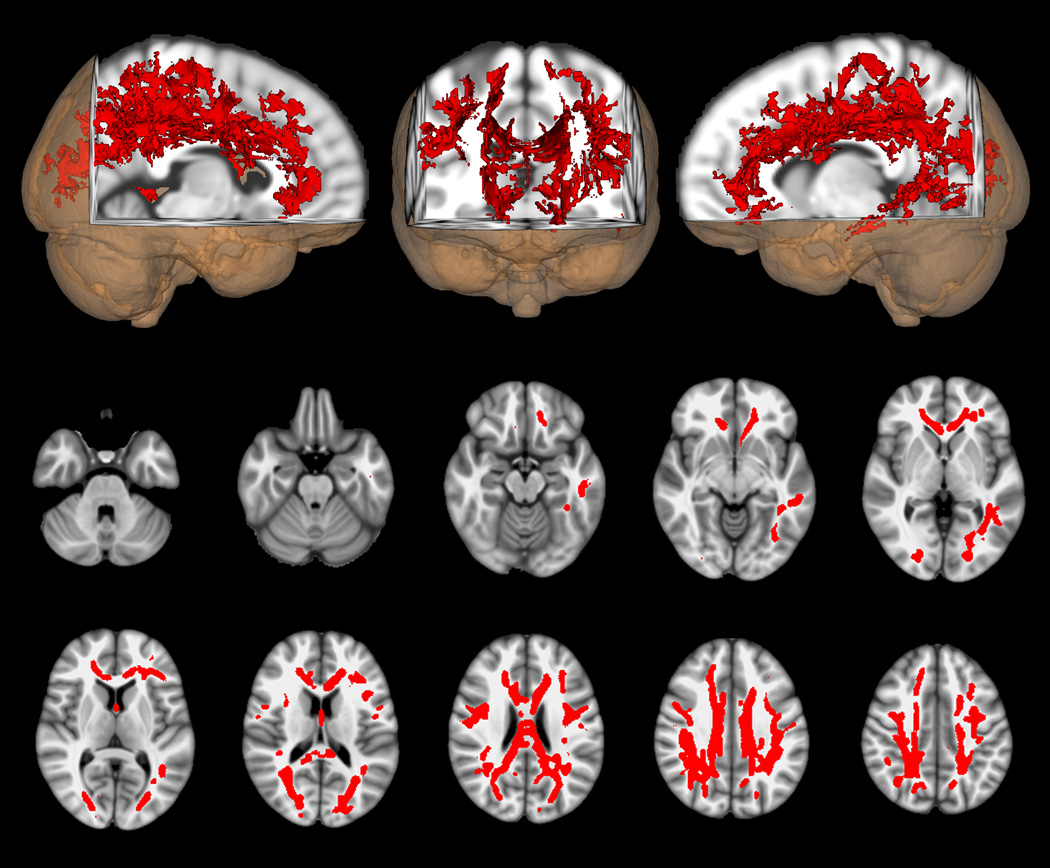
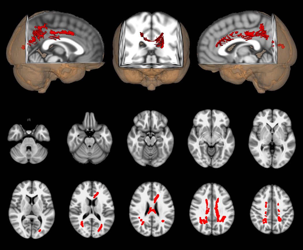
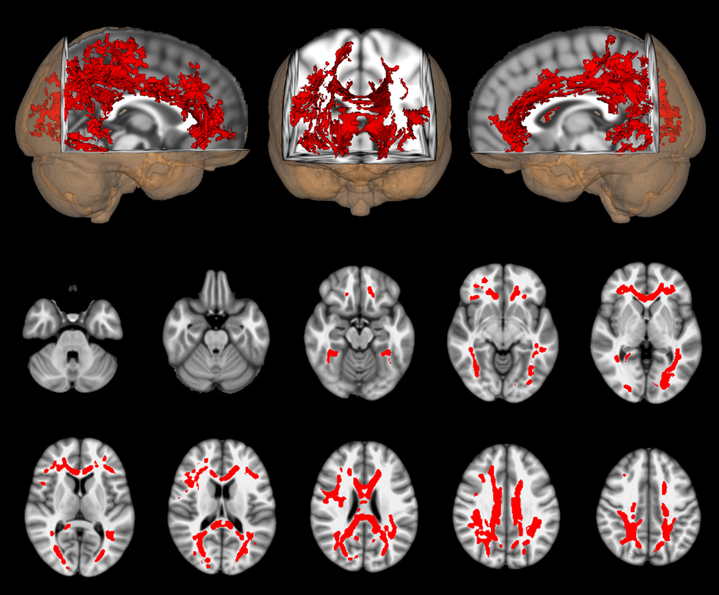
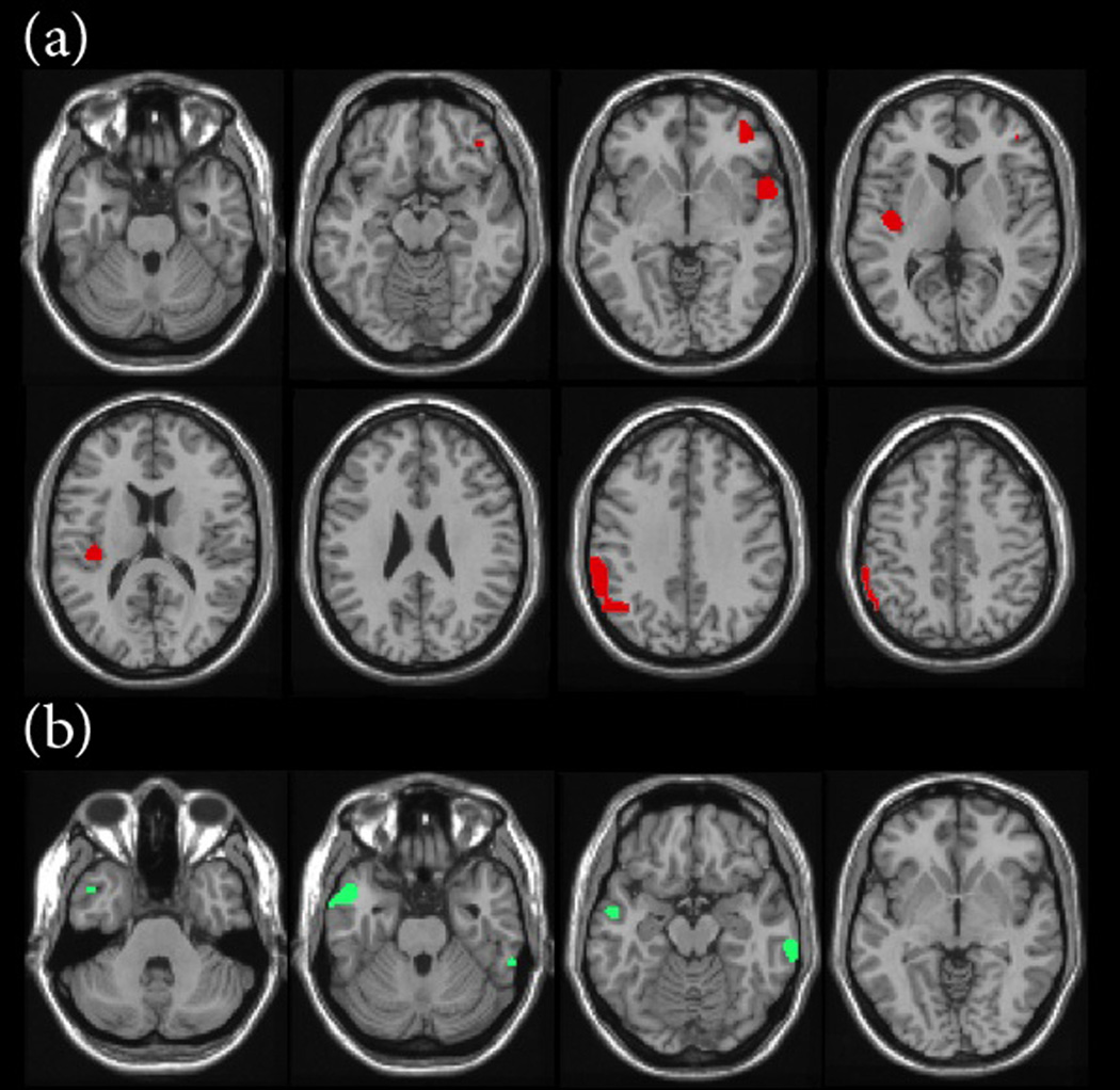
OBJECTIVES To assess cognitive impairment and depression in aging former professional football (National Football League [NFL]) players and to identify neuroimaging correlates of these dysfunctions. DESIGN We compared former NFL players with cognitive impairment and depression, cognitively normal retired players who were not depressed, and matched healthy control subjects. SETTING Research center in the North Texas region of the United States. PATIENTS Cross-sectional sample of former NFL players with and without a history of concussion recruited from the North Texas region and age-, education-, and IQ-matched controls. Thirty-four retired NFL players (mean age, 61.8 years) underwent neurological and neuropsychological assessment. A subset of 26 players also underwent detailed neuroimaging; imaging data in this subset were compared with imaging data acquired in 26 healthy matched controls. MAIN OUTCOME MEASURES Neuropsychological measures, clinical diagnoses of depression, neuroimaging mea-sures of white matter pathology, and a measure of cerebral blood flow. RESULTS Of the 34 former NFL players, 20 were cognitively normal. Four were diagnosed as having a fixed cognitive deficit; 8, mild cognitive impairment; 2, dementia; and 8, depression. Of the subgroup in whom neuroimaging data were acquired, cognitively impaired participants showed the greatest deficits on tests of naming, word finding, and visual/verbal episodic memory. We found significant differences in white matter abnormalities in cognitively impaired and depressed retired players compared with their respective controls. Regional blood flow differences in the cognitively impaired group (left temporal pole, inferior parietal lobule, and superior temporal gyrus) corresponded to regions associated with impaired neurocognitive performance (problems with memory, naming, and word finding). CONCLUSIONS Cognitive deficits and depression appear to be more common in aging former NFL players compared with healthy controls. These deficits are correlated with white matter abnormalities and changes in regional cerebral blood flow.
Conflict of interest statement
The authors report no conflict of interest in the publication of this work, financial or otherwise.
Figures
Comment in
-
Cognitive dysfunction and contact sports.JAMA Neurol. 2013 Mar 1;70(3):301-2. doi: 10.1001/jamaneurol.2013.1930. JAMA Neurol. 2013. PMID: 23303238 No abstract available.
References
-
- McAllister TW, Saykin AJ, Flashman LA, et al. Brain activation during working memory 1 month after mild traumatic brain injury: a functional MRI study. Neurology. 1999;53(6):1300–1308. - PubMed
-
- McAllister TW, Flashman LA, McDonald BC, Saykin AJ. Mechanisms of working memory dysfunction after mild and moderate TBI: evidence from functional MRI and neurogenetics. J Neurotrauma. 2006;23(10):1450–1467. - PubMed
-
- Chen JK, Johnston KM, Frey S, Petrides M, Worsley K, Ptito A. Functional abnormalities in symptomatic concussed athletes: an fMRI study. Neuroimage. 2004;22(1):68–82. - PubMed
-
- Lovell MR, Collins MW, Iverson GL, et al. Recovery from mild concussion in high school athletes. J Neurosurg. 2003;98(2):296–301. - PubMed
-
- Reddy CC, Collins MW. Sports concussion: management and predictors of outcome. Curr Sports Med Rep. 2009;8(1):10–15. - PubMed
Publication types
MeSH terms
Grants and funding
LinkOut - more resources
Full Text Sources
Other Literature Sources
Medical
