. 2013 Apr;27(4):1519-31.
doi: 10.1096/fj.12-219105. Epub 2013 Jan 9.

11β-hydroxysteroid dehydrogenase type 1 deficiency in bone marrow-derived cells reduces atherosclerosis

Affiliations

11β-hydroxysteroid dehydrogenase type 1 deficiency in bone marrow-derived cells reduces atherosclerosis

Tiina Kipari et al. FASEB J. 2013 Apr.

Abstract

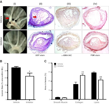
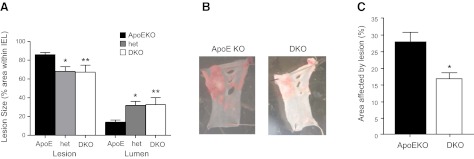
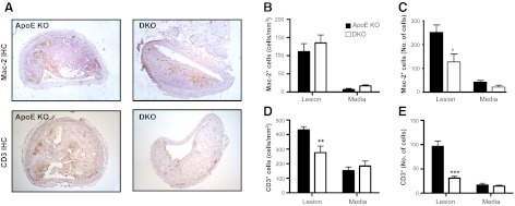
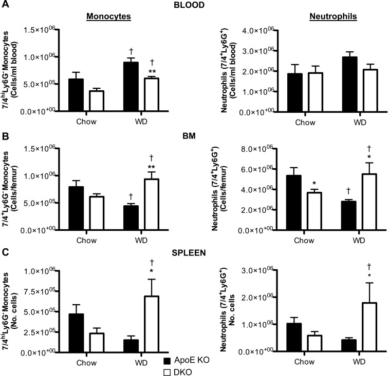
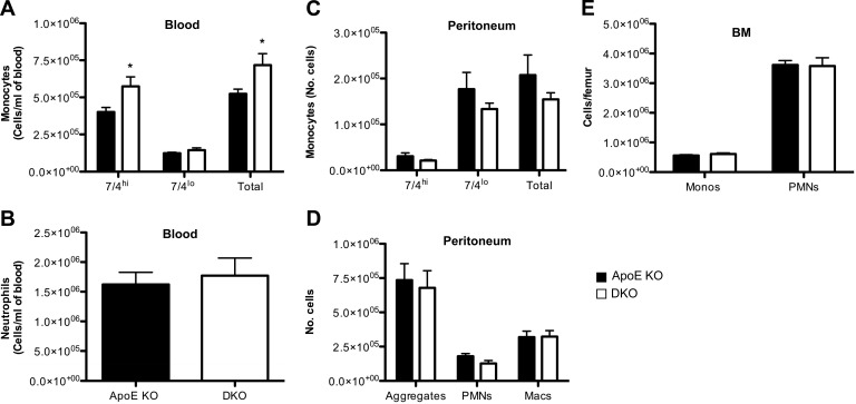
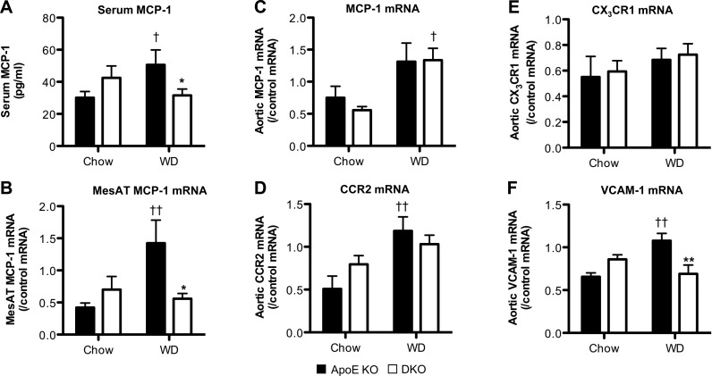
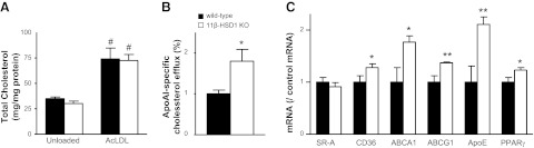
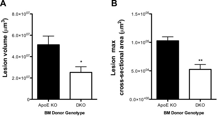
11β-Hydroxysteroid dehydrogenase type-1 (11β-HSD1) converts inert cortisone into active cortisol, amplifying intracellular glucocorticoid action. 11β-HSD1 deficiency improves cardiovascular risk factors in obesity but exacerbates acute inflammation. To determine the effects of 11β-HSD1 deficiency on atherosclerosis and its inflammation, atherosclerosis-prone apolipoprotein E-knockout (ApoE-KO) mice were treated with a selective 11β-HSD1 inhibitor or crossed with 11β-HSD1-KO mice to generate double knockouts (DKOs) and challenged with an atherogenic Western diet. 11β-HSD1 inhibition or deficiency attenuated atherosclerosis (74-76%) without deleterious effects on plaque structure. This occurred without affecting plasma lipids or glucose, suggesting independence from classical metabolic risk factors. KO plaques were not more inflamed and indeed had 36% less T-cell infiltration, associated with 38% reduced circulating monocyte chemoattractant protein-1 (MCP-1) and 36% lower lesional vascular cell adhesion molecule-1 (VCAM-1). Bone marrow (BM) cells are key to the atheroprotection, since transplantation of DKO BM to irradiated ApoE-KO mice reduced atherosclerosis by 51%. 11β-HSD1-null macrophages show 76% enhanced cholesterol ester export. Thus, 11β-HSD1 deficiency reduces atherosclerosis without exaggerated lesional inflammation independent of metabolic risk factors. Selective 11β-HSD1 inhibitors promise novel antiatherosclerosis effects over and above their benefits for metabolic risk factors via effects on BM cells, plausibly macrophages.


Figures

Figure 1.
Figure 1.
Selective 11β-HSD1 inhibition reduces atherosclerotic lesions in WD-fed ApoE-KO mice. A) i) On gross inspection, lesions were evident on the lesser curvature of the aortic arch (black arrow), in the innominate artery (red arrow) and at the origins of the left carotid and left subclavian arteries (black arrowheads). Lesions appeared to be smaller following 11β-HSD1 inhibition. ii) UST demonstrated large complex lesions in the innominate artery. iii) Immunohistochemistry with α-SMA identified smooth muscle cells. iv) PSR staining identified collagen. B) Image analyses of these sections confirmed that short-term selective 11β-HSD1 inhibition reduced innominate artery atherosclerosis in ApoE-KO mice. C) 11β-HSD1 inhibition did not alter the proportion of smooth muscle cells in atherosclerotic lesions in the innominate artery of ApoE-KO mice. However, collagen content was increased following 11β-HSD1 inhibition, and smaller areas devoid of cells or collagen, probably reflecting extracellular lipid pools, were reduced, compared with ApoE-KO controls (n=6/group). Original view ×40. *P < 0.05 vs. vehicle control.
Figure 2.
Figure 2.
11β-HSD1 deficiency reduces atherosclerotic lesions in WD-fed ApoE-KO mice. A) Atherosclerotic lesions were smaller in the innominate arteries of 4.5-mo-old DKO mice (n=10) fed high-cholesterol WD for 14 wk. Age-matched 11β-HSD1+/−, ApoE-KO het mice (n=3) also showed significant atheroprotection compared with age-matched ApoE-KO control mice (n=11). *P < 0.05, B) Lipid incorporation was also measured by en face Sudan IV staining of sections of the aortic arch. C) Quantification of Sudan IV staining showed that DKO mice had less lipid incorporation than ApoE-KO controls. (n=10–11/group). *P < 0.05, **P < 0.01 vs. ApoE-KO.
Figure 3.
Figure 3.
11β-HSD1 deletion attenuates macrophage and T-cell infiltration in atherosclerotic lesions. A) ApoE-KO and DKO mice (4.5 mo old) were fed WD for 14 wk, and Mac-2 or CD3 immunoreactivity was detected in the intimal lesion and the underlying media of innominate arteries. B) Cell numbers were quantified absolutely or normalized to the area of the lesion and media to give cell density (cells/mm2). Macrophage infiltration was unaltered in the lesions and media of DKO mice, when matched for lesion area. C) However, the overall numbers of macrophages were reduced in the lesions of DKO mice. D, E) T-cell infiltration (cells/mm2; D) or absolute T-cell numbers (E) were reduced into the lesions in DKO mice, but were unaltered in the media (n=5–7/group). Original view ×40. *P < 0.05, **P < 0.01, ***P < 0.001 compared with ApoE-KO.
Figure 4.
Figure 4.
Effect of 11β-HSD1 deficiency on inflammatory monocytes (7/4hiLy6GCD11b+) and neutrophils (7/4+Ly6G+CD11b+) in blood, BM, and spleen. DKO and ApoE-KO mice (4 wk old) were fed chow diet or WD for 14 wk, and then monocyte and neutrophil numbers in the blood (A), BM (B), and spleen (C) were quantified by flow cytometric analysis (n=5–21/group). *P < 0.05, **P < 0.01 for effects of genotype; P < 0.05 for effects of diet.
Figure 5.
Figure 5.
DKO monocytes and neutrophils are recruited to sites of inflammation during sterile peritonitis induced in chow-fed mice by thioglycollate. At 72 h postinjection of thioglycollate, peritoneal cells were lavaged and analyzed by flow cytometry. Quantitative analysis of blood monocyte subsets (inflammatory 7/4hiLy6GCD11b+ monocytes, resident 7/4loLy6GCD11b+ monocytes, and total monocytes; A); blood neutrophils (PMNs; 7/4+Ly6G+CD11b+; B), monocyte subsets in the peritoneum (C); peritoneal PMNs, macrophages (macs), and neutrophil:macrophage aggregate (F4/80+Ly6G+CD11b+) monocytes (monos; D); and PMNs in the BM (E); n = 9–11/group. *P < 0.05 vs. ApoE-KO.
Figure 6.
Figure 6.
Effect of 11β-HSD1 deficiency on circulating MCP-1 and mesenteric adipose tissue MCP-1 mRNA expression and mRNA expression of MCP-1, CCR2, CX3CR1, and VCAM-1 in the ascending aortas. ApoE-KO and DKO mice (4 wk old) were fed chow diet or WD for 14 wk. A, B) Serum MCP-1 level (A) and MCP-1 mRNA expression levels in MesAT (B) were elevated by WD in ApoE-KO control mice, but not in DKO mice. C, D) Furthermore, although aortic MCP-1 (C), and CCR2 (D) mRNA levels were elevated by WD, there was no difference by genotype. E) CX3CR1 mRNA transcript levels in the aorta were unaltered by genotype and diet. F) Aortic VCAM-1 mRNA levels were similar in ApoE-KO and DKO mice fed chow diet. However, while WD increased VCAM-1 mRNA in ApoE-KO mice, there was no change in DKO tissue (n=5–8/group). *P < 0.05, **P < 0.01 for effects of genotype; P < 0.05, ††P < 0.01 for effects of diet.
Figure 7.
Figure 7.
11β-HSD1 deficiency increases macrophage cholesterol efflux. A) BM-derived macrophages from 11β-HSD1-KO and C57BL/6J (wild-type) controls showed no difference in cholesterol content either basally (unloaded) or following loading with 50 mg/ml AcLDL for 24 h. Data are representative of 2 independent experiments each performed in triplicate. B) ApoAI-stimulated cholesterol efflux was increased in AcLDL-loaded macrophages from 11β-HSD1-KO mice. C) expression of mRNAs encoding cholesterol efflux transporters ApoE, ABCA1, and ABCG1 was increased in 11β-HSD1-KO macrophages, plausibly because of increased peroxisome proliferator-activated receptor γ (PPARγ). In contrast, SR-A mRNA was unaltered, though CD36 antigen was also increased. *P < 0.05, **P < 0.01 vs. wild type; #P < 0.05 vs. unloaded cells.
Figure 8.
Figure 8.
11β-HSD1 deletion in BM cells reduces atherosclerotic lesion size. Lethally irradiated ApoE-KO recipient mice were reconstituted with BM from ApoE-KO mice or DKO mice and fed WD for 12 wk. Formalin-fixed innominate arteries underwent OPT scanning. Analysis of lesion volumes (A) and lesion cross-sectional area (B) in the innominate arteries demonstrated that ApoE-KO recipient mice reconstituted with DKO BM cells exhibited significantly smaller lesions when compared to ApoE-KO mice reconstituted with ApoE-KO BM cells (n=5/group). *P < 0.05, **P < 0.01 vs. ApoE-KO mice reconstituted with ApoE-KO BM cells.

Similar articles

Cited by

References

    1. Libby P. (2002) Inflammation in atherosclerosis. Nature 420, 868–874 - PubMed
    1. Hansson G. K., Libby P. (2006) The immune response in atherosclerosis: a double-edged sword. Nat. Rev. Immunol. 6, 508–519 - PubMed
    1. Hansson G. K., Hermansson A. The immune system in atherosclerosis. Nat. Immunol. 12, 204–212 - PubMed
    1. Taleb S., Tedgui A., Mallat Z. Adaptive T cell immune responses and atherogenesis. Curr. Opin. Pharmacol. 10, 197–202 - PubMed
    1. Packard R. R., Lichtman A. H., Libby P. (2009) Innate and adaptive immunity in atherosclerosis. Semin. Immunopathol. 31, 5–22 - PMC - PubMed

Publication types

MeSH terms

Substances