

Attenuation of choroidal neovascularization by β(2)-adrenoreceptor antagonism
- PMID: 23303344
- PMCID: PMC3652586
- DOI: 10.1001/jamaophthalmol.2013.1476
Attenuation of choroidal neovascularization by β(2)-adrenoreceptor antagonism
Abstract
Objectives: To determine whether β-adrenergic blockade inhibits choroidal neovascularization (CNV) in a mouse model of laser-induced CNV and to investigate the mechanism by which β-adrenoreceptor antagonism blunts CNV.
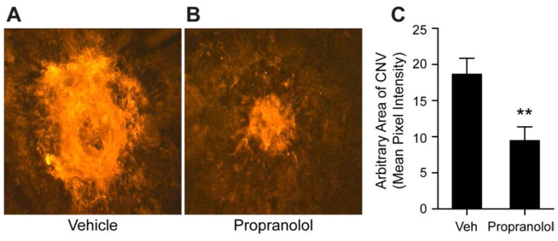
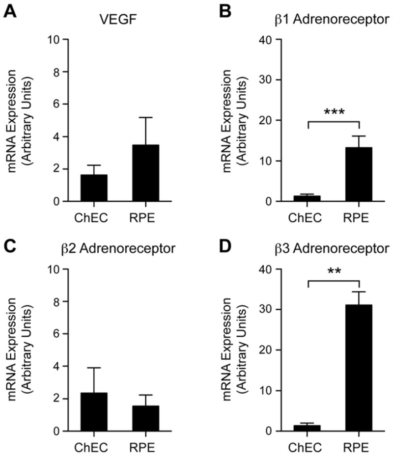
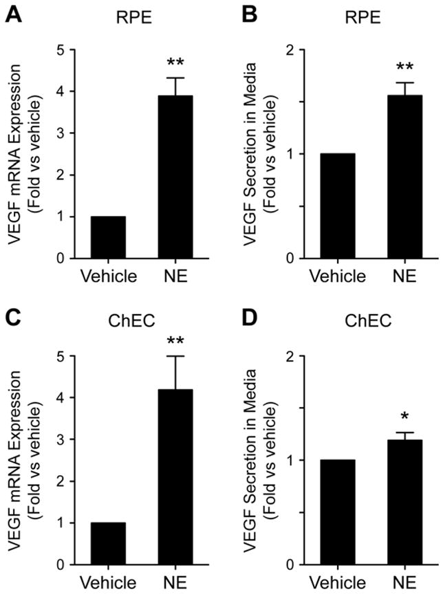
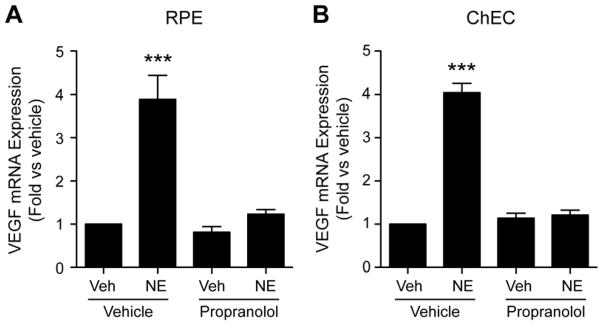
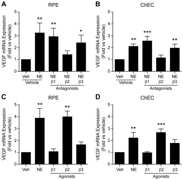
Design: Mice were subjected to laser burns, inducing CNV, and were treated with daily intraperitoneal injections of propranolol hydrochloride. Neovascularization was measured on choroidal-scleral flat mounts using intercellular adhesion molecule 2 immunofluorescence staining. The effect of β-adrenoreceptor signaling on expression of vascular endothelial growth factor (VEGF) was investigated using primary mouse choroidal endothelial cells (ChECs) and retinal pigment epithelial (RPE) cells. These cells were incubated with β-adrenoreceptor agonists and/or antagonists and assayed for Vegf messenger RNA and protein levels.
Setting: University of Wisconsin School of Medicine and Public Health.
Participants: Wild-type 6-week-old female C57BL/6j mice.
Main outcome measures: Inhibition of CNV after propranolol treatment and Vegf messenger RNA and protein expression after treatment with β-adrenoreceptor agonists and antagonists.
Results: Propranolol-treated mice demonstrated a 50% reduction in laser-induced CNV. Treatment with norepinephrine bitartrate stimulated Vegf messenger RNA expression and protein secretion in ChECs and RPE cells. This effect was blocked by β2-adrenoreceptor antagonism and mimicked by β2-adrenoreceptor agonists.
Conclusions: Attenuation of CNV is achieved by β-adrenergic blockade. The β2-adrenoreceptors regulate VEGF expression in ChECs and RPE cells.
Clinical relevance: Antagonists of β-adrenoreceptors are safe and well tolerated in patients with glaucoma and cardiovascular disease. Thus, blockade of β-adrenoreceptors may provide a new avenue to inhibit VEGF expression in CNV.
Figures
References
-
- Lim LS, Mitchell P, Seddon JM, Holz FG, Wong TY. Age-related macular degeneration. The Lancet. 2012;379(9827):1728–1738. - PubMed
-
- Lopez PF, Grossniklaus HE, Lambert HM, et al. Pathologic features of surgically excised subretinal neovascular membranes in age-related macular degeneration. American Journal of Ophthalmology. 1991;112(6):647–656. - PubMed
-
- Grossniklaus HE, Martinez JA, Brown VB, et al. Immunohistochemical and histochemical properties of surgically excised subretinal neovascular membranes in age-related macular degeneration. American Journal of Ophthalmology. 1992;114(4):464–472. - PubMed
-
- Thomas JW, Grossniklaus HE, Lambert HM, Aaberg TM, L’Hernault N. Ultrastructural features of surgically excised idiopathic subfoveal neovascular membranes. Retina (Philadelphia, Pa ) 1993;13(2):93–98. - PubMed
-
- Lopez PF, Lambert HM, Grossniklaus HE, Sternberg P. Well-defined subfoveal choroidal neovascular membranes in age-related macular degeneration. Ophthalmology. 1993;100(3):415– 422. - PubMed
Publication types
MeSH terms
Substances
Grants and funding
LinkOut - more resources
Full Text Sources
Other Literature Sources
