. 2013 Mar;11(3):219-29.
doi: 10.1158/1541-7786.MCR-12-0547-T. Epub 2013 Jan 9.

A modified HSP70 inhibitor shows broad activity as an anticancer agent

Affiliations

A modified HSP70 inhibitor shows broad activity as an anticancer agent

Gregor M Balaburski et al. Mol Cancer Res. 2013 Mar.

Abstract

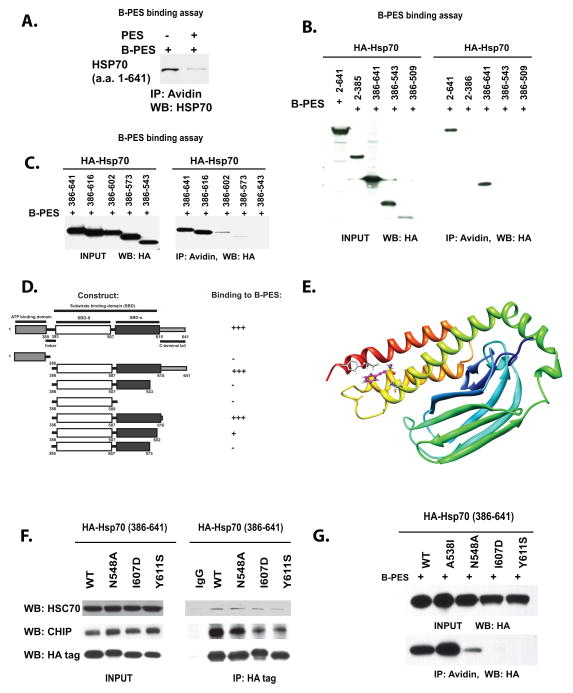
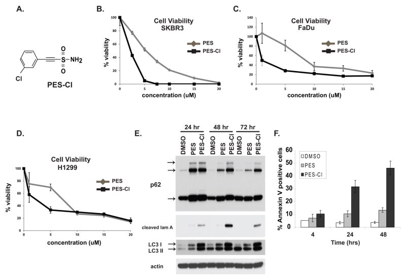
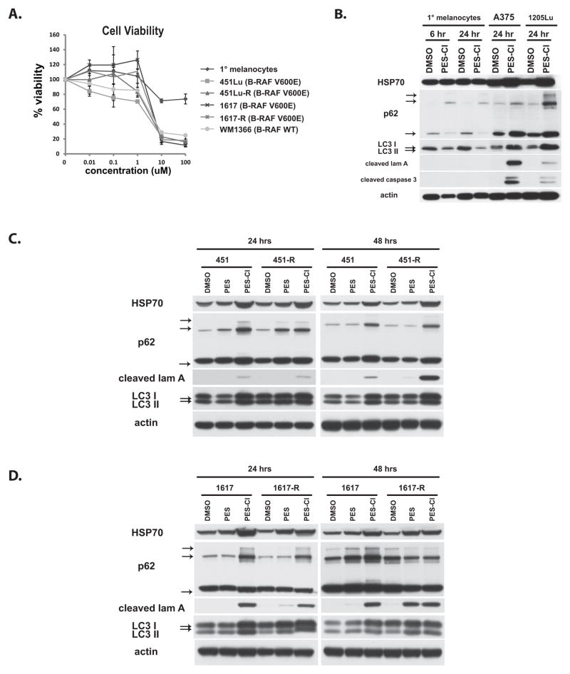
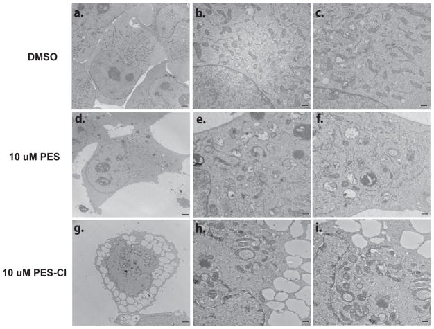
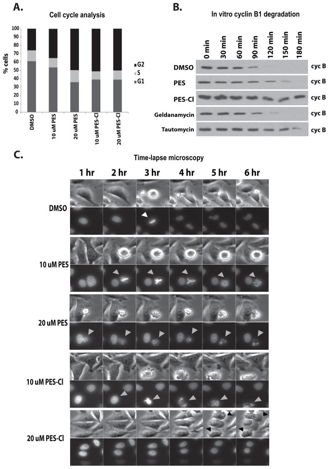
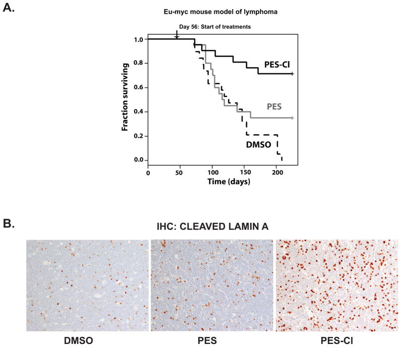
The stress-induced HSP70 is an ATP-dependent molecular chaperone that plays a key role in refolding misfolded proteins and promoting cell survival following stress. HSP70 is marginally expressed in nontransformed cells, but is greatly overexpressed in tumor cells. Silencing HSP70 is uniformly cytotoxic to tumor but not normal cells; therefore, there has been great interest in the development of HSP70 inhibitors for cancer therapy. Here, we report that the HSP70 inhibitor 2-phenylethynesulfonamide (PES) binds to the substrate-binding domain of HSP70 and requires the C-terminal helical "lid" of this protein (amino acids 573-616) to bind. Using molecular modeling and in silico docking, we have identified a candidate binding site for PES in this region of HSP70, and we identify point mutants that fail to interact with PES. A preliminary structure-activity relationship analysis has revealed a derivative of PES, 2-(3-chlorophenyl) ethynesulfonamide (PES-Cl), which shows increased cytotoxicity and ability to inhibit autophagy, along with significantly improved ability to extend the life of mice with pre-B-cell lymphoma, compared with the parent compound (P = 0.015). Interestingly, we also show that these HSP70 inhibitors impair the activity of the anaphase promoting complex/cyclosome (APC/C) in cell-free extracts, and induce G2-M arrest and genomic instability in cancer cells. PES-Cl is thus a promising new anticancer compound with several notable mechanisms of action.


Conflict of interest statement

The authors declare there are no conflicts of interest

Figures

Figure 1
Figure 1. Binding to PES requires residues within a C-terminal helical bundle of HSP70
A) Pull-down assay of purified, recombinant HSP70 with biotinylated PES (B-PES). 238 nM of purified HSP70 protein was incubated with 0.25 mM biotinylated PES (B-PES), in the absence or the presence of 0.125 mM untagged PES. Immunoprecipitation-western blot (IP-WB) analysis reveals direct interaction between B-PES and HSP70. B) Pull-down assays of H1299 cells transfected with HA-tagged full-length and deletion mutants of HSP70, followed by treatment with 20 uM biotinylated Biotin-PES (B-PES). The HA-tagged mutants were precipitated with avidin beads, eluted and detected with an anti-HA antibody following imunoblotting. Input is shown on the left panel; immunoprecipitation (IP) with avidin is depicted on the right. C) Pull-down assays of additional C-terminal deletion mutants of HA-tagged HSP70, performed as in B. Input is shown in the left panel; immunoprecipitation with avidin beads is depicted in the right panel. D) Summary of the results of multiple independent B-PES binding assays performed as in B. E) Molecular model of the substrate-binding domain of human HSP70; depiction of one pose for PES binding from an in silico analysis of potential PES docking sites. The depicted pose highlights three potential interaction residues; clockwise from top these are Y611, I607 and N548. F) Immunoprecipitation-western analysis depicting the interaction of HSP70 point mutants with the co-chaperones CHIP and Hsc70. Input is shown on the left, and the co-precipitated CHIP and Hsc70 are depicted on the right. Mouse immunoglobulin (IgG) is the negative control. G) PES-binding analysis of C-terminal point mutants of HA-tagged HSP70, performed as in (B). Input is shown on the top panel, immunoprecipitation with avidin beads (IP) is on the bottom.
Figure 2
Figure 2. PES-Cl shows increased cytotoxicity and superior ability to inhibit autophagy
A) Chemical structure of PES-Cl. B, C, D) Cell viability analysis (MTT) in the breast carcinoma line SKBR3 (B), the head and neck squamous cell carcinoma line FaDu (C), and the lung adenocarcinoma line H1299 (D), treated with PES or PES-Cl. Cells were treated with the indicated dose for forty eight hours. Data are the averaged results from three independent experiments, and error bars mark standard deviations. E) Immunoblot analysis for the autophagy markers p62SQSTM1 and LC3 I and II, as well as caspase-cleaved lamin A (cleaved lam A) in H1299 cells treated with 10 uM PES or PES-Cl for the indicated timepoints. The arrows mark monomeric and oligomeric forms of p62, as well as cleaved and lipidated forms of LC3 I and II. F) Quantification of Annexin V positive H1299 cells following treatment with 10 uM PES or PES-Cl for the indicated timepoints. Data are the averaged results from three independent experiments, and error bars mark standard deviations.
Figure 3
Figure 3. Efficacy of PES-Cl in melanoma
A) Cell viability analysis (MTT) of primary neonatal epidermal melanocytes (black diamonds), and melanoma lines 451Lu (BRAF V600E mutant), 1617 (BRAF V600E mutant) and WM1366 (wt BRAF, mutant NRas) as well as two melanoma lines that are resistant to the BRAF inhibitor SB-590885 (451Lu-R and 1617-R). Cells were treated with the indicated doses for forty eight hours. Data are the averaged results from three independent experiments, and error bars mark standard deviations. B) Immunoblot analysis of autophagy (p62SQSTM1, LC3 I and II) and apoptosis (cleaved lamin A, cleaved caspase 3) markers in primary (1°) melanocytes and melanomas A375 and 1205Lu treated with 10 uM PES-Cl for the indicated times. The arrows mark monomeric and oligomeric forms of p62, as well as LC3 I and II. C, D) Immunoblot analysis of autophagy (p62SQSTM1, LC3 I and II) and apoptosis (cleaved lamin A) markers in parental 451 and BRAF-inhibitor resistant 451-R melanoma lines (C) as well as 1617 parental and BRAF-inhibitor resistant 1617-R melanoma lines (D) treated with 10 uM PES or PES-Cl for the indicated times. The arrows mark monomeric and oligomeric forms of p62, as well as LC3 I and II.
Figure 4
Figure 4. Enhanced autophagosome accumulation in cells treated with PES-Cl
Electron micrographs of H1299 cells treated with 10 uM PES or PES-Cl for twenty four hours. Scale bar in a, d and g is 2 microns. Scale bar in b, c, e, f, h and i is 500 nm.
Figure 5
Figure 5. Treatment with PES or PES-Cl causes G2/M cell cycle arrest and inhibition of APC/C function
A) Asynchronous HeLa cells were treated with DMSO, PES or PES-Cl at the doses indicated and analyzed for cell cycle stage by propidium iodide staining. Data are the average results from three independent experiments. B) The degradation of cyclin B in synchronized HeLa cell extracts was monitored by immunoblot analysis. Equal amounts of extract were analyzed at the timepoints indicated following treatment of extracts with DMSO or 10 uM PES or PES-Cl. Treatment with 10 uM Geldanamycin or 150 nM Tautomycin served as positive controls. C) Time-lapse video microscopy of HeLa cells that were released from synchronization in S phase following thymidine block and treated with the indicated concentrations of DMSO, PES or PES-Cl for 3 hours. Top panels: phase contrast images. Bottom panels: GFP-H2B fluorescence. Gray arrowheads: treated cells with chromosome abnormalities. Representative images at the indicated time points are shown.
Figure 6
Figure 6. PES-Cl significantly promotes survival in the Eμ-myc mouse model of lymphoma
A) Eight week old Eμ-myc mice were treated once per week with intra-peritoneal injections of DMSO (n=19), 20 mg/kg PES (n=20) or 20 mg/kg PES-Cl (n=21), for a total of 20 treatments. All treatments were terminated at day 209. Log rank analysis of data: DMSO vs. PES (p = 0.0839); PES vs. PES-Cl (p = 0.0151) and DMSO vs. PES-Cl (p = 4.7 × 10⁻⁶). B) Immunohistochemistry for cleaved lamin A in lymph nodes from tumor-bearing Eμ-myc mice twenty-four hours after treatment with DMSO, PES or PES-Cl (10% DMSO in PBS, or 20 mg/kg PES or PES-Cl in 10% DMSO). Samples shown are from equal-sized tumors from 2 mice each and are representative of multiple views from each tumor.

Similar articles

Cited by

References

    1. Daugaard M, Rohde M, Jäättelä M. The heat shock protein 70 family: Highly homologous proteins with overlapping and distinct functions. FEBS Lett. 2007;581:3702–10. - PubMed
    1. Hunt CR, Dix DJ, Sharma GG, Pandita RK, Gupta A, Funk M, Pandita TK. Genomic instability and enhanced radiosensitivity in Hsp70.1- and Hsp70.3-deficient mice. Mol Cell Biol. 2004;24:899–911. - PMC - PubMed
    1. Powers MV, Jones K, Barillari C, Westwood I, van Montfort RL, Workman P. Targeting HSP70: the second potentially druggable heat shock protein and molecular chaperone? Cell Cycle. 2010;9:1542–50. - PubMed
    1. Rohde M, Daugaard M, Jensen MH, Helin K, Nylandsted J, Jäättelä M. Members of the heat-shock protein 70 family promote cancer cell growth by distinct mechanisms. Genes Dev. 2005;19:570–82. - PMC - PubMed
    1. Schmitt E, Maingret L, Puig PE, Rerole AL, Ghiringhelli F, Hammann A, Solary E, Kroemer G, Garrido C. Heat shock protein 70 neutralization exerts potent antitumor effects in animal models of colon cancer and melanoma. Cancer Res. 2006;66:4191–7. - PubMed

Publication types

MeSH terms