Comparative Study
. 2013 Mar 15;304(6):F770-80.
doi: 10.1152/ajprenal.00646.2012. Epub 2013 Jan 9.

Role of RACK1 in the differential proliferative effects of neuropeptide Y(1-36) and peptide YY(1-36) in SHR vs. WKY preglomerular vascular smooth muscle cells

Affiliations
Comparative Study

Role of RACK1 in the differential proliferative effects of neuropeptide Y(1-36) and peptide YY(1-36) in SHR vs. WKY preglomerular vascular smooth muscle cells

Dongmei Cheng et al. Am J Physiol Renal Physiol. .

Abstract

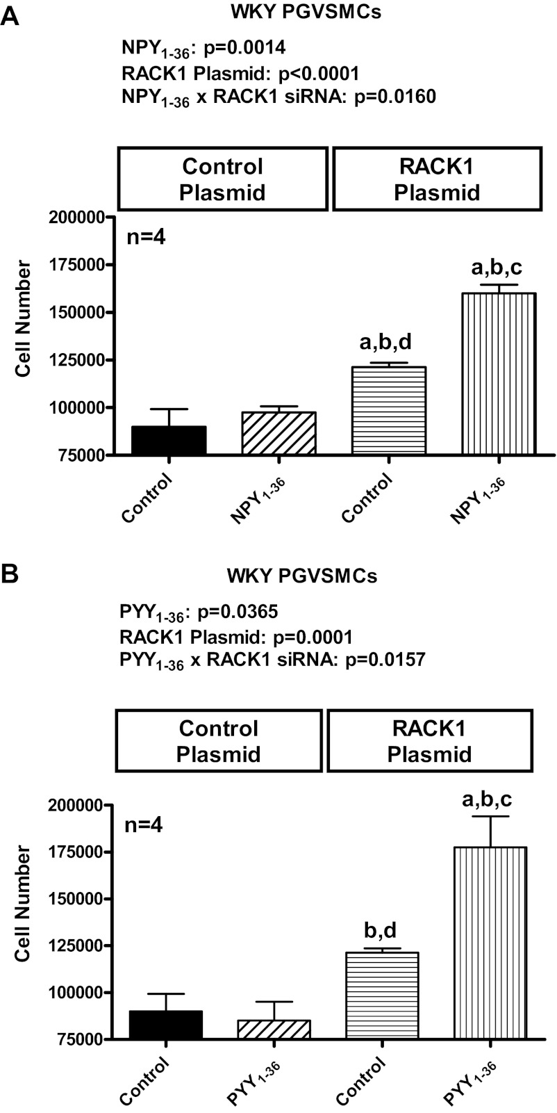
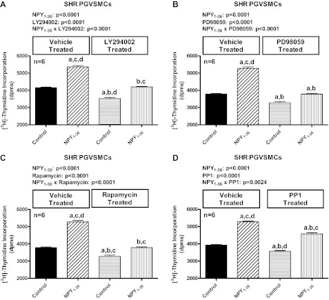
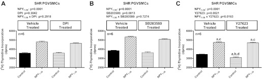
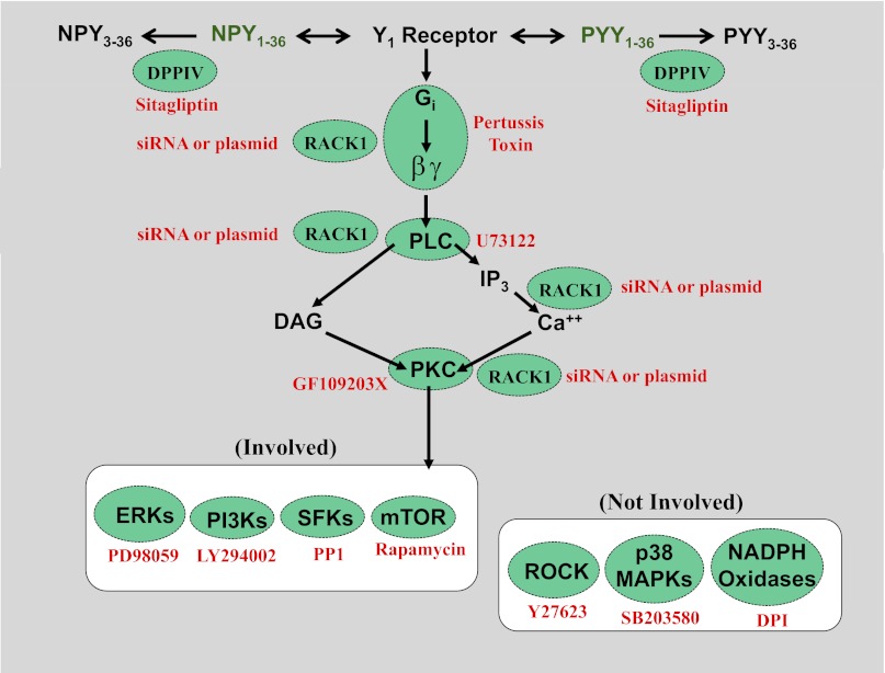
Previous studies show that neuropeptide Y(1-36) (NPY(1-36)) and peptide YY(1-36) (PYY(1-36)), by engaging Y1 receptors, stimulate proliferation of spontaneous hypertensive rat (SHR) preglomerular vascular smooth muscle cells (PGVSMCs). In contrast, these peptides have little effect on proliferation of Wistar-Kyoto (WKY) PGVSMCs. Why SHR and WKY PGVSMCs differ in this regard is unknown. Because receptor for activated C kinase 1 (RACK1) can modulate cell proliferation, we tested the hypothesis that differences in RACK1 levels/localization may explain the differential response of SHR vs. WKY PGVSMCs to NPY(1-36) and PYY(1-36). Western blotting for RACK1 in subcellular fractions of cultured SHR and WKY PGVSMCs demonstrated increased levels of RACK1 in the membrane and cytoskeletal subcellular fractions of SHR vs. WKY PGVSMCs. NPY(1-36) and PYY(1-36) stimulated proliferation of SHR PGVSMCs, and siRNA knockdown of RACK1 abrogated this effect. Neither NPY(1-36) nor PYY(1-36) stimulated the proliferation of WKY PGVSMCs. However, in WKY PGVSMCs treated with a RACK1 plasmid, both NPY(1-36) and PYY(1-36) stimulated proliferation. In SHR PGVSMCs, inhibitors of the G(i)/phospholipase C/PKC pathway (a pathway known to be organized by RACK1) attenuated the ability of NPY(1-36) to stimulate the proliferation of SHR PGVSMCs. Our results suggest that RACK1 modulates the ability of PGVSMCs to respond to the proliferative actions of NPY(1-36) and PYY(1-36)and differences in RACK1 levels/localization account for, in part, differential proliferative responses to NPY(1-36) and PYY(1-36) in SHR vs. WKY PGVSMCs. Because dipeptidyl peptidase IV inhibitors increase NPY(1-36) and PYY(1-36) levels, our findings have implications for the use of such drugs in diabetic patients.


Figures

Fig. 1.
Fig. 1.
A: Western blots depict subcellular localization of receptor for activated C kinase 1 (RACK1) (green bands; 36 kDa) expression in preglomerular vascular smooth muscle cells (PGVSMCs) from spontaneously hypertensive rats (SHR) vs. Wistar-Kyoto normotensive rats (WKY). Also shown are the signals for β-actin (red bands), which were used as loading controls. Subcellular fractions from SHR vs. WKY were processed on the same gel with the same amount of total protein. Bar graphs depict RACK1 expression in the membrane (B), cytoskeletal (C), nuclear (D), and cytosolic (E) fractions in SHR vs. WKY PGVSMCs. P values are from Student's unpaired t-test. Values are expressed as means ± SE.
Fig. 2.
Fig. 2.
Bar graphs depict effects of RACK1 siRNA vs. negative control siRNA (control) on RACK1 mRNA (A) and protein (B) expression in PGVSMCs from SHR. For both negative control siRNA-treated cells (control) and RACK1 siRNA-treated cells (RACK1 siRNA), expression levels of RACK1 mRNA and protein were normalized to β-actin mRNA or β-actin protein, respectively. For mRNA, this was performed by subtracting the threshold cycle (Ct) for β-actin mRNA from the Ct for RACK1 mRNA to calculate 2ΔCt. For protein, this was performed by dividing the optical density of the RACK1 band by the optical density of the β-actin band. For both mRNA and protein, the readings in the RACK1 siRNA cells were expressed as a % of the average reading for the control cells. Values are expressed as means ± SE, and P values are from unpaired Student's t-test.
Fig. 3.
Fig. 3.
Bar graphs illustrate the effects of either neuropeptide Y1–36 (NPY1–36; 10 nmol/l) (A) or peptide YY1–36 (PYY1–36; 10 nmol/l) (B) on cell number in PGVSMCs from SHR in the absence and presence of RACK1 siRNA. The PP-fold peptides were coadministered with sitagliptin (1 μM) to block their metabolism and inactivation. P values in panels are from two-factor ANOVA, and letters above bars (a, b, c, d) indicate significantly different [Fisher's least significant difference (LSD) test] from group without RACK1 siRNA and without NPY1–36 or PYY1–36 (a), group without RACK1 siRNA but with NPY1–36 or PYY1–36 (b), group with RACK1 siRNA but without NPY1–36 or PYY1–36 (c), or group with siRNA and with NPY1–36 or PYY1–36 (d). “Control siRNA” indicates nontargeting siRNA. Values are expressed as means ± SE.
Fig. 4.
Fig. 4.
The cDNA fragment encoding rat RACK1 was obtained by PCR using rat PGVSMC cDNA as a template with primer set: sense, 5′-ctaagctatccggtgccatc-3′ and antisense, 5′-gcgggtaccaatagtcacctg-3′. The RACK1 cDNA was then cloned into pcDNA 3.1/V5-His-TOPO vector by using pcDNA3.1/V5-His TOPO TA expression kit (Life Technologies) as per the manufacturer's instructions. The clone was verified by DNA sequencing. Western blot depicts expression of V5-tagged RACK1 in WKY PGVSMCs treated with RACK1-expressing plasmid vs. and empty (control) plasmid.
Fig. 5.
Fig. 5.
Bar graphs illustrate the effects of either NPY1–36 (10 nmol/l; A) or peptide YY1–36 (PYY1–36; 10 nmol/l; B) on cell number in PGVSMCs from WKY in the absence and presence of a RACK1-expressing plasmid. The polypeptide (PP)-fold peptides were coadministered with sitagliptin (1 μM) to block their metabolism and inactivation. P values in panels are from two-factor ANOVA, and letters above bars indicate significantly different (Fisher's LSD test) from group without RACK1-expressing plasmid and without NPY1–36 or PYY1–36 (a), group without RACK1-expressing plasmid but with NPY1–36 or PYY1–36 (b), group with RACK1-expressing plasmid but without NPY1–36 or PYY1–36 (c), or group with RACK1-expressing plasmid and with NPY1–36 or PYY1–36 (d). “Control plasmid” indicates empty plasmid not expressing RACK1. Values are expressed as means ± SE.
Fig. 6.
Fig. 6.
Bar graphs illustrate the effects of neuropeptide Y1–36 (NPY1–36; 10 nmol/l) on [3H]thymidine incorporation in PGVSMCs from SHR in the absence and presence of pertussis toxin (A; 100 ng/ml), U73122 (B; 10 μmol/l), and GF109203X (C; 10 μmol/l). NPY1–36 was coadministered with sitagliptin (1 μM) to block its metabolism and inactivation. P values in panels are from two-factor ANOVA, and letters above bars indicate significantly different (Fisher's LSD test) from group without inhibitor and without NPY1–36 (a), group without inhibitor but with NPY1–36 (b), group with inhibitor but without NPY1–36 (c), or group with inhibitor and with NPY1–36 (d). Values are expressed as means ± SE.
Fig. 7.
Fig. 7.
Bar graphs illustrate the effects of neuropeptide Y1–36 (NPY1–36; 10 nmol/l) on [3H]thymidine incorporation in PGVSMCs from SHR in the absence and presence of LY294002 (A; 10 μmol/l), PD98059 (B; 10 μmol/l), rapamycin (C; 0.2 μmol/l), and PP1 (D; 0.1 μmol/l). NPY1–36 was coadministered with sitagliptin (1 μM) to block its metabolism and inactivation. P values in panels are from two-factor ANOVA, and letters above bars indicate significantly different (Fisher's LSD test) from group without inhibitor and without NPY1–36 (a), group without inhibitor but with NPY1–36 (b), group with inhibitor but without NPY1–36 (c), or group with inhibitor and with NPY1–36 (d). Values are expressed as means ± SE.
Fig. 8.
Fig. 8.
Bar graphs illustrate the effects of NPY1–36 (10 nmol/l) on [3H]thymidine incorporation in PGVSMCs from SHR in the absence and presence of diphenyleneiodonium (A; DPI, 10 μmol/l), SB203580 (B; 10 μmol/l), and Y27623 (C; 10 μmol/l). NPY1–36 was coadministered with sitagliptin (1 μM) to block its metabolism and inactivation. P values in panels are from two-factor ANOVA, and letters above bars indicate significantly different (Fisher's LSD test) from group without inhibitor and without NPY1–36 (a), group without inhibitor but with NPY1–36 (b), group with inhibitor but without NPY1–36 (c), or group with inhibitor and with NPY1–36 (d). Values are expressed as means ± SE.
Fig. 9.
Fig. 9.
Summary of signaling pathway by which PP-fold peptides increase cell proliferation. Components in green circles indicate sites of interventions (noted in red next to the green circles) conducted in this study. DPPIV, dipeptidyl peptidase IV; NPY1–36, neuropeptide Y1–36; PYY1–36, peptide YY1–36; NPY3–36, neuropeptide Y3–36; PYY3–36, peptide YY3–36; Gi, inhibitory G-protein; βγ, βγ subunits of G protein; PLC, phospholipase C; PKC, protein kinase C; DAG, diacyglycerol; IP3, inositol trisphosphate; Ca2+, calcium; RACK1, receptor for activated C kinase 1; ERKs, extracellular-signal-regulated kinases; PI3Ks, phosphatidylinositol 3-kinases; DPI, diphenyleneiodonium; SFKs, src family kinases; mTOR, mammalian target of rapamycin; ROCK, Rho-associated protein kinase; p38 MAPKs, p38 mitogen-activated protein kinases; NADPH oxidase, nicotinamide adenine dinucleotide phosphate oxidases.

Similar articles

Cited by

References

    1. Alessi DR, Cuenda A, Cohen P, Dudley DT, Saltiel AR. PD 098059 is a specific inhibitor of the activation of mitogen-activated protein kinase kinase in vitro and in vivo. J Biol Chem 270: 27489– 27494, 1995 - PubMed
    1. Ambasta RK, Ai X, Emerson CP., Jr Quail Sulf1 function requires asparagine-linked glycosylation. J Biol Chem 282: 34492– 34499, 2007 - PubMed
    1. Anini Y, Fu-Cheng X, Cuber JC, Kervran A, Chariot J, Roz C. Comparison of the postprandial release of peptide YY and proglucagon-derived peptides in the rat. Pflügers Arch 438: 299– 306, 1999 - PubMed
    1. Armstrong DN, Ballantyne GH, Adrian TE, Bilchik AJ, McMillen MA, Modlin IM. Adaptive increase in peptide YY and enteroglucagon after proctocolectomy and pelvic ileal reservoir construction. Dis Colon Rectum 34: 119– 125, 1991 - PubMed
    1. Barnett A. DPP-4 inhibitors and their potential role in the management of type 2 diabetes. Int J Clin Pract 60: 1454– 1470, 2006 - PubMed

Publication types

MeSH terms

LinkOut - more resources