Review
. 2013 Feb;34(1):130-62.
doi: 10.1210/er.2012-1043. Epub 2013 Jan 9.

Progesterone action in endometrial cancer, endometriosis, uterine fibroids, and breast cancer

Affiliations
Review

Progesterone action in endometrial cancer, endometriosis, uterine fibroids, and breast cancer

J Julie Kim et al. Endocr Rev. 2013 Feb.

Abstract

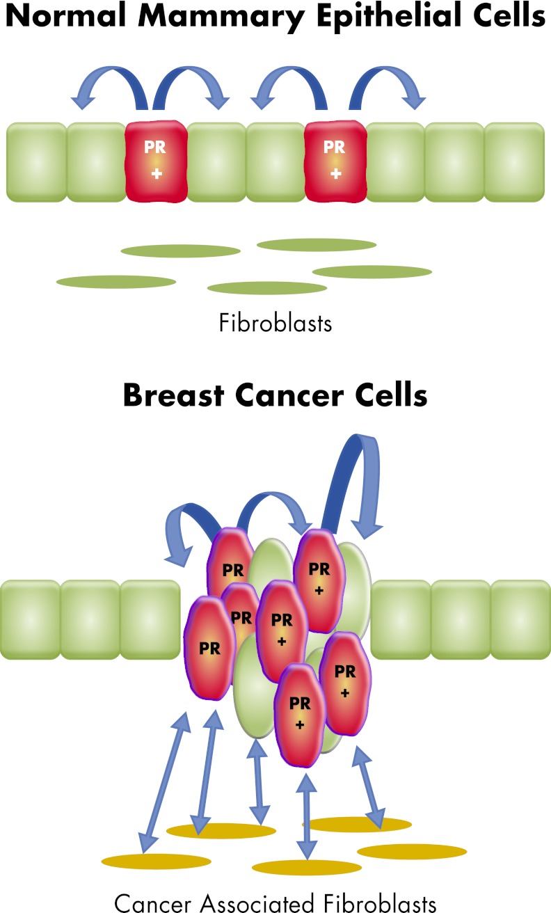
Progesterone receptor (PR) mediates the actions of the ovarian steroid progesterone, which together with estradiol regulates gonadotropin secretion, prepares the endometrium for implantation, maintains pregnancy, and differentiates breast tissue. Separation of estrogen and progesterone actions in hormone-responsive tissues remains a challenge. Pathologies of the uterus and breast, including endometrial cancer, endometriosis, uterine fibroids, and breast cancer, are highly associated with estrogen, considered to be the mitogenic factor. Emerging evidence supports distinct roles of progesterone and its influence on the pathogenesis of these diseases. Progesterone antagonizes estrogen-driven growth in the endometrium, and insufficient progesterone action strikingly increases the risk of endometrial cancer. In endometriosis, eutopic and ectopic tissues do not respond sufficiently to progesterone and are considered to be progesterone-resistant, which contributes to proliferation and survival. In uterine fibroids, progesterone promotes growth by increasing proliferation, cellular hypertrophy, and deposition of extracellular matrix. In normal mammary tissue and breast cancer, progesterone is pro-proliferative and carcinogenic. A key difference between these tissues that could explain the diverse effects of progesterone is the paracrine interactions of PR-expressing stroma and epithelium. Normal endometrium is a mucosa containing large quantities of distinct stromal cells with abundant PR, which influences epithelial cell proliferation and differentiation and protects against carcinogenic transformation. In contrast, the primary target cells of progesterone in the breast and fibroids are the mammary epithelial cells and the leiomyoma cells, which lack specifically organized stromal components with significant PR expression. This review provides a unifying perspective for the diverse effects of progesterone across human tissues and diseases.


Figures

Figure 1.
Figure 1.
Paracrine regulation of mouse endometrial epithelial proliferation by progesterone (P4). Four types of chimeric uteri were generated with stroma (S) and epithelium (E) from wild-type (Wt) and PRKO uteri. In Wt-E + Wt-S uterus, P4 acts on epithelium through PR in both epithelium and stroma, whereas P4 has no effect on PRKO-E + PRKO-S uterus. P4 action is mediated only by epithelial or stromal PR in the Wt-E + PRKO-S and PRKO-E + Wt-S uteri, respectively. Immunohistochemistry for PR (brown staining) confirms the epithelial/stromal-specific expression of PR. As assessed by incorporation of [3H]thymidine (black dots), estradiol (E2) equally stimulates epithelial proliferation in all four types of uteri. In contrast, P4 inhibits epithelial proliferation only in uterus in which PR was expressed in stroma. Epithelial PR is not necessary for the antiproliferative action of P4 on epithelial cells. [Modified from T. Kurita et al.: Stromal progesterone receptors mediate the inhibitory effects of progesterone on estrogen-induced uterine epithelial cell deoxyribonucleic acid synthesis. Endocrinology 139:4708–4713, 1998 (13), with permission. © The Endocrine Society.]
Figure 2.
Figure 2.
The normal and neoplastic endometrium. Normal proliferative phase endometrium, hyperplasia, grade 1 and grade 3 endometrial adenocarcinoma are shown. Endometrial hyperplasia is characterized by an increase in the epithelium-stroma ratio, irregularities in gland shape, multiple epithelial cell layers, and variation in gland size. Endometrial adenocarcinoma is exhibited by a confluent glandular epithelial pattern in which individual glands appear “back-to-back” with an altered fibroblastic stroma (desmoplastic stromal response). Grade 3 endometrial cancer exhibits more solid growth with a loss in gland-like architecture. Only sheets of malignant epithelial cells are present.
Figure 3.
Figure 3.
Ovarian endometriosis. Ectopic endometriotic tissue in this ovarian hilar region shows endometrioid glands and stroma with hemorrhage and hemosiderin deposition, consistent with endometriosis.
Figure 4.
Figure 4.
Proposed model for progesterone and retinoid action in endometrium and endometriosis. The deficient genes and pathways in endometriosis compared with normal endometrium were indicated using arrows and dotted lines. In endometrium, progesterone action is mediated by PR in stromal cells. Blood vessels that transport progesterone (P4) are adjacent to stromal cells. Stromal PR activated by circulating P4 produces a number of paracrine factors including RA. RA and other paracrine factors stimulate differentiation and oppose estradiol (E2)-dependent proliferation in neighboring epithelial cells. Moreover, RA stimulates the enzyme, HSD17B2, which converts biologically active E2 to estrogenically weak estrone (E1). On the other hand, significantly lower stromal PR expression causes deficient formation of RA and other paracrine factors. Consequently, epithelial cells differentiate poorly and do not express HSD17B2, leading to accumulation of E2. The mechanisms for retinoid transport between these two cell types are not well understood. STRA6 in endometrial stromal cells serve as a receptor for the uptake of RBP-bound retinol from the circulation. Retinol is converted to RA, which is then transported to nuclear RARs by the shuttling protein named cellular RA binding protein-2 (CRABP2). RA-RAR enhances differentiation and apoptosis in endometrial stromal cells. PR, in a ligand-independent fashion, induces stromal STRA6 and CRABP2. Deficiency of PR, STRA6, and CRABP2 disrupts this pathway in endometriosis. Moreover, expression of the RA-metabolizing enzymes CYP26B1 and CYP26A1 in stromal and epithelial compartments are perturbed in endometriosis. 4OH-RA, 4-Hydroxy-RA.
Figure 5.
Figure 5.
Myometrium and leiomyoma. Myometrium exhibits organized smooth muscle cell bundles with dedicated and enriched vasculatures. Leiomyoma is composed of smooth muscle cells that are disorganized in storiform and whorl-appearing patterns with abundant extracellular collagen and significantly reduced vessel density, in comparison to matched myometrium.
Figure 6.
Figure 6.
Expression of ER and PR in fibroid xenografts and its correlation with proliferation. Fibroid xenografts were treated with estradiol (E2) plus progesterone (P4) for 2 wk. DAPI (blue), Ki67 (red), and ERα (upper panels) or PR (lower panels) immunofluorescent staining was done. Ki67-positive cells (arrows) are also positive for ERα and PR. E2 induces expression of PR, and P4 action via PR increases the size of fibroid through accumulation of ECM, proliferation, and hypertrophy of fibroid cells. [Modified from H. Ishikawa et al.: Progesterone is essential for maintenance and growth of uterine leiomyoma. Endocrinology 151:2433–2442, 2010 (7), with permission. © The Endocrine Society.]
Figure 7.
Figure 7.
Progesterone action in the normal and malignant breast epithelium. In the normal mammary tissue and breast cancer, the primary target cell of progesterone action is in the glands or malignant epithelial cells, with no evidence of specifically organized stromal components that express PR. Only a small population of normal epithelial cells express PR. Proliferation in response to progesterone occurs in the cells that are negative for PR as a result of paracrine actions, whereas PR-expressing cells do not proliferate. In breast cancer cells, progesterone acts directly on PR-expressing cells and promotes proliferation. The role of surrounding stromal fibroblasts in mediating the progesterone-drive proliferation is unclear.
Figure 8.
Figure 8.
Diverse actions of progesterone. Estradiol (E2) action via stromal ERα is critical for the development of endometrial cancer. By the same token, E2-ERα-dependent stimulation of stromal PR expression is the key protective mechanism against E2-induced carcinogenesis. Progesterone (P4) acts via stromal PR to oppose this carcinogenic effect of E2. Deficient P4 synthesis or defective stromal cell function may play key roles in the development of endometrial cancer. Interestingly, there is a progressive decline in stromal cell numbers from premalignant endometrial hyperplasia to high grade cancer, which is completely devoid of any stromal cells. In endometriosis, stromal cells are deficient in ERα but rich in ERβ. ERβ suppresses PR expression. In endometriosis, levels of PR are dramatically lower and response to P4 is blunted, resulting in promotion of survival and deficient epithelial differentiation. In breast cancer and uterine leiomyomas (fibroids), P4 acts directly via PR in malignant epithelial cells or leiomyoma smooth muscle cells with little evidence of specifically organized stromal components that mediate P4 action. In both tissues, P4 and PR are tumorigenic. In uterine leiomyoma smooth muscle cells, the primary role of E2 is to maintain PR expression.

Similar articles

Cited by

References

    1. Lee K, Jeong J, Tsai MJ, Tsai S, Lydon JP, DeMayo FJ. 2006. Molecular mechanisms involved in progesterone receptor regulation of uterine function. J Steroid Biochem Mol Biol 102:41–50 - PMC - PubMed
    1. Lydon JP, DeMayo FJ, Funk CR, Mani SK, Hughes AR, Montgomery CA, Jr, Shyamala G, Conneely OM, O'Malley BW. 1995. Mice lacking progesterone receptor exhibit pleiotropic reproductive abnormalities. Genes Dev 9:2266–2278 - PubMed
    1. Graham JD, Clarke CL. 1997. Physiological action of progesterone in target tissues. Endocr Rev 18:502–519 - PubMed
    1. Mulac-Jericevic B, Lydon JP, DeMayo FJ, Conneely OM. 2003. Defective mammary gland morphogenesis in mice lacking the progesterone receptor B isoform. Proc Natl Acad Sci USA 100:9744–9749 - PMC - PubMed
    1. Mulac-Jericevic B, Mullinax RA, DeMayo FJ, Lydon JP, Conneely OM. 2000. Subgroup of reproductive functions of progesterone mediated by progesterone receptor-B isoform. Science 289:1751–1754 - PubMed

Publication types