. 2013 Feb 1;41(4):2202-15.
doi: 10.1093/nar/gks1365. Epub 2013 Jan 8.

Gene-specific factors determine mitotic expression and bookmarking via alternate regulatory elements

Affiliations

Gene-specific factors determine mitotic expression and bookmarking via alternate regulatory elements

Panagiota Arampatzi et al. Nucleic Acids Res. .

Abstract

Transcriptional silencing during mitosis is caused by inactivation of critical transcriptional regulators and/or chromatin condensation. Inheritance of gene expression patterns through cell division involves various bookmarking mechanisms. In this report, we have examined the mitotic and post-mitotic expression of the DRA major histocompatibility class II (MHCII) gene in different cell types. During mitosis the constitutively MHCII-expressing B lymphoblastoid cells showed sustained occupancy of the proximal promoter by the cognate enhanceosome and general transcription factors. In contrast, although mitotic epithelial cells were depleted of these proteins irrespectively of their MHCII transcriptional activity, a distal enhancer selectively recruited the PP2A phosphatase via NFY and maintained chromatin accessibility. Based on our data, we propose a novel chromatin anti-condensation role for this element in mitotic bookmarking and timing of post-mitotic transcriptional reactivation.


Figures

Figure 1.
Figure 1.
Enhanceosome components are dynamically associated with mitotic chromatin. (A) Immunostaining of HeLa cells using antibodies against CREB (upper panel) and RFX5 (lower panel). (B) Subnuclear localization of pECFP-CREB (upper panel) and pEGFP-RFX5 (lower panel) in interphase and mitotic HeLa cells. Nuclear-ID Red (red pseudo-colour) was used for DNA counterstaining. (C and D) FRAP experiments with transiently expressed ECFP-CREB (C), EGFP-RFX5 and Monomeric DsRed-H2A (D) in different stages of the cell cycle. Mitotic cells were chosen by confocal misroscopy. Results show mean values and standard deviation from at least six cells.
Figure 2.
Figure 2.
MHCII expression state-related maintenance of the enhanceosome (MCE) and activation-associated histone PTMs in asynchronous or mitotically arrested B lymphoblastoid and epithelial cells. (A) Flow cytometric analysis of DNA content upon propidium iodide staining of B lymphoblastoid parental Raji and its CIITA negative derivative RJ2.2.5 cell line (left panels), or epithelial parental HeLa and a CIITA transfectant HeLa (CIITA+) cell line (right panels), growing asynchronously (upper panels) or mitotically arrested (lower panels) in prometaphase with nocodazole. (B–E) Chromatin from the above cell lines was used for ChIP-qPCR analysis with antibodies against CREB, RFX5, NFYA, NFYB, CIITA, TBP, RNA PolII and IgG (B, D) or acetyl-H3, acetyl-H4, H3K4me2, H3K4me3 (C, E) on the DRA promoter. Factor occupancy is expressed as % of input chromatin (B, D) or as relative occupancy of histone H3 or H4 PTMs normalized against total histone H3 or H4 (not shown) respectively (C, E). A value of 1 set for acH3/H3 in asynchronous Raji (C) or HeLa CIITA (E) cells represents 7.5%/0.93% and 11%/4% acH3 and total H3, respectively. Triplicate means and standard deviations are shown.
Figure 3.
Figure 3.
DRA locus accessibility in mitotic B lymphoblastoid cells correlates with MCE maintenance. (A) Schematic representation of SspI restriction enzyme site and the flanking primer target sites relative to the DRA gene and its regulatory elements. (B–E). Restriction endonuclease accessibility assay and qPCR analysis to measure the undigested DNA was performed on asynchronous growing and mitotically arrested populations of Raji (B), RJ2.2.5 (C), HeLa CIITA (CIITA+) (D) and HeLa (E) cell lines. Cells were treated with increasing amounts of SspI enzyme (0, 10, 50, 100 and 200 units). SJO is an RFX5-deficient cell line, which is used as a negative control cell line with inaccessible SspI site on the DRA promoter. To normalize for template loading, a primer set spanning a region in the DRA exon 5 was also used that was devoid of the SspI recognition sequence. Map is not to scale.
Figure 4.
Figure 4.
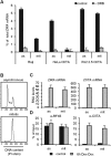
Cell type-specific variation of mitotic DRA transcriptional activity. (A) Total RNA was extracted from asynchronous and mitotic Raji, HeLa CIITA and RJ2.2.5 CIITA cells and active transcription of the DRA gene was analysed with qRT-PCR using two primer sets (DRA exon5–exon5 and DRA exon1–intron1). RNA from untreated and DRB-treated cells was used to measure net active transcription. These primer sets that monitor unspliced (nascent) or spliced (mature/total) transcripts were normalized for differences of their amplification efficiencies using a standard curve of a genomic DNA template. Newly synthesized RNA is presented as percent of mature DRA mRNA. (B) DNA content analysis by PI and flow cytometry of RJ2.2.5 cells expressing CIITA under the control of rtTA-GBD and Dex-Dox treatment (see materials and methods). Cells were blocked by nocodazole prior to CIITA induction by a 6-h treatment with Dex–Dox. (C) RNA was extracted from control and induced, asynchronous and mitotic RJ2.2.5 tetCIITA cells. DRA and CIITA gene expression was analysed by qRT-PCR. (D) Chromatin was extracted from the same samples as in (C), and ChIP assays against RFX5 and CIITA were performed.
Figure 5.
Figure 5.
The DRA-LCR/XL4 mediates PP2A recruitment via a direct interaction with NFYA in mitosis. (A) DRA-LCR/XL4 factor occupancy using the same chromatin samples, antibodies and primer sets from asynchronous and mitotic HeLa CIITA (CIITA+) and HeLa cell lines shown in Figure 2D and E. The inset shows the relative fold change of occupancy of LCR/XL4 over promoter for the indicated factors. Mean and error bars are derived as in Figure 2B. (B) Whole cell extracts of HEK293T cells expressing myc-tagged PP2Ac and the indicated full-length or truncated GFP- or Jelly Red-fusion proteins were used for protein immunoprecipitation experiments using an α-myc antibody. Western blotting was performed using α-JellyRed, α-GFP or α-myc antibodies. Input represents 10% of the lysate used for the immunoprecipitation. (C) Whole cell extracts of asynchronous or nocodazole-treated HeLa cells were subjected to immunoprercipitation by α-NFYA and immunoblotted against α-PP2Ac. Shown are the input (5% of immunoprecipitated samples), the normal IgG control and the verification of efficient NFYA immunoprecipitation.
Figure 6.
Figure 6.
NFYA loss compromises recruitment of PP2A to chromatin. (A) Western blotting of asynchronous or mitotic HeLa cell extracts following lentiviral infection with sh-scrambled control (shSCR) or shNFYA. Equivalent protein amounts were probed with antibodies against NFYA, NFYB, PP2Ac and β-tubulin. (B) Flow cytometric analysis of propidium iodide-stained DNA from shSCR or shNFYA infected HeLa cells, untreated or mitotically arrested. (C) ChIP from shSCR or shNFYA infected, asynchronous or mitotic HeLa cells, probing the indicated regions (DRA promoter, DRA-LCR/XL4, DRB1 promoter, XL7 and XL9) with antibodies against NFYA, PP2A, H3 and CREB. Mean and error bars are derived as in Figure 2.
Figure 7.
Figure 7.
PP2A bookmarks the DRA gene for transcription timing upon entry into the G1 phase. (A) RNA was extracted from mitotic HeLa CIITA cells in which siRNA transfection was carried out to knock down PP2Ac gene expression and samples were analysed with qRT-PCR. A scrambled siRNA (siSCR) was used as a negative control. (B) HeLa CIITA cells transfected with siPP2Ac or siSCR blocked in mitotis with nocodazole. Upon release the samples were collected at the indicated time points (0, 2 and 5 h). Flow cytometric analysis of DNA content upon propidium iodide staining is shown on distribution plots. (C) RNA was extracted from the same samples as in (B) and active transcription (DRA and MYC) or total mRNA (CIITA) were measured by qRT-PCR analysis. (D and E) HeLa CIITA cells were mitotically arrested by nocodazole—and then treated with 50 nM okadaic acid for 3 h. DNase I hypersensitivity assay was performed and digestion was measured by qPCR with primers for five different regions on the DRA locus (D), H4 and CD4 promoters (E) in the absence or presence of okadaic acid. Results only from 20 units DNase I treatment are depicted. (F) Schematic representation of the DRA-LCR/XL4 mitotic bookmarking mechanism. Maps are not to scale.

Similar articles

Cited by

References

    1. Reith W, Mach B. The bare lymphocyte syndrome and the regulation of MHC expression. Annu. Rev. Immunol. 2001;19:331–373. - PubMed
    1. DeSandro A, Nagarajan UM, Boss JM. The bare lymphocyte syndrome: molecular clues to the transcriptional regulation of major histocompatibility complex class II genes. Am. J. Hum. Genet. 1999;65:279–286. - PMC - PubMed
    1. Durand B, Sperisen P, Emery P, Barras E, Zufferey M, Mach B, Reith W. RFXAP, a novel subunit of the RFX DNA binding complex is mutated in MHC class II deficiency. Embo. J. 1997;16:1045–1055. - PMC - PubMed
    1. Nagarajan UM, Louis-Plence P, DeSandro A, Nilsen R, Bushey A, Boss JM. RFX-B is the gene responsible for the most common cause of the bare lymphocyte syndrome, an MHC class II immunodeficiency. Immunity. 1999;10:153–162. - PubMed
    1. Steimle V, Durand B, Barras E, Zufferey M, Hadam MR, Mach B, Reith W. A novel DNA-binding regulatory factor is mutated in primary MHC class II deficiency (bare lymphocyte syndrome) Genes Dev. 1995;9:1021–1032. - PubMed

Publication types

MeSH terms