. 2013 Jan;25(1):102-14.
doi: 10.1105/tpc.112.104331. Epub 2013 Jan 9.

An evolutionarily conserved signaling mechanism mediates far-red light responses in land plants

Affiliations

An evolutionarily conserved signaling mechanism mediates far-red light responses in land plants

Anja Possart et al. Plant Cell. 2013 Jan.

Erratum in

  • Correction.
    [No authors listed] [No authors listed] Plant Cell. 2015 Jul;27(7):2075. doi: 10.1105/tpc.15.00527. Epub 2015 Jun 19. Plant Cell. 2015. PMID: 26091692 Free PMC article. No abstract available.

Abstract

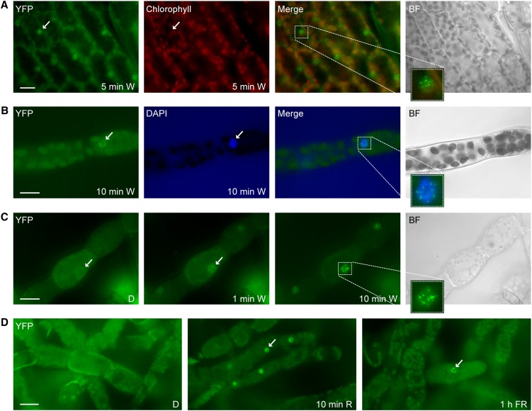
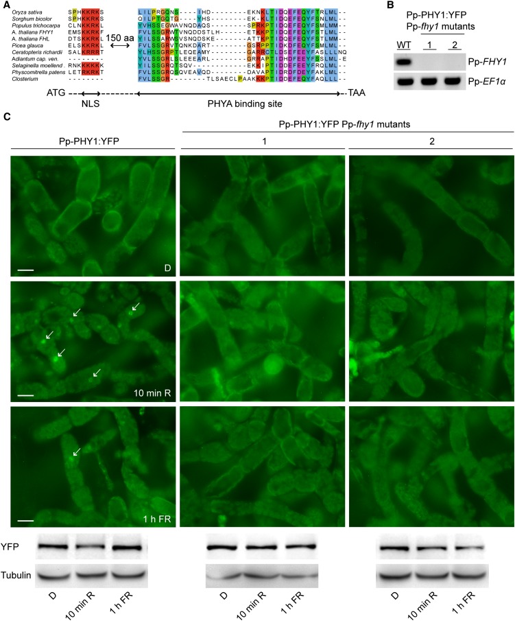
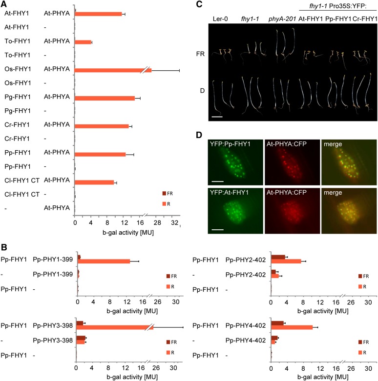
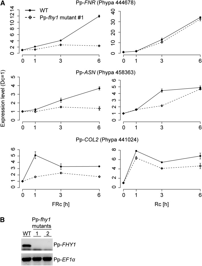
Phytochromes are plant photoreceptors important for development and adaptation to the environment. Phytochrome A (PHYA) is essential for the far-red (FR) high-irradiance responses (HIRs), which are of particular ecological relevance as they enable plants to establish under shade conditions. PHYA and HIRs have been considered unique to seed plants because the divergence of seed plants and cryptogams (e.g., ferns and mosses) preceded the evolution of PHYA. Seed plant phytochromes translocate into the nucleus and regulate gene expression. By contrast, there has been little evidence of a nuclear localization and function of cryptogam phytochromes. Here, we identified responses to FR light in cryptogams, which are highly reminiscent of PHYA signaling in seed plants. In the moss Physcomitrella patens and the fern Adiantum capillus-veneris, phytochromes accumulate in the nucleus in response to light. Although P. patens phytochromes evolved independently of PHYA, we have found that one clade of P. patens phytochromes exhibits the molecular properties of PHYA. We suggest that HIR-like responses had evolved in the last common ancestor of modern seed plants and cryptogams and that HIR signaling is more ancient than PHYA. Thus, other phytochromes in seed plants may have lost the capacity to mediate HIRs during evolution, rather than that PHYA acquired it.


Figures

Figure 1.
Figure 1.
Rapid Light-Induced Nuclear Transport of P. patens PHY1. (A) Light-regulated nuclear accumulation of Pp-PHY1 in gametophores. Dark-adapted gametophores of transgenic P. patens plants expressing Pp-PHY1:YFP were exposed to W light for 5 min and used for fluorescence microscopy. (B) DAPI staining. Dark-adapted protonema filaments of Pp-PHY1:YFP expressing P. patens plants were exposed to W light for 10 min, fixed with formaldehyde, stained with DAPI, and analyzed by fluorescence microscopy. (C) Rapid light-induced nuclear transport of Pp-PHY1 in protonema filaments. Dark-adapted protonema filaments of P. patens plants expressing YFP-tagged Pp-PHY1 were used for time series fluorescence microscopy. Images were acquired before (dark control [D]) and 1 and 10 min after the onset of irradiation with W light. (D) Nuclear accumulation of P. patens PHY1 is induced by R and FR light. Protonema filaments of P. patens plants expressing Pp-PHY1:YFP were dark adapted and used for fluorescence microscopy. Images were acquired before and after irradiation with R light (10 min, 22 μmol m−2 s−1) or FR light (1 h, 18 μmol m−2 s−1). The samples were fixed with formaldehyde before microscopy analysis. Arrows indicate nuclei; insets show enlargements of nuclei. Merge, merge of YFP and chlorophyll/DAPI channels; BF, bright field. Bars = 20 μm.
Figure 2.
Figure 2.
Pp-FHY1 Is Essential for Pp-PHY1 Nuclear Transport in P. patens. (A) Cryptogams contain FHY1-like proteins. Sequence alignment of FHY1-like proteins from monocots, dicots, gymnosperms, and cryptogams. Only regions of high sequence similarity are shown. The dashed line indicates nonaligned regions. aa, amino acids. (B) RT-PCR analysis of Pp-fhy1 mutants. Pp-FHY1 was deleted in Pp-PHY1:YFP-expressing lines. Two independent Pp-fhy1 mutants were used for RT-PCR analysis and Pp-EF1α was used as a control. WT, the wild type. (C) Light-induced nuclear transport of Pp-PHY1 depends on Pp-FHY1. Dark-adapted protonema filaments of P. patens wild type or Pp-fhy1 mutants expressing Pp-PHY1:YFP were fixed before microscopy analysis. Images were acquired before (dark control [D]) and after irradiation with either R light (10 min, 22 μmol m−2 s−1) or FR light (1 h, 18 μmol m−2 s−1). Arrows indicate nuclei. Immunoblot analysis shows Pp-PHY1:YFP levels in the different light conditions. Bars = 20 μm.
Figure 3.
Figure 3.
Cryptogam and Seed Plant FHY1 Are Functional Homologs. (A) Cryptogam FHY1 proteins contain a PHYA binding motif. AD plasmids containing the coding sequence for the C-terminal phytochrome binding motif of FHY1 from Closterium sp (Cl; green algae) or full-length FHY1 from Arabidopsis (At), dandelion (Taraxacum officinale; To), rice (Oryza sativa; Os), white spruce (Picea glauca; Pg), Ceratopteris richardii (Cr; fern), or P. patens (Pp) fused to the GAL4 activation domain were used for yeast two-hybrid analysis with Arabidopsis PHYA fused to the GAL4 DNA binding domain. To convert PHYA to the Pfr or Pr form, yeast cultures were irradiated for 5 min with R (12 μmol m−2 s−1) or FR light (12 μmol m−2 s−1) and incubated for 4 h in the dark before measuring the β-galactosidase activity. MU, Miller Units; Error bars represent se; n = 3. (B) P. patens phytochromes interact with Pp-FHY1 in a light regulated fashion. N-terminal fragments of P. patens phytochromes fused to the binding domain were used for yeast two-hybrid assays with AD:Pp-FHY1 as described in (A). MU, Miller Units; Error bars represent se; n = 3. (C) P. patens and Ceratopteris FHY1 are functional in Arabidopsis. Landsberg erecta-0 (Ler-0), fhy1-1, and phyA-201 as well as fhy1-1 seedlings expressing 35S promoter–driven YFP:At-FHY1, YFP:Pp-FHY1, or YFP:Cr-FHY1 were grown for 5 d in darkness (D) or FR (12 μmol m−2 s−1). Bar = 5 mm. (D) Pp-FHY1 and At-PHYA colocalize in light-induced nuclear bodies. Etiolated mustard seedlings were transformed by particle bombardment with constructs coding for Pro35S:PHYA:CFP and either Pro35S:YFP:At-FHY1 or Pro35S:YFP:Pp-FHY1. After transformation, the seedlings were incubated for 2 d in darkness and used for microscopy. The images were acquired after 5 min irradiation with W light. Bars = 10 μm.
Figure 4.
Figure 4.
Pp-FHY1 Is Essential for FR Light–Induced Gene Expression. (A) Protonemata cultures of P. patens wild type (WT) and Pp-fhy1 mutant lines were dark adapted and exposed to either R light (28 μmol m−2 s−1) or FR light (16 μmol m−2 s−1). Samples for quantitative RT-PCR analyses were harvested after 1, 3, and 6 h of light treatment or darkness. The expression levels of FNR, ASN, and COL2 were normalized to the levels of 26S rRNA. Expression levels in darkness were set to 1. Error bars represent se of technical replicates, n = 3. An independent biological replicate is shown in Supplemental Figure 7 online. (B) RT-PCR analysis of Pp-fhy1 mutants. Pp-fhy1 knockout lines were generated using gene targeting. Two independent Pp-fhy1 mutant lines were used for RT-PCR analysis with primers specific for either Pp-FHY1 or Pp-EF1α.
Figure 5.
Figure 5.
HIR-Like Responses to High Fluence Rate FR Light in P. patens. (A) Spore germination in FR light requires continuous irradiation. P. patens spores were irradiated for 3 d with continuous FR light (FRc; 3.5 μmol m−2 s−1) or with 3-min FR light pulses (FRp; 70 μmol m−2 s−1) of the same total fluence, interrupted by 57-min dark periods. To ensure that spores irradiated with FR pulses were viable, they were irradiated for an additional 3 d with continuous FR light (FRp + FRc). The bar plot shows significantly reduced germination rate in FRp (Fisher’s exact test P < 2.2e-16). Bar = 100 μm. (B) Protonemata growth in FR light depends on continuous irradiation. P. patens cultures were grown for 20 d in continuous FR light (3.5 μmol m−2 s−1) or irradiated with 3-min FR light pulses (70 μmol m−2 s−1) of the same total fluence, interrupted by 57-min dark periods. For dark controls, see Supplemental Figure 8B online. Bar = 100 μm. (C) Continuous irradiation is essential for FR light–induced gametophore growth. P. patens gametophores were grown for 9 d in continuous FR light (3.5 μmol m−2 s−1) or irradiated with 3-min FR light pulses (70 μmol m−2 s−1) of the same total fluence, interrupted by 57-min dark periods. For dark controls, see Supplemental Figure 8C online. Bar = 500 μm. (D) Pfr-dependent degradation of Pp-PHY1. Dark-adapted protonemata cultures of P. patens lines expressing YFP-tagged Pp-PHY1 were irradiated with R light (22 μmol m−2 s−1) or FR light (18 μmol m−2 s−1) for different time periods. Total protein was isolated and analyzed by SDS-PAGE and immunoblotting with anti-YFP antibody. Protein extracts from dark-adapted wild-type P. patens cultures were used as negative controls. Tubulin is shown as a loading control. D, darkness; WT, the wild type. (E) Pfr-dependent degradation of Pp-PHY3. Dark-adapted protonemata cultures of P. patens lines expressing Pp-PHY3:YFP were irradiated with either R or FR light and used for SDS-PAGE and immunoblot analysis as described in (D). [See online article for color version of this figure.]
Figure 6.
Figure 6.
Pp-FHY1 Is Essential for HIR-Like Responses to High Fluence Rate FR Light. (A) Spore germination in FR light depends on Pp-FHY1. Spores from wild-type (WT) P. patens plants and two independent Pp-fhy1 mutant lines were irradiated for 8 d with continuous FR light (18 μmol m−2 s−1). To ensure that Pp-fhy1 mutant spores irradiated with FR light were viable, they were irradiated for an additional 5 d with W light after the FR light treatment. The bar plot shows significantly reduced germination rate in Pp-fhy1 mutants (Fisher’s exact test P < 3.4e-05). Bar = 100 μm. (B) Pp-FHY1 is essential for protonemata growth in FR light. Wild-type and Pp-fhy1 mutant P. patens cultures were grown for 13 d in FR light (18 μmol m−2 s−1). Bar = 100 μm. (C) FR light–induced gametophore growth requires Pp-FHY1. Wild-type and Pp-fhy1 mutant P. patens gametophores were grown for 11 d in FR (18 μmol m−2 s−1). Bar = 500 μm. [See online article for color version of this figure.]

Similar articles

Cited by

References

    1. Block M.D., Botterman J., Vandewiele M., Dockx J., Thoen C., Gosselé V., Movva N.R., Thompson C., Montagu M.V., Leemans J. (1987). Engineering herbicide resistance in plants by expression of a detoxifying enzyme. EMBO J. 6: 2513–2518 - PMC - PubMed
    1. Böse G., Schwille P., Lamparter T. (2004). The mobility of phytochrome within protonemal tip cells of the moss Ceratodon purpureus, monitored by fluorescence correlation spectroscopy. Biophys. J. 87: 2013–2021 - PMC - PubMed
    1. Botto J.F., Sanchez R.A., Whitelam G.C., Casal J.J. (1996). Phytochrome A mediates the promotion of seed germination by very low fluences of light and canopy shade light in Arabidopsis. Plant Physiol. 110: 439–444 - PMC - PubMed
    1. Burgin M.J., Casal J.J., Whitelam G.C., Sanchez R.A. (1999). A light-regulated pool of phytochrome and rudimentary high-irradiance responses under far-red light in Pinus elliottii and Pseudotsuga menziesii. J. Exp. Bot. 50: 831–836
    1. Chen M., Tao Y., Lim J., Shaw A., Chory J. (2005). Regulation of phytochrome B nuclear localization through light-dependent unmasking of nuclear-localization signals. Curr. Biol. 15: 637–642 - PubMed

Publication types

MeSH terms