. 2013 Jan;25(1):22-37.
doi: 10.1105/tpc.112.105643. Epub 2013 Jan 9.

Lessons on RNA silencing mechanisms in plants from eukaryotic argonaute structures

Affiliations

Lessons on RNA silencing mechanisms in plants from eukaryotic argonaute structures

Christian Poulsen et al. Plant Cell. 2013 Jan.

Abstract

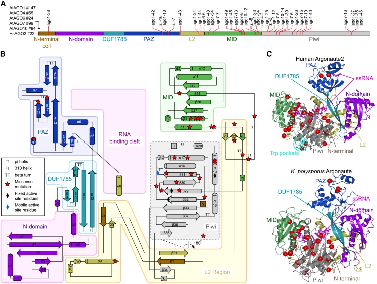
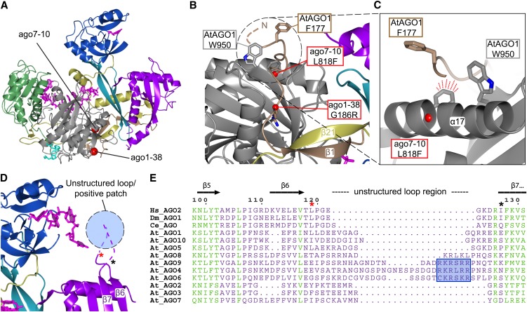
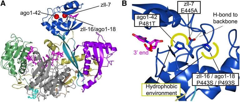
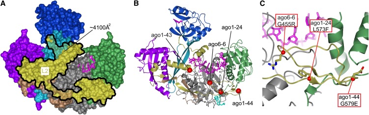
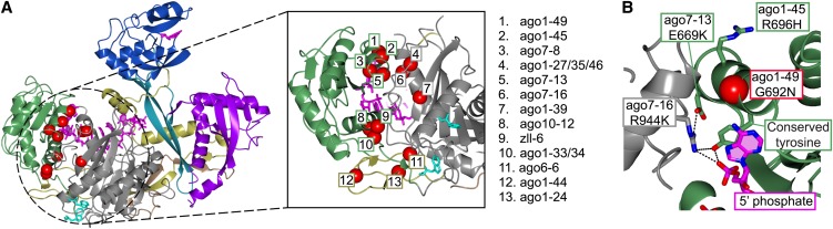
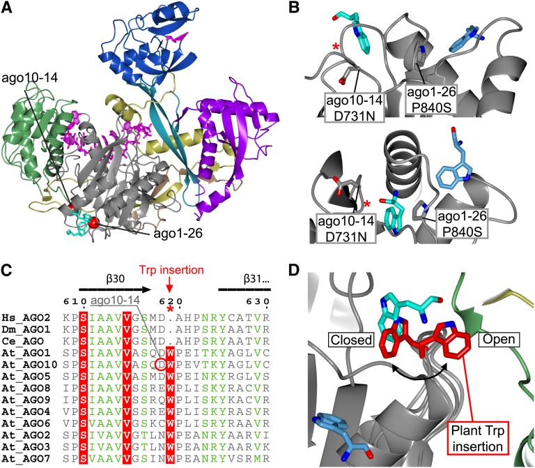
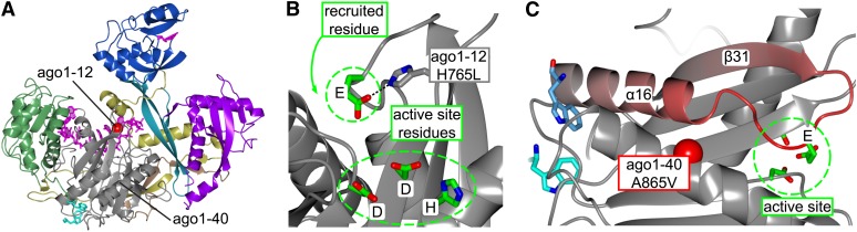
RNA silencing refers to a collection of gene regulatory mechanisms that use small RNAs for sequence specific repression. These mechanisms rely on ARGONAUTE (AGO) proteins that directly bind small RNAs and thereby constitute the central component of the RNA-induced silencing complex (RISC). AGO protein function has been probed extensively by mutational analyses, particularly in plants where large allelic series of several AGO proteins have been isolated. Structures of entire human and yeast AGO proteins have only very recently been obtained, and they allow more precise analyses of functional consequences of mutations obtained by forward genetics. To a large extent, these analyses support current models of regions of particular functional importance of AGO proteins. Interestingly, they also identify previously unrecognized parts of AGO proteins with profound structural and functional importance and provide the first hints at structural elements that have important functions specific to individual AGO family members. A particularly important outcome of the analysis concerns the evidence for existence of Gly-Trp (GW) repeat interactors of AGO proteins acting in the plant microRNA pathway. The parallel analysis of AGO structures and plant AGO mutations also suggests that such interactions with GW proteins may be a determinant of whether an endonucleolytically competent RISC is formed.


Figures

Figure 1.
Figure 1.
Structural Mapping of AGO Missense Mutations. (A) Schematic diagram of common elements of the primary structure of the AGO protein family depicting the location of known plant missense alleles. The length of N-terminal extensions in each AGO protein is shown to the left of the diagram. (B) Topology diagram of the human Ago2 structure colored according to domains and structural regions. The RNA binding cleft is located between one lobe containing the N, DUF1785, and PAZ domains and a second lobe containing the MID and Piwi domains. The L2 connects the two lobes and forms secondary structure elements with several domains and the structured N terminus. The plant ago missense mutations are marked with red stars. (C) The tertiary structures of human Ago2 and K. polysporus Ago as determined by crystallography. Note the overall structural similarity between human and yeast Ago, except in the PAZ domain that exists in closed (human Ago2) and open (K. polysporus Ago) conformations. The positions of plant ago missense mutations are marked with red spheres. ssRNA, single-stranded RNA.
Figure 2.
Figure 2.
The Structurally Fixed N-Terminal Coil and the N Domain. (A) Ribbon diagram of human Ago2 pointing out the locations of the two missense mutations ago7-10 and ago1-38. (B) The structured N terminus (brown) is tightly interacting with the Piwi domain (gray) as an extended coil-like structure, and the β1-strand is fixed between β21 (L2; gold) and β34 (Piwi; not seen). The G186R mutation in ago1-38 (red sphere; modeled) is expected to locally dislocate the N-terminal coil since the Arg side chain is predicted to heavily clash with the Piwi domain. The L818F mutation in ago7-10 located in the α17-helix is likewise expected to dislocate the extended N-terminal coil. (C) Close-up view of the α17-helix containing the modeled L818F ago7-10 mutation. The bulkier Phe is predicted to clash with the peptide backbone of the N-terminal region. The side chains of two conserved hydrophobic and solvent exposed residues, At-AGO1 F177 and At-AGO1 W950, are presented. (D) The close proximity between the 3′end of the guide strand and the unstructured region connecting β6-β7 is displayed in the human Ago2 structure loaded with miR-20a (Protein Data Bank entry 4F3T). The first and last residues in the unstructured loop are indicated with asterisks matching those in (E). (E) The primary sequence alignment of human (Hs), Drosophila (Dm), C. elegans (Ce), and Arabidopsis AGOs shows how the loop region connecting β6-β7 varies in length and sequence. A conserved positively charged patch in AGO4, 6, and 9 (blue box) is suggested to interact with the 3′end of the guide strand.
Figure 3.
Figure 3.
Structural Integrity of the PAZ Domain Is Important for Function. (A) Ribbon diagram of human Ago2 pointing out the locations of three plant missense mutations in the PAZ domain. (B) Three mutations in zll-16/ago1-18 and ago1-42 change two conserved Pro residues constituting important hydrophobic interactions (illustrated in yellow) needed for proper folding of the domain. The zll-7 mutation E445A disrupts H-bonds to two backbone amides also resulting in destabilization of the PAZ domain.
Figure 4.
Figure 4.
L2 Is a Significant Structural Unit. (A) Surface representation of the human Ago2 highlighting the large footprint of L2. Note that all domains are interacting with L2. (B) Structural overview of the missense mutations in L2 with orientation as in (A). The ago1-43 missense mutation is likely to reduce the stability of the interface between the N- and C-lobes. (C) View of three modeled L2 missense mutations located between the MID (green) and Piwi (gray) domains. The G455R (ago6-6) and L573F (ago1-24) could have an indirect effect on the RNA binding, whereas the destabilization caused by the G579E mutation in ago1-44 is less clear.
Figure 5.
Figure 5.
The MID and Piwi Domains Are Highly Sensitive to Mutations. (A) Structural overview highlighting the density of missense mutations on or in close proximity to the MID-Piwi interface. A close-up view of the region shows the location of the 13 missense mutations in this region. (B) Presentation of mutations directly involved in the 5′RNA binding. Conserved residues are part of a H-bond network involved in 5′phosphate coordination.
Figure 6.
Figure 6.
Hs-Ago2 Trp Binding Pockets Are Likely to Be Conserved in Plants. (A) Structural overview of Hs-Ago2 pointing out the close proximity between the ago10-14 and ago1-26 mutations and the ligand-bound Trp residues (cyan), suggesting a functional connection to the Trp binding sites. (B) Top: Side view of the two Trp binding pockets. Pro-840 mutated in ago1-26 constitutes parts of one of the pockets (Trp; light blue), but the mutation could interfere with both pockets due to its central location (cyan and light blue). The ago10-14 D731N mutation shows how changing the nonconserved environment involved in Trp binding in Hs-Ago2 has an effect in plants. Bottom: Different view of top panel. The red asterisks indicate the location of the Trp insertion pointed out in (C). (C) The primary sequence alignment of Human (Hs), Drosophila (Dm), C. elegans (Ce), and Arabidopsis AGOs reveals insertion of a conserved Trp in the loop connecting β30-β31 in plant AGOs. (D) Backbone modeling of the At-AGO1 loop connecting β30-β31. The model shows the conformation of the plant-specific Trp (red) inserted at the Hs-Ago2 Trp binding site (cyan). The model suggests two possible conformations: one excluding additional Trp binding (closed) and one allowing binding of hydrophobic elements from an external source (open).
Figure 7.
Figure 7.
The ago1-12 Allele Provides Evidence for a Catalytic Tetrad in Plants. (A) Overview showing the location of the ago1-12 mutation. (B) Close-up view of the Hs-Ago2 active site region showing the catalytic tetrad (EDDH). The ago1-12 H765L mutation disrupts the H-bond to the deeply conserved Glu (Glu-736 in Hs-Ago2), with the active site residue suggested to be recruited for slicing. (C) The proximity of the Trp binding sites and the E736 loop (in red). Interactors bound to the Trp binding pockets could reach the E736 loop and thereby modulate its active site recruitment. Note that A865V missense mutation in ago1-40 is located in this area expected to be included in the GW interaction surface.

References

    1. Agorio A., Vera P. (2007). ARGONAUTE4 is required for resistance to Pseudomonas syringae in Arabidopsis. Plant Cell 19: 3778–3790 - PMC - PubMed
    1. Allen E., Xie Z., Gustafson A.M., Carrington J.C. (2005). MicroRNA-directed phasing during trans-acting siRNA biogenesis in plants. Cell 121: 207–221 - PubMed
    1. Azevedo J., Garcia D., Pontier D., Ohnesorge S., Yu A., Garcia S., Braun L., Bergdoll M., Hakimi M.A., Lagrange T., Voinnet O. (2010). Argonaute quenching and global changes in Dicer homeostasis caused by a pathogen-encoded GW repeat protein. Genes Dev. 24: 904–915 - PMC - PubMed
    1. Baumberger N., Baulcombe D.C. (2005). Arabidopsis ARGONAUTE1 is an RNA Slicer that selectively recruits microRNAs and short interfering RNAs. Proc. Natl. Acad. Sci. USA 102: 11928–11933 - PMC - PubMed
    1. Bohmert K., Camus I., Bellini C., Bouchez D., Caboche M., Benning C. (1998). AGO1 defines a novel locus of Arabidopsis controlling leaf development. EMBO J. 17: 170–180 - PMC - PubMed

Publication types

MeSH terms

LinkOut - more resources