. 2013 Jan 9;33(2):485-94.
doi: 10.1523/JNEUROSCI.3483-12.2013.

Physostigmine and galanthamine bind in the presence of agonist at the canonical and noncanonical subunit interfaces of a nicotinic acetylcholine receptor

Affiliations

Physostigmine and galanthamine bind in the presence of agonist at the canonical and noncanonical subunit interfaces of a nicotinic acetylcholine receptor

Ayman K Hamouda et al. J Neurosci. .

Abstract

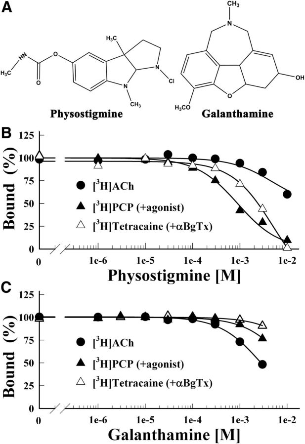
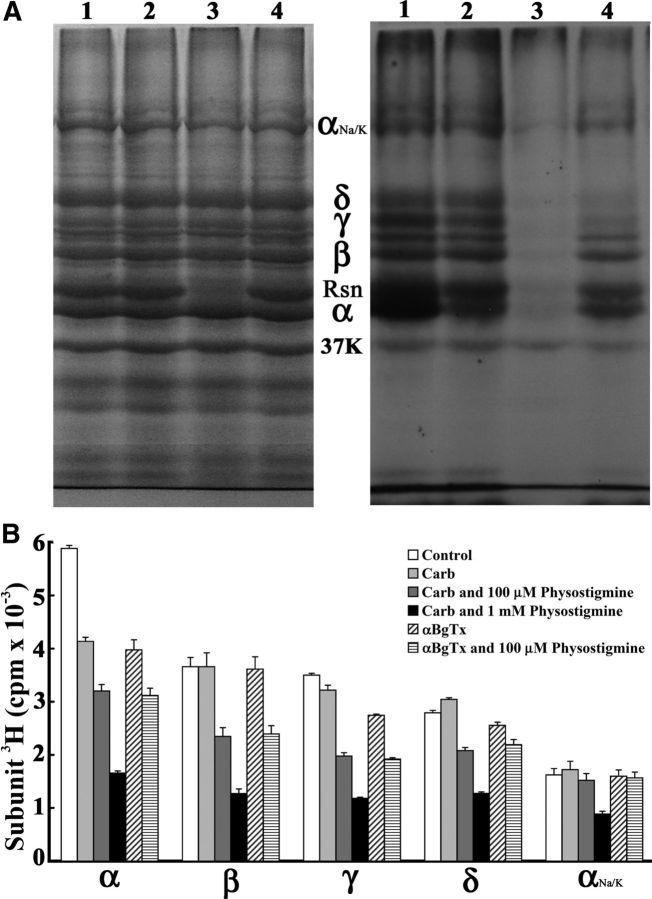
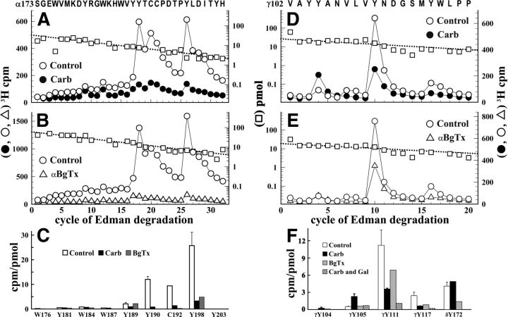
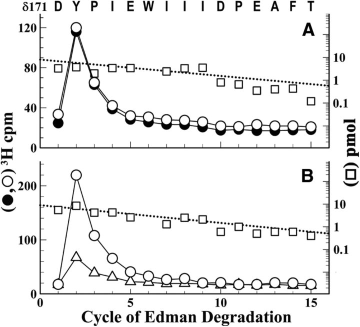
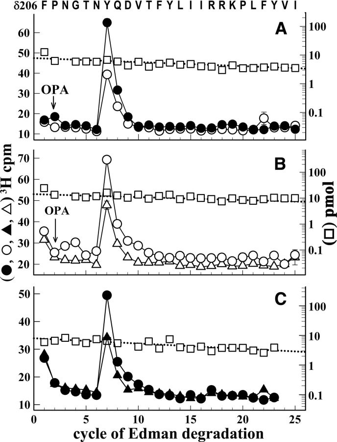
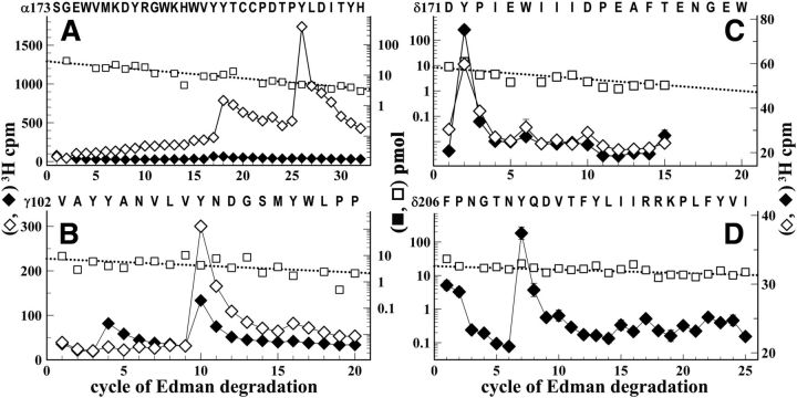
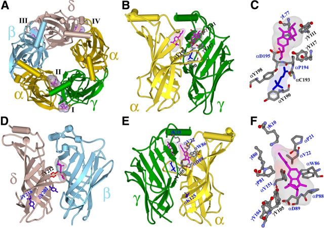
Galanthamine and physostigmine are clinically used cholinomimetics that both inhibit acetylcholinesterase and also interact directly with and potentiate nAChRs. As with most nAChR-positive allosteric modulators, the location and number of their binding site(s) within nAChRs are unknown. In this study, we use the intrinsic photoreactivities of [(3)H]physostigmine and [(3)H]galanthamine upon irradiation at 312 nm to directly identify amino acids contributing to their binding sites in the Torpedo californica nAChR. Protein sequencing of fragments isolated from proteolytic digests of [(3)H]physostigmine- or [(3)H]galanthamine-photolabeled nAChR establish that, in the presence of agonist (carbamylcholine), both drugs photolabeled amino acids on the complementary (non-α) surface of the transmitter binding sites (γTyr-111/γTyr-117/δTyr172). They also photolabeled δTyr-212 at the δ-β subunit interface and γTyr-105 in the vestibule of the ion channel, with photolabeling of both residues enhanced in the presence of agonist. Furthermore, [(3)H]physostigmine photolabeling of γTyr-111, γTyr-117, δTyr-212, and γTyr-105 was inhibited in the presence of nonradioactive galanthamine. The locations of the photolabeled amino acids in the nAChR structure and the results of computational docking studies provide evidence that, in the presence of agonist, physostigmine and galanthamine bind to at least three distinct sites in the nAChR extracellular domain: at the α-γ interface (1) in the entry to the transmitter binding site and (2) in the vestibule of the ion channel near the level of the transmitter binding site, and at the δ-β interface (3) in a location equivalent to the benzodiazepine binding site in GABA(A) receptors.


Figures

Figure 1.
Figure 1.
Physostigmine and galanthamine inhibition of the equilibrium binding of [3H]ACh, [3H]tetracaine, and [3H]PCP to the Torpedo nAChR. A, The chemical structures of physostigmine and galanthamine. B, C, The equilibrium binding of [3H]ACh (●), [3H]tetracaine (+α-BgTx, ▵), or [3H]PCP (+Carb, ▴) was determined using centrifugation assays in the absence or presence of increasing concentrations of physostigmine (B) or galanthamine (C). For each competition experiment, the data were normalized to the specific binding in the absence of competitor. Total binding for [3H]ACh was 1313 ± 5 cpm, with 88 ± 10 cpm nonspecific (+ 1 mm Carb); for [3H]tetracaine, 24,400 ± 120 cpm, with 5583 ± 16 cpm nonspecific (+ 100 μm tetracaine); for [3H]PCP, 6854 ± 52 cpm, with 437 ± 6 cpm nonspecific (+ 100 μm Proadifen). Physostigmine inhibited [3H]tetracaine and [3H]PCP binding with IC50s of 5 ± 1 mm and 0.9 ± 0.1 mm, respectively. Galanthamine inhibited [3H]ACh binding with IC50 = 2.8 ± 0.1 mm.
Figure 2.
Figure 2.
[3H]Physostigmine photoincorporation into the Torpedo nAChR. Torpedo nAChR-rich membranes at 2 mg protein/ml (150 pmol ACh binding site/condition) were photolabeled with 1 μm [3H]physostigmine in the absence or presence of cholinergic ligands, and the membrane polypeptides were resolved on two parallel SDS-PAGE gels. Gels were stained with Coomassie Blue, and one gel was processed for fluorography. A, Coomassie Blue staining (left) and fluorogram (right, 2 weeks exposure) of a gel with membranes in lanes 1–4 photolabeled by [3H]physostgimine: (1) in the absence of other drugs; (2) + 1 mm Carb; (3) + 1 mm nonradioactive physostigmine; or (4) + 1 mm galanthamine. The electrophoretic mobilities of the α, β, γ, and δ nAChR subunits, rapsyn (Rsn), the Na+/K+-ATPase α subunit (αNa/K), and calectrin (37K) are indicated. B, 3H incorporation (average and range) in the polypeptide bands excised from two gels containing nAChR-rich membranes photolabeled with [3H]physostigmine in the absence or presence of Carb, α-BgTx, or physostigmine.
Figure 3.
Figure 3.
[3H]Physostigmine photolabeling at the α-γ extracellular interface in the presence or absence of Carb or α-BgTx. A, B, D, E, 3H (○, ●, ▵) and PTH-amino acids (□) released during sequence analysis of fragments beginning at αSer-173 (A, B) and γVal-102 (D, E) isolated from Torpedo nAChR photolabeled with [3H]physostigmine in the absence of other drugs (A, B, D, E, ○, □), in the presence of 1 mm Carb (A, D, ●), or in the presence of 8 μm α-BgTx (B, E, ▵). C, F, Quantification of [3H]physostigmine photoincorporation (in cpm/pmol) into aromatic amino acid residues within the primary structure of fragment beginning at αSer-173 (C) or γVal-102 (F). For the control and +Carb conditions, the values plotted are the mean ± SD from two independent photolabeling experiments, whereas data for +α-BgTx are from a single experiment. To calculate the efficiency of labeling at αCys-192 (cycle 20) from sequencing data shown in A, the background 3H release in cycle 20 originating from the photolabeling of αTyr-190 in cycle 18 was estimated by fitting the observed 3H releases in cycles 18, 19, and 25 to an exponential decay. F, Also included are values from a third [3H]physostigmine preparative photolabeling in the presence of Carb or Carb and galanthamine and the efficiency of photolabeling of δTyr-172, derived from sequencing data presented in Figure 4.
Figure 4.
Figure 4.
[3H]Physostigmine photolabels δTyr-172 in the absence and presence of Carb or α-BgTx. 3H (○, ●, ▵) and PTH-amino acids (□) released during sequence analysis of a δ subunit fragment beginning at δAsp-171 that was isolated from nAChRs photolabeled with [3H]physostigmine in the absence of other drugs (○, □), or presence of 1 mm Carb (●) or 8 μm α-BgTx (▵). A, B, The peaks of 3H release at cycle 2 indicate photolabeling of δTyr-172 that was potentiated in the presence of Carb (A, −/+ Carb, 3.4/4.9 cpm/pmol) but inhibited in the presence of α-BgTx (B, −/+ α-BgTx, 5/1.6 cpm/pmol). The subunit fragment beginning at δAsp-171was isolated by rpHPLC fractionation of an Endo Lys-C digest of δ subunit.
Figure 5.
Figure 5.
[3H]Physostigmine photolabels δTyr-212 at the δ-β interface in the nAChR extracellular domain. A–C, 3H (○, ●, ▵, ▴) and PTH-amino acids (□) released during sequence analysis of a fragment beginning at δPhe-206 isolated from nAChRs photolabeled with [3H]physostigmine in the absence of other drugs (A, B, ○, □), or in the presence of 1 mm Carb (A, C, ●,), or 8 μm α-BgTx (B, ▵) or 1 mm Carb and 300 μm galanthamine (C, ▴). This fragment was isolated by rpHPLC purification of polypeptides eluted from a 10 to 14 kDa band from a Tricine SDS-PAGE fractionation of EndoLys-C digests of the δ subunit. The calculated efficiencies of [3H]physostigmine photolabeling of δTyr-212 (cycle 7) in the different conditions are shown in Table 1. The arrow and OPA at cycle 2 indicate that sequencing was interrupted and the filter was treated with OPA before resuming sequencing (see Materials and Methods for protein sequencing).
Figure 6.
Figure 6.
[3H]Galanthamine photolabels amino acids at the α-γ, α-δ, and δ-β extracellular interfaces in the absence and presence of agonist. A–D, 3H (♦, ◊) and PTH-amino acids (□) released during sequence analysis of subunit fragments beginning at αSer-173 (A), γVal-102 (B), δAsp-171 (C), and δPhe-206 (D) isolated from Torpedo nAChRs photolabeled with [3H]galanthamine in the absence (◊) or presence of Carb (♦). These fragments were isolated as described in the text and figure legends for the corresponding [3H]physostigmine-photolabeled fragments. The efficiencies of amino acid photolabeling (−Carb/+Carb, in cpm/pmol) calculated from the peaks of 3H release and the PTH-amino acid mass levels were as follows: A, in cycles 18 and 26, αTyr-190 (11/0.8) and αTyr-198 (45/0.1); B, in cycles 5, 10 and 15, γTyr-105 (0.3/2.1), γTyr-111 (12.6/5), and γTyr-117 (1.2/0.2); C, in cycle 2, δTyr-172 (1.1/1.6 cpm/pmol); D, in cycle 7, δTyr-212 (+Carb, 0.2 cpm/pmol).
Figure 7.
Figure 7.
The binding sites for physostigmine in the nAChR extracellular domain. A–F, A view from the top (A) and side views from the exterior (B–D) and from the inside of the channel vestibule (E, F) of the extracellular domain of the Torpedo nAChR (α, gold; β, cyan; γ, green; and δ, brown) from a homology model based on the x-ray structure of the L-AChBP in the presence of Carb (PDB#1UV6 (Celie et al., 2004)). Physostigmine is shown in stick format (magenta, volume, 197 Å3) bound in its lowest energy orientation in four sites, with Connolly surface representations of the ensembles of the 30 lowest energy-docking orientations: in the presence of Carb at the α-γ interface at Site I on the outer surface of the nAChR at the entry to the transmitter binding site (B; expanded in C; volume, 280 Å3) and at Site II in the vestibule of the ion channel (E; expanded in F; volume, 390 Å3), and in the absence of Carb at Site III at the δ-β subunit interface (D, volume, 270 Å3) and at the α-δ interface in the transmitter binding site (Site IV) (A, volume, 250 Å3). In the absence of Carb, physostigmine is also predicted to bind in the transmitter binding site at the α-γ interface equivalent to Site IV (data not shown). Binding to Site IV is identified by the photolabeling of αTyr-93, αTyr-190, and αTyr-198 that is inhibited by >90% in the presence of Carb or α-BgTx. Carb bound at the α-γ transmitter binding site is included in stick format (B, C, E; blue). Amino acids predicted to contribute to each binding site are shown in stick format colored by elements, except in D where δTyr-212 is highlighted in red to contrast with the nonphotolabeled δTyr-202 and δTyr-218 (in blue). The amino acids photolabeled in this study are identified in black by single letter code and subunit position. αLys-125, which was previously reported to be photolabeled by [3H]physostigmine (Schrattenholz et al., 1993; Luttmann et al., 2009), is shown in blue (E).

Similar articles

Cited by

References

    1. Akk G, Steinbach JH. Galantamine activates muscle-type nicotinic acetylcholine receptors without binding to the acetylcholine-binding site. J Neurosci. 2005;25:1992–2001. - PMC - PubMed
    1. Arias HR. Positive and negative modulation of nicotinic receptors. Adv Prot Chem Struct Biol. 2010;80:153–203. - PubMed
    1. Bertrand D, Gopalakrishnan M. Allosteric modulation of nicotinic acetylcholine receptors. Biochem Pharmacol. 2007;74:1155–1163. - PubMed
    1. Blanton MP, Cohen JB. Identifying the lipid-protein interface of the Torpedo nicotinic acetylcholine receptor: secondary structure implications. Biochemistry. 1994;33:2859–2872. - PubMed
    1. Brauer AW, Oman CL, Margolies MN. Use of ophthalaldehyde to reduce background during automated Edman degradation. Anal Biochem. 1984;137:134–142. - PubMed

Publication types

MeSH terms

LinkOut - more resources