. 2013 Jan 9;33(2):595-610.
doi: 10.1523/JNEUROSCI.2583-12.2013.

Enhanced recruitment of endosomal Na+/H+ exchanger NHE6 into Dendritic spines of hippocampal pyramidal neurons during NMDA receptor-dependent long-term potentiation

Affiliations

Enhanced recruitment of endosomal Na+/H+ exchanger NHE6 into Dendritic spines of hippocampal pyramidal neurons during NMDA receptor-dependent long-term potentiation

Emily C Deane et al. J Neurosci. .

Abstract

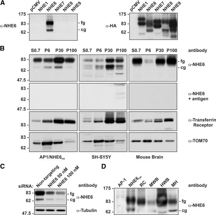
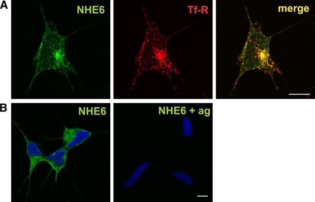
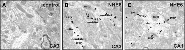
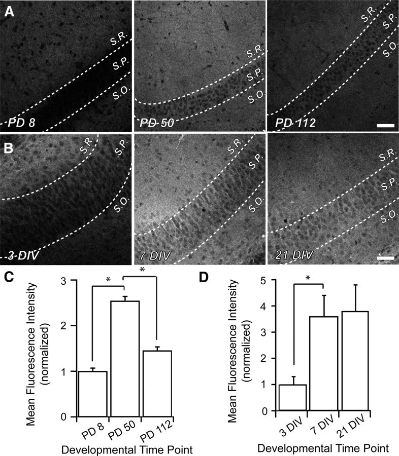
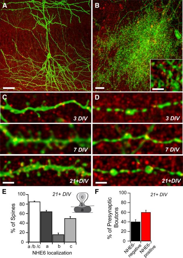
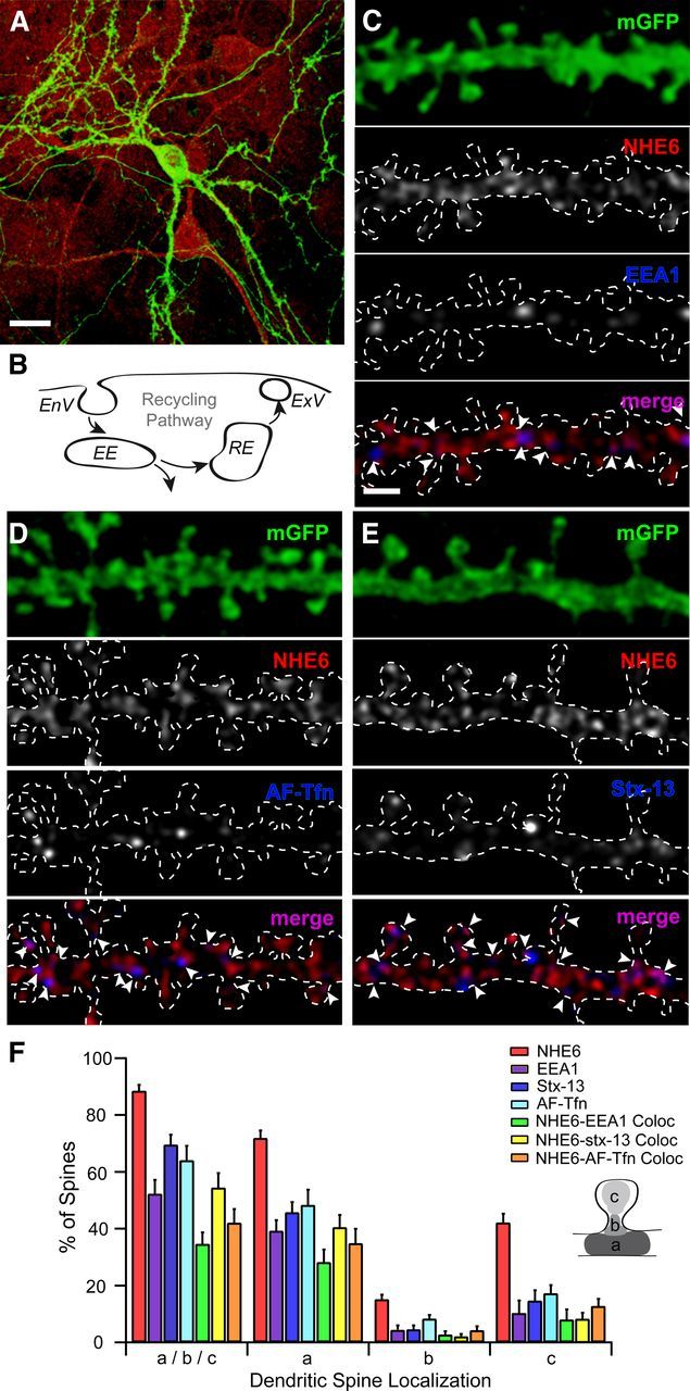
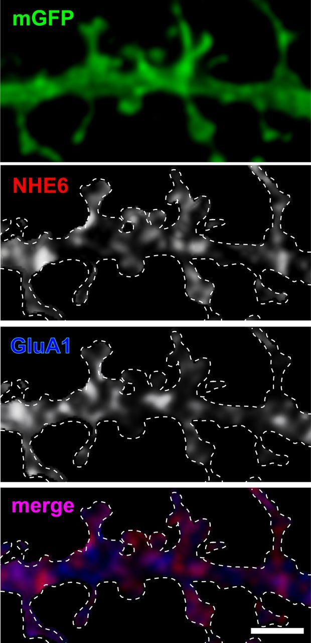
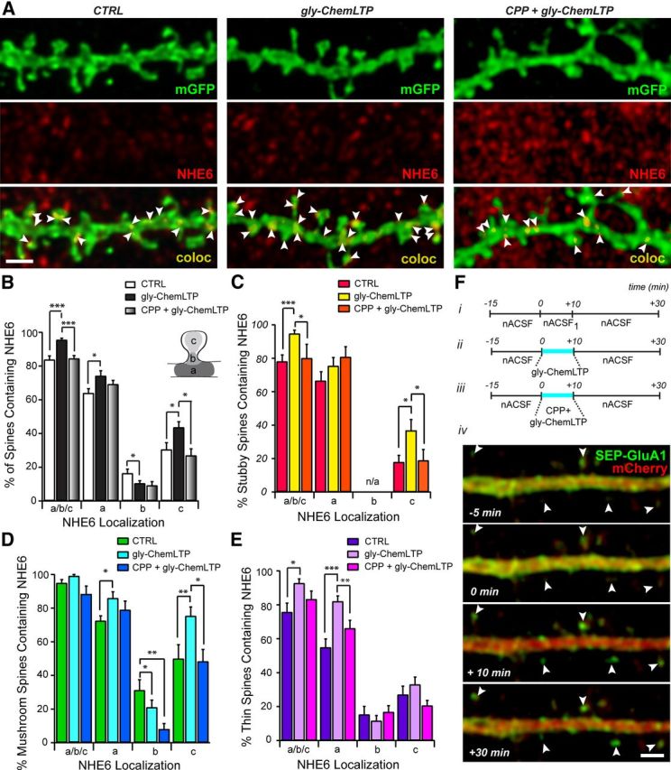
Postsynaptic endosomal trafficking has emerged as a principal regulatory mechanism of structural and functional plasticity of glutamatergic synapses. Recycling endosomes perform activity-dependent transport of AMPA receptors (AMPARs) and lipids to the postsynaptic membrane, activities that are known to contribute to long-term synaptic potentiation and hypothesized to subserve learning and memory processes in the brain. Recently, genetic defects in a widely expressed vesicular pH-regulating transporter, the Na(+)/H(+) exchanger NHE6 isoform, have been implicated in neurodevelopmental disorders including severe X-linked mental retardation and autism. However, little information is available regarding the cellular properties of this transporter in the CNS. Here, we show by quantitative light microscopy that the protein abundance of NHE6 is developmentally regulated in area CA1 of the mouse hippocampus. Within pyramidal neurons, NHE6 was found to localize to discrete puncta throughout the soma and neurites, with noticeable accumulation at dendritic spines and presynaptic terminals. Dual immunolabeling of dendritic spines revealed that NHE6 partially colocalizes with typical markers of early and recycling endosomes as well as with the AMPAR subunit GluA1. Significantly, NHE6-containing vesicles exhibited enhanced translocation to dendritic spine heads during NMDA receptor (NMDAR)-dependent long-term potentiation. These data suggest that NHE6 may play a unique, previously unrecognized, role at glutamatergic synapses that are important for learning and memory.


Figures

Figure 1.
Figure 1.
Characterization of an NHE6 isoform-specific rabbit polyclonal antibody by immunoblotting. A, Lysates of AP-1 cells transiently expressing rat NHE1HA, human NHE6HA, NHE7HA, NHE8HA, and NHE9HA were subjected to SDS-PAGE and immunoblot analysis. When the blot was probed with the affinity-purified anti-NHE6 antibody (α-NHE6), a signal was detected only in cells transfected with NHE6HA (left). The immunoblot was reprobed with a mouse monoclonal anti-HA antibody (α-HA), showing expression of all NHE isoforms (right). B, Homogenates of AP-1/NHE6HA cells, SH-SY5Y cells, and mouse whole-brain were fractionated by differential gravity centrifugation to enrich for the following subcellular compartments: S0.7 (total cell supernatant lacking nuclei), P6 (mitochondria), P30 (microsomes), and P100 (plasma membrane) (see Materials and Methods for details), followed by SDS-PAGE and immunoblot analysis with the anti-NHE6 antibody. NHE6 was enriched in the P30 microsomal and P100 plasma membrane fractions. When the NHE6 antibody was pre-incubated with the immunizing antigen, no bands were detected. The membranes were also immunoblotted with antibodies that recognize established compartment markers; i.e., the Tf-R, which is enriched in the endosomal P30 and the plasma membrane P100 fractions, and TOM70, which is enriched in the P6 mitochondrial fraction. C, In AP-1 cells stably expressing human NHE6HA, our NHE6 antibody detected reduced expression of NHE6 only in cells treated with the specific anti-NHE6 siRNA and not in cells treated with a scrambled siRNA (top). As a control, tubulin levels were not affected by the depletion of NHE6 by siRNA (bottom). D, Detection of native NHE6 in brain tissue homogenates. Lysates of AP-1 cells transiently transfected with empty vector (AP-1) or NHE6HA, as well as homogenates of rat cerebrum (RC), mouse whole-brain (MWB), human whole-brain (HWB), and mouse hippocampus (MH) were subjected to SDS-PAGE and immunoblot analysis with the anti-NHE6 antibody. The fully glycosylated (fg), mature form of NHE6, as well as the core-glycosylated form (cg) are visible.
Figure 2.
Figure 2.
Characterization of an NHE6 isoform-specific rabbit polyclonal antibody by ICC. A, Colocalization of native NHE6 with the Tf-R in the soma and neurites of cultured human SH-SY5Y neuroblastoma cells. Scale bar, 5 μm. B, Control experiment showing that the signal detected with the NHE6 antibody is specific in SH-SY5Y neuroblastoma cells, as no fluorescent signal is observed in the presence of immunizing antigen (ag). Nuclei were stained with DAPI. The green Alexa Fluor 488-conjugated secondary goat anti-rabbit antibody was applied to both samples. Scale bar, 10 μm.
Figure 3.
Figure 3.
Subcellular localization of native NHE6 in areas CA3 and CA1 of mouse hippocampus by TEM. A, In control experiments, no signals were observed in hippocampal area CA3 in the absence of primary antibody, demonstrating the specificity of the signal produced by the NHE6 antibody. B, NHE6-positive puncta were present mostly in dendrites (sometimes close to PSDs) of hippocampal neurons in area CA3, but are occasionally associated with presynaptic boutons. C, NHE6 immunogold-positive puncta were detected in dendrites in hippocampal area CA1. Magnification: ×24400. SV, Synaptic vesicles; PSD, postsynaptic density; mito, mitochondria.
Figure 4.
Figure 4.
NHE6 is increased in area CA1 of the mouse hippocampus during synaptogenesis. Representative low-magnification confocal micrographs of NHE6 immunostaining in area CA1 of the mouse hippocampus. A, Cryostat-sectioned coronal brain slices from early postnatal (PD8; left), young adult (PD50; middle), and mature animals (PD112; right). During development, NHE6 puncta are diffuse throughout all strata of area CA1. Magnification in all parts (A, B) is identical. Scale bar, 50 μm. B, Organotypic mouse hippocampal slice cultures exhibited a similar expression profile as was detected in vivo. Throughout slice culture development (3 DIV, left; 7 DIV, middle; 21–28 DIV, right), NHE6 puncta are diffuse throughout all strata of area CA1. C, NHE6 significantly increases by 2.5-fold in area CA1 between PD8 and PD50 as measured by MFI. This period has been shown to be critical for elimination of superfluous connections in the brain and establishing refined synaptic circuits. There is also a significant decrease of NHE6 in area CA1 to ∼1.5× of early postnatal levels by PD112, by which time circuitry in the brain has stabilized. n = 5 slices for each time point; normalized MFI ± SEM. *p < 0.05 by Student's t test. D, NHE6, as measured by MFI, significantly increases in area CA1 during the period of synaptic pruning and refinement in organotypic hippocampal slice cultures, but does not return to early postnatal levels. n = 8 slices for each time point; normalized MFI ± SEM. *p < 0.05 by Student's t test. S.R., Stratum radiatum; S.P., stratum pyramidalis; S.O., stratum oriens; PD, postnatal day.
Figure 5.
Figure 5.
NHE6 localizes to somatodendritic and axonal compartments of hippocampal neurons in mouse organotypic slice cultures. A, Representative confocal micrograph of area CA1 in an organotypic mouse hippocampal slice culture (21+ DIV) processed for NHE6 immunofluorescence. A subset of CA1 pyramidal neurons express mGFP demonstrating complex arborization. NHE6 is found throughout all strata of area CA1 in mature cultures in diffuse puncta, intensified in a subset of pyramidal cell bodies and neuronal processes. Scale bar, 50 μm. B, mGFP-positive astrocyte in area CA1 of a mouse hippocampal slice culture with NHE6 immunolabeling. NHE6 puncta can be seen throughout the cell body and cytoplasmic processes (high-magnification inset) of this glial support cell. mGFP-positive axons from neighboring hippocampal neurons are also visible. Scale bar, 10 μm; inset, 2 μm. C, NHE6 localizes to mGFP-positive CA1 pyramidal cell dendrites and to the base, neck, and head of some dendritic spines during development. During spinogenesis (3 DIV, top), few NHE6 puncta are visible within dendrites and at the tips of growing spines. Increased levels of NHE6 can be found within the dendrite and at the base, neck, and head of dendritic spines as development proceeds (7 DIV, middle; 21+ DIV, bottom). Magnification in all parts is identical. Scale bar, 2 μm. D, NHE6 puncta are visible at a subset of putative presynaptic boutons along mGFP-positive CA1 pyramidal cell axons at all developmental time points. Magnification in all parts is identical. Scale bar, 2 μm. E, Summary of NHE6 localization in mature CA1 pyramidal cell dendrites; inset schematic depicts dendritic spine subregions used for quantification. NHE6 localizes to ∼85% of mature dendritic spines, primarily in association with the spine base (a) and/or head (c), and to a lesser extent at the spine neck (b). Note: All-immunopositive spines are described as a/b/c. Percentage values for NHE6 localization at dendritic spine areas (i.e., a, b, and c) do not add up to 100%, as individual spines could fall into more than one category. n = 326 dendritic spines along 268.3 μm apical and basal dendrite lengths from eight pyramidal neurons. Mean ± SEM localization per dendrite segment. F, Summary of NHE6 localization in mature CA1 pyramidal cell axons; ∼60% of putative synaptic boutons of mature axons show NHE6 localization. n = 131 putative presynaptic terminals along 315.2 μm pyramidal cell axon lengths from eight pyramidal neurons. Mean ± SEM localization per axon segment.
Figure 6.
Figure 6.
NHE6 colocalizes with early and recycling endosomal markers at dendritic spines of mouse primary hippocampal neurons. A, C–E, Representative confocal micrographs show immunofluorescent localization of NHE6 and its colocalization with early and recycling endosomes positive for EEA1, AF-Tfn, or Stx-13 in mouse primary hippocampal neurons (14+ DIV). Arrowheads denote protein localization at different regions of dendritic spines. A, NHE6 localizes to the somatodendritic compartment of an mGFP-positive neuron. It also localizes to neighboring unlabeled neurons and to cells of the surrounding glial cell bed. Scale bar, 20 μm. B, Simplified schematic representation of the endosomal recycling pathway at the dendritic spine surface. C, NHE6-EEA1 colocalization occurs at a subset of NHE6-positive dendritic spines in primary hippocampal neurons. EEA1 is almost exclusively found in close proximity to or partially overlapping NHE6 puncta. Magnification in all parts (C–E) is identical. Scale bar, 2 μm. D, mGFP-positive dendrite following incubation with AF-Tfn (100 μg/ml) for 1 h at 37°C to label recycling endosomes. Puncta of internalized AF-Tfn are frequently adjacent to or overlapping NHE6. E, NHE6-Stx-13 colocalization is seen only at a subset of NHE6-positive dendritic spines in primary hippocampal neurons. Stx-13 is almost exclusively found in close proximity or partially overlapping NHE6 puncta. F, Quantitative summary of NHE6 colocalization with recycling endosomal markers in dendritic spines of primary hippocampal neurons. NHE6 localizes to 90% of mature dendritic spines, showing colocalization with EEA1 at 35% of spines, AF-Tfn at 50% of spines, and with Stx-13 at 40% of spines. By the Mander's coefficient, the majority of Stx-13 and EEA1-positive puncta are significantly overlapped by NHE6 (Stx-13-NHE6: 0.8564, p < 0.05; EEA1-NHE6: 0.9611, p < 0.05), with approximately half of AF-Tfn overlapping with NHE6 (AF-Tfn-NHE6: 0.5733, p < 0.05). NHE6 colocalization with recycling endosomes occurs primarily at the spine base (a) and/or head (c), and to a minimal extent at the spine neck (b) (see inset schematic). Note: All immunopositive spines are described as a/b/c. Percentage values for dendritic spine areas (i.e., a, b, and c) do not add up to 100%, as individual spines could fall into more than one category. n = 163 dendritic spines from 172.78 μm dendrite from six neurons, nAF-Tfn = 221 dendritic spines from 228.70 μm dendrite from six neurons, 266 dendritic spines from 323.53 μm dendrite from eight neurons. Mean ± SEM localization per dendrite segment. White dashed line denotes secondary dendrite of an mGFP-positive dendrite. EnV, Endocytic vesicle; EE, early endosome; RE, recycling endosome; ExV, exocytic vesicle; EEA1, early endosome antigen 1; AF-Tfn, Alexa Fluor 568-conjugated transferrin; Stx-13, syntaxin-13; mGFP, membrane-tagged enhanced green fluorescent protein.
Figure 7.
Figure 7.
NHE6 significantly overlaps with AMPAR subunit GluA1 in dendrites and dendritic spines of primary hippocampal neurons. Representative confocal micrographs show immunofluorescent localization of NHE6 and GluA1 and their colocalization in mouse primary hippocampal neurons (14+ DIV). NHE6 andGluA1 partially colocalize in dendrites and at dendritic spines, but they are found to be significantly overlapping with one another by the Mander's coefficient (NHE6:GluA1, 0.7916; GluA1:NHE6, 0.845; p < 0.05). Magnification in all panels is identical. Scale bar, 2 μm. White dashed line denotes secondary dendrite of an mGFP-positive dendrite.
Figure 8.
Figure 8.
gly-ChemLTP induction leads to changes in NHE6 localization at dendritic spines. A, Representative high-magnification confocal micrographs of mGFP-positive dendrites in untreated (CTRL; left), glycine-treated (gly-ChemLTP; middle), and (RS)-CPP-pretreated/glycine-treated (CPP + glyChemLTP; right) conditions (F, i-iii), processed for NHE6 ICC. Arrowheads denote NHE6 localization at different regions of dendrite spines. Magnification in all panels is identical. Scale bar, 2 μm. B, Summary of NHE6 localization at dendritic spines of primary hippocampal neurons in CTRL, gly-ChemLTP, and CPP + glyChemLTP conditions. There is a significant increase in the percentage of mature dendritic spines positive for NHE6 following glycine treatment compared with control and CPP-pretreated neurons. At positive spines, NHE6 is found primarily in association with the spine base (a) and to a lesser extent at the spine head (c) and neck (b) (inset, dendritic spine schematic). NHE6 undergoes a significant increase at the base and heads of dendritic spines, and a significant decrease at the spine neck following glycine application. This effect is inhibited by CPP-mediated NMDAR block. Note: Percentage values for dendritic spine areas (i.e., a, b, and c) do not add up to 100%, as individual spines could fall into more than one category. C–E, Summary of NHE6 localization at dendritic spines of primary hippocampal neurons in CTRL, gly-ChemLTP, and CPP + glyChemLTP conditions according to spine type. Changes in NHE6 localization are specific to individual spine varieties; there are significant increases in NHE6 localization at the base of thin and mushroom spines with glycine treatment, significant decreases at the neck of mushroom-shaped spines, and finally, significant increases at the head of both stubby and mushroom-shaped spines. The increase at mushroom and stubby spine heads is dependent upon NMDAR activation as CPP-pretreatment blocks this effect. Note: All immunopositive spines are described as a/b/c. Stubby spines do not possess distinct neck regions and thus b is indicated as n/a. Percentage values for dendritic spine areas (i.e., a, b, and c) do not add up to 100%, as individual spines could fall into more than one category. nCTRL = 267 spines from 264.18 μm dendrite in seven neurons; ngly-ChemLTP = 391 spines from 397.49 μm dendrite in nine neurons; and nCPP + gly-ChemLTP = 183 spines from 225.59 μm dendrite in five neurons. Mean ± SEM localization per dendrite segment. *p < 0.05, **p < 0.01, ***p < 0.005 by Student's t test. F, i-iii, Experimental paradigms in gly-ChemLTP protocol; CTRL, gly-ChemLTP, and CPP + gly-ChemLTP (see Materials and Methods). iv, Synaptic incorporation of SEP-GluA1-containing AMPARs was detected immediately after gly-ChemLTP (+10 min) and remained stable for up to 20 min after (+30 min) in neurons cotransfected with mCherry and the SEP-GluA1 construct. Arrowheads denote dendritic spines exhibiting GluA1 insertion. Magnification in all parts is identical. Scale bar, 2 μm.

Similar articles

Cited by

References

    1. Blanpied TA, Scott DB, Ehlers MD. Dynamics and regulation of clathrin coats at specialized endocytic zones of dendrites and spines. Neuron. 2002;36:435–449. - PubMed
    1. Bourne J, Harris KM. Do thin spines learn to be mushroom spines that remember? Curr Opin Neurobiol. 2007;17:381–386. - PubMed
    1. Brett CL, Wei Y, Donowitz M, Rao R. Human Na+/H+ exchanger isoform 6 is found in recycling endosomes of cells, not in mitochondria. Am J Physiol Cell Physiol. 2002;282:C1031–C1041. - PubMed
    1. Brown TC, Tran IC, Backos DS, Esteban JA. NMDA Receptor-dependent activation of the small GTPase Rab5 drives the removal of synaptic AMPA Receptors during hippocampal LTD. Neuron. 2005;45:81–94. - PubMed
    1. Brown TC, Correia SS, Petrok CN, Esteban JA. Functional compartmentalization of endosomal trafficking for the synaptic delivery of AMPA receptors during long-term potentiation. J Neurosci. 2007;27:13311–13315. - PMC - PubMed

Publication types

MeSH terms