. 2013 Jan 9;33(2):824-39.
doi: 10.1523/JNEUROSCI.2229-12.2013.

RIM3γ and RIM4γ are key regulators of neuronal arborization

Affiliations

RIM3γ and RIM4γ are key regulators of neuronal arborization

Elena Alvarez-Baron et al. J Neurosci. .

Abstract

The large isoforms of the Rab3 interacting molecule (RIM) family, RIM1α/β and RIM2α/β, have been shown to be centrally involved in mediating presynaptic active zone function. The RIM protein family contains two additional small isoforms, RIM3γ and RIM4γ, which are composed only of the RIM-specific C-terminal C2B domain and varying N-terminal sequences and whose function remains to be elucidated. Here, we report that both, RIM3γ and RIM4γ, play an essential role for the development of neuronal arborization and of dendritic spines independent of synaptic function. γ-RIM knock-down in rat primary neuronal cultures and in vivo resulted in a drastic reduction in the complexity of neuronal arborization, affecting both axonal and dendritic outgrowth, independent of the time point of γ-RIM downregulation during dendrite development. Rescue experiments revealed that the phenotype is caused by a function common to both γ-RIMs. These findings indicate that γ-RIMs are involved in cell biological functions distinct from the regulation of synaptic vesicle exocytosis and play a role in the molecular mechanisms controlling the establishment of dendritic complexity and axonal outgrowth.


Figures

Figure 1.
Figure 1.
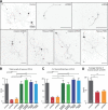
Overlapping but distinct expression patterns of RIM3γ and RIM4γ in adult rat brain. A, ISH micrographs showing RIM3γ and RIM4γ mRNA distribution in the whole brain. Negative controls with excess unlabeled oligonucleotides were devoid of signal (results not shown). Scale bar, 5 mm. B, Higher resolution pictures of emulsion-dipped sections of the hippocampus, the cerebellum, the olfactory bulb, and the cortex for RIM3γ (left) and RIM4γ (right). Scale bar, 300 μm. C, Homogenates of HEK-293T cells transfected with the indicated full-length RIM expression plasmids were analyzed by immunoblotting with affinity-purified antisera against RIM3γ and RIM4γ. The two antibodies were specific for the respective isoforms (peptides) they were raised against. D, Western blot analysis of adult rat tissues. Specific bands corresponding to the molecular weight of RIM3γ (32 kDa) and RIM4γ (27 kDa) were only detected in brain. E, Whole-brain homogenates from rats of the indicated ages (P0–P30) analyzed by immunoblotting with specific antibodies against RIM3γ and RIM4γ. Expression of both isoforms increased during postnatal brain development. F, Homogenates from the specified brain regions were analyzed by immunoblotting with isoform-specific antibodies and specificity of the antibody controlled by peptide blocking. Incubation of the blot with RIM3γ antibody revealed an unspecific 26 kDa band (*) in the thalamus. RIM3γ and RIM4γ proteins were ubiquitously expressed in the brain. However, the expression levels differed between the isoforms in various brain regions. GL, glomerular layer; EPL, external plexiform layer; MC, mossy cell; GC, granular cell; DG, dentate gyrus; I–VI, cortical layers.
Figure 2.
Figure 2.
RIM3γ and RIM4γ proteins are components of the presynaptic and postsynaptic cytomatrix. A, Rat brain homogenates were fractionated into the crude synaptosomal fraction (S1), the synaptosomal cytosol fraction (S2), the crude synaptosomal pellet fraction (P2), and the lysed synaptosomal membrane fraction (LP1, LS1), which consists of synaptosomal cytosol and SV-enriched fraction: the crude SV, the SPM, and myelin. The SPM was extracted twice with increasing Triton X-100 concentrations yielding the supernatant of the 0.5% (w/v) or 1% (w/v) Triton X-100 soluble fraction (TX1 SUPP and TX2 SUPP, respectively) and the Triton X-100 insoluble fraction of the SPM (TX1 and TX2). Fractions were analyzed using antibodies against RIM3γ and RIM4γ, as well as against Rab3A and PSD-95. Even though a fraction of RIM3γ and RIM4γ is extracted by Triton X-100, a substantial amount of the two proteins is still associated with the Triton X-100 insoluble fraction after the second extraction, resembling the pattern observed with PSD-95. B, Confocal micrographs of vertical rat retina sections labeled with antibodies against RIM3γ or RIM4γ. ONL, outer nuclear layer; OPL, outer plexiform layer; INL, inner nuclear layer; IPL, inner plexiform layer. Scale bar, 10 μm. RIM3γ exhibits a specific labeling in both synaptic layers, whereas RIM4γ is also present at these synapses but more broadly distributed. C–E, Double immunolabelings of hippocampal neurons (DIV 14) with the presynaptic marker Synapsin (C), the dendritic marker MAP2 (D), and the postsynaptic marker PSD-95 (E). Scale bars: (in C) 30 μm; (in D, E)10 μm.
Figure 3.
Figure 3.
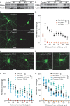
Knock-down of RIM3γ and RIM4γ results in altered neuronal morphology. A, B, Immunoblotting of cellular lysates from hippocampal primary neurons (DIV 14) transduced on DIV 1 with lentiviral particles expressing various RIM3γ-specific (A) or RIM4γ-specific (B) shRNAs (SH#1–4) and the empty vector (Control) revealed that shRIM3 results in a strong reduction of RIM3γ, and shRIM4 strongly reduces RIM4γ protein levels. Staining against tubulin was used as loading control. C, Hippocampal neurons were transduced at DIV 1 with lentiviral particles expressing GFP and either the empty vector (Control) or the RIM3γ/RIM4γ shRNA (shRIM3/4). All neurons were analyzed at DIV 14 using confocal microscopy. D, Sholl analysis revealed a loss of neuronal processes. E, F, The specificity of the observed phenotype was examined by transfecting either mutated shRNAs (mutated shRIM3/4) or functional shRNAs together with resistant RIM3γ/RIM4γ containing silent mutations (Rescue RIM3/Rescue RIM4). G, H, Quantification of the experiments in E and F by Sholl analysis for RIM3γ (G) and RIM4γ (H). Together, these control experiments show that the observed phenotype caused a reduction in the levels of RIM3γ and RIM4γ. One-way ANOVA, ***p < 0.001.
Figure 4.
Figure 4.
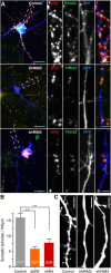
Absence of dendritic spines and reduction in synapse density in RIM3γ and RIM4γ knock-down neurons. A, Hippocampal neurons transfected at DIV 3 with either a vector-expressing GFP (Control) or GFP and the shRNA against RIM3γ (shRIM3) or RIM4γ (shRIM4). All neurons were immunostained using anti-Synapsin (SYN) and anti-PSD-95 (PSD-95) antibodies and analyzed at DIV 14 by confocal microscopy. Scale bar, 30 μm, * = 5 μm. B, Quantification of PSD-95/Synapsin colabeled synaptic punctae on RIM3γ and RIM4γ knock-down dendrites. RIM3γ-shRNA (shR3) and RIM4γ-shRNA (shR4) neurons exhibit a decreased synapse density compared with control. Quantification of Synapsin punctae density was performed using ImageJ software (n: # branches/# cells, one-way ANOVA, ***p < 0.001). C, Confocal image of a dendrite from a control and a RIM3γ-knock-down and a RIM4γ-knock-down neuron showing that substantially fewer dendritic spines can be found on knock-down dendrites (representative image of 5 independent cultures with >5 cells per condition each). Scale bar, 10 μm.
Figure 5.
Figure 5.
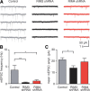
RIM3γ and RIM4γ knock-down decreases miniature excitatory synaptic activity. A, Representative recordings of mEPSCs from a control neuron (gray), and neurons with reduced levels of either RIM3γ (RIM3γ shRNA; black) or RIM4γ (RIM4γ shRNA; red). B, C, Analysis of mEPSC frequency and amplitude after RIM3γ or RIM4γ knock-down. B, Both RIM3γ and RIM4γ knock-down cause significant reduction in mEPSC frequency (p < 0.01 and p < 0.05, respectively; one-way ANOVA with Dunnett's post-test). C, RIM3γ knock-down causes a significant decrease of the mean amplitude (p < 0.05; one-way ANOVA with Dunnett's post-test; n = 7, 6, and 5 for RIM3γ and RIM4γ knock-down and control neurons, respectively).
Figure 6.
Figure 6.
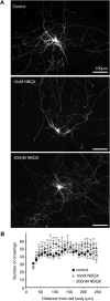
Synaptic silencing has no effect on neuronal morphology. A, Hippocampal neurons were transfected with a plasmid-expressing GFP at DIV 1 to visualize dendrites and axons. From DIV 2 to DIV 14 the cells were exposed to 10 or 200 nm NBQX. Control cells were incubated in normal media. All neurons were analyzed at DIV 14. Scale bar, 100 μm. B, Sholl analysis indicated no difference in neurite branching after synaptic silencing. To detect even small changes in distal dendrites Sholl analysis was performed up to 250 μm from the center of the neuron.
Figure 7.
Figure 7.
Development of neuronal processes is also affected by RIM3γ and RIM4γ knock-down at later time points. A, Hippocampal neurons transduced at DIV 1, 3, and 7, with shRNA and GFP (control) expressing lentiviral particles, and were analyzed by confocal microscopy at DIV 14. Neurons infected later exhibited a more complex dendritic tree than those in which the knock-down had been performed earlier. However, at all time points there was an obvious reduction in the number of processes in the shRNA-treated neurons as compared with control. Scale bar, 30 μm. B, Time-lapse imaging of hippocampal neurons transfected on DIV 7 with a plasmid-expressing shRIM3, shRIM4, or GFP alone (control) at 24, 48, 72, and 96 h after transfection. Already 24 h after transfection the dendritic tree of knock-down neurons appeared less complex and stopped growing after ∼50 h. Scale bar, 100 μm. C, Quantification of the length of the dendritic tree at 5 h intervals from the time-lapse experiment described in B. Dendrites were traced using ImageJ with NeuronJ plug-in; data depicted as mean± SEM were analyzed with Prism GraphPad 4 using one-way ANOVA followed by Tukey's multiple-comparison test, ***p < 0.001.
Figure 8.
Figure 8.
RIM3γ and RIM4γ knock-down affects early axonal outgrowth. A, Hippocampal neurons transfected DIV 1 expressing GFP (Control), shRNAs against RIM3γ (shRIM3) and RIM4γ (shRIM4), shRNAs with few nucleotide exchanges (mutated shRIM3 and mutated shRIM4) and coexpressing the shRNAs with the respective resistant cDNAs (Rescue RIM3 and Rescue RIM4) were fixed and analyzed by confocal microscopy at DIV 5. B, C, Quantitative analysis of total axonal length (B) and the number of axonal branches (C) showed that axonal outgrowth and branching are strongly reduced after knock-down of RIM3γ (shRIM3) and RIM4γ (shRIM4) as compared with controls. Both parameters are unaffected after cotransfection of shRIM3 or shRIM4 and a resistant version of the respective RIM variant (Rescue RIM3 and RIM4) or using mutated shRNAs against RIM3γ (mutated shRIM3) and RIM4γ (mutated shRIM4). D, Measurements of axonal growth of hippocampal neurons at DIV 8–DIV 9 from the time-lapse experiment of Figure 7B revealed that neurons transfected with shRNAs grow with reduced velocity 24–48 h after transfection. Significance: one-way ANOVA with Tukey's multiple-comparison test, ***p < 0.001, **p < 0.01.
Figure 9.
Figure 9.
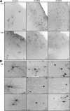
In vivo knock-down of RIM3γ and RIM4γ reproduces effect on neuronal morphology. A, Lentiviral particles expressing GFP alone (Control) or together with shRNAs against shRIM3 or shRIM4 were injected into the ventricle of P0 rat brains. Brains were analyzed at P14 (top row) and P21 (bottom row) by immunohistochemistry with an antibody against GFP. Cortical control neurons displayed a normal morphology (left), showing regular dendritic growth. In contrast, neurons transduced with the shRNA sequences exhibited a strong deficit in the number of neurites, indicating a greatly compromised neuronal branching (middle and right). Scale bar, 200 μm. B, Higher magnification images of control and knock-down cortical neurons (P21) revealed a striking loss in the dendritic arbor of neurons with decreased levels of RIM3γ and RIM4γ as compared with control (left). Scale bar, 50 μm.
Figure 10.
Figure 10.
RIM3γ in vivo rescue of neuronal morphology, reduction in spine density after loss of RIM3γ in hippocampus and cortex. A, Hippocampal neurons in P21 rats show that neurons expressing the shRNA against RIM3γ exhibit a reduced number of spines compared with control cells expressing only RFP. B, Quantification revealed a significant loss in spine density after knock-down of RIM3γ in hippocampal and cortical neurons (t test, hippocampus ***p = 0.0003, cortex **p = 0.0042). C, Lentiviral particles expressing RFP (Control) and the shRNA against RIM3γ alone (shRIM3) or together with a green fluorescent-resistant variant of RIM3 (Rescue RIM3) were injected into P0 rat brains. At P21, RIM3 knock-down cortical neurons exhibited the expected loss in arborization, while neurons expressing both shRNA and the resistant RIM3 were undistinguishable from control neurons expressing RFP. Scale bar, 50 μm.
Figure 11.
Figure 11.
The C2B domain present in both RIM3γ and RIM4γ is sufficient to rescue the knock-down phenotype. A, Hippocampal neurons were transduced at DIV 1 with viral particles expressing GFP (Control) or shRIM3 together with either RIM3γ (nonresistant) or RIM4γ. Neurons transduced at DIV 1 with viral particles expressing GFP (Control) or shRIM4 in combination with either RIM4γ (nonresistant) or RIM3γ. B, C, Quantification shows that the phenotype caused by knock-down of either RIM3γ (B) or RIM4γ (C) can be rescued by overexpression of the respective other isoform. (A, right column) Neurons were transduced with viral particles expressing shRIM3 or shRIM4 in combination with the respective RIM3/4γ-C2B domain. D, The quantification revealed that the C2B domain is sufficient to restore dendritic complexity almost to control levels. All neurons were analyzed at DIV 14 using confocal microscopy. Sholl analysis was performed to quantify the loss of neuronal processes. One-way ANOVA; ***p < 0.001.
Figure 12.
Figure 12.
Structural alteration of the Golgi apparatus in neurons lacking RIM3γ/4γ. A, Confocal images of GM130-labeled cultured hippocampal neurons, transfected at DIV 3 with either shRNAs against RIM3γ (shRIM3) or RIM4γ (shRIM4), or mutated variants of the both shRNAs (mutated shRIM3, mutated shRIM4), or GFP alone (control). Cells were fixed at DIV 14 and stained against the Golgi marker GM130 (red). Scale bar, 50 μm; * 20 μm. B, Quantification of Golgi dispersion. While Golgi dispersion in RIM4γ knock-down cells is indistinguishable from control, RIM3γ knock-down leads to increased fragmentation and dispersion. C, Quantification of Golgi size shows that knock-down of RIM4γ leads to a smaller, more condensed Golgi apparatus as compared with controls. B, C, These structural alterations were abolished using the mutated shRNAs against RIM3 or RIM4. Significance: one-way ANOVA followed by Tukey's multiple-comparison test, ***p < 0.0001.

Similar articles

Cited by

References

    1. Beffert U, Dillon GM, Sullivan JM, Stuart CE, Gilbert JP, Kambouris JA, Ho A. Microtubule plus-end tracking protein CLASP2 regulates neuronal polarity and synaptic function. J Neurosci. 2012;32:13906–13916. - PMC - PubMed
    1. Betz A, Thakur P, Junge HJ, Ashery U, Rhee JS, Scheuss V, Rosenmund C, Rettig J, Brose N. Functional interaction of the active zone proteins Munc13-1 and RIM1 in synaptic vesicle priming. Neuron. 2001;30:183–196. - PubMed
    1. Calakos N, Schoch S, Südhof TC, Malenka RC. Multiple roles for the active zone protein RIM1alpha in late stages of neurotransmitter release. Neuron. 2004;42:889–896. - PubMed
    1. Coppola T, Magnin-Luthi S, Perret-Menoud V, Gattesco S, Schiavo G, Regazzi R. Direct interaction of the Rab3 effector RIM with Ca2+ channels, SNAP-25, and synaptotagmin. J Biol Chem. 2001;276:32756–32762. - PubMed
    1. Deng L, Kaeser PS, Xu W, Südhof TC. RIM proteins activate vesicle priming by reversing autoinhibitory homodimerization of Munc13. Neuron. 2011;69:317–331. - PMC - PubMed

Publication types

MeSH terms

LinkOut - more resources