

Macrophage-mediated inflammation and disease: a focus on the lung
- PMID: 23304058
- PMCID: PMC3530802
- DOI: 10.1155/2012/140937
Macrophage-mediated inflammation and disease: a focus on the lung
Abstract
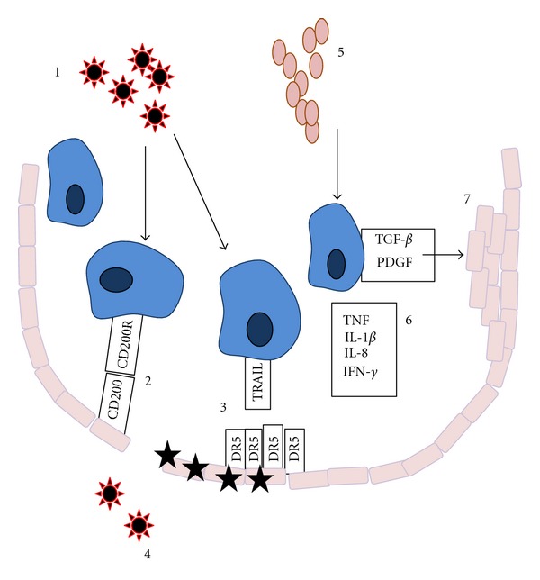
The lung is exposed to a vast array of inhaled antigens, particulate matter, and pollution. Cells present in the airways must therefore be maintained in a generally suppressive phenotype so that excessive responses to nonserious irritants do not occur; these result in bystander damage to lung architecture, influx of immune cells to the airways, and consequent impairment of gas exchange. To this end, the resident cells of the lung, which are predominantly macrophages, are kept in a dampened state. However, on occasion the suppression fails and these macrophages overreact to antigenic challenge, resulting in release of inflammatory mediators, induction of death of lung epithelial cells, deposition of extracellular matrix, and development of immunopathology. In this paper, we discuss the mechanisms behind this macrophage-mediated pathology, in the context of a number of inflammatory pulmonary disorders.
Figures
References
-
- Lin KL, Suzuki Y, Nakano H, Ramsburg E, Gunn MD. CCR2+ monocyte-derived dendritic cells and exudate macrophages produce influenza-induced pulmonary immune pathology and mortality. Journal of Immunology. 2008;180(4):2562–2572. - PubMed
-
- Vermaelen K, Pauwels R. Accurate and simple discrimination of mouse pulmonary dendritic cell and macrophage populations by flow cytometry: methodology and new insights. Cytometry A. 2004;61(2):170–177. - PubMed
Publication types
MeSH terms
Substances
Grants and funding
LinkOut - more resources
Full Text Sources
Other Literature Sources
Medical
