

A preliminary study on the potential of manuka honey and platelet-rich plasma in wound healing
- PMID: 23304152
- PMCID: PMC3523149
- DOI: 10.1155/2012/313781
A preliminary study on the potential of manuka honey and platelet-rich plasma in wound healing
Abstract
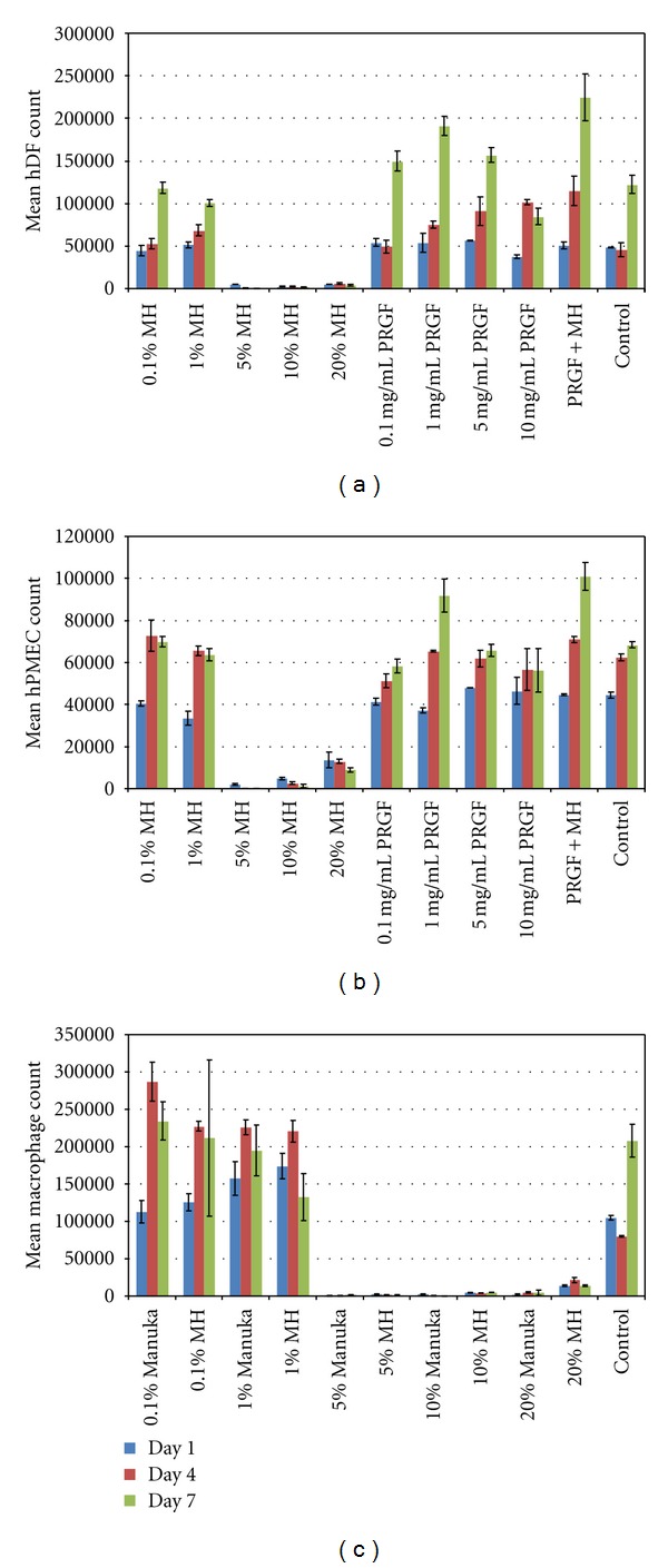
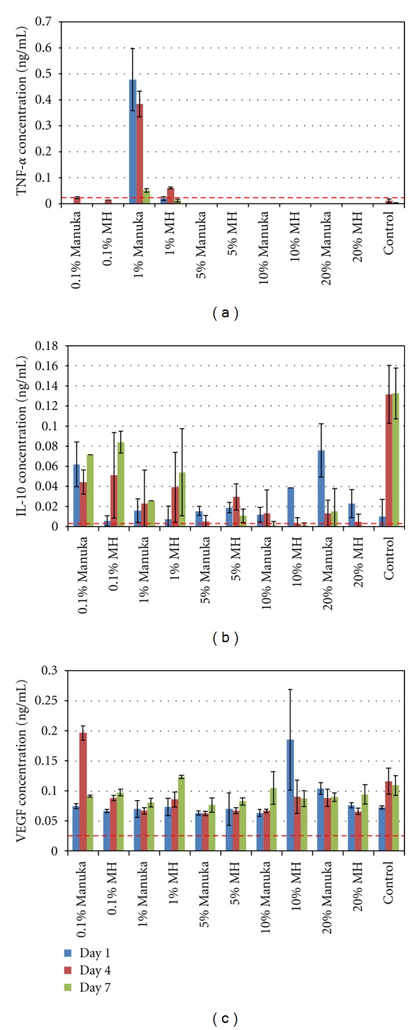
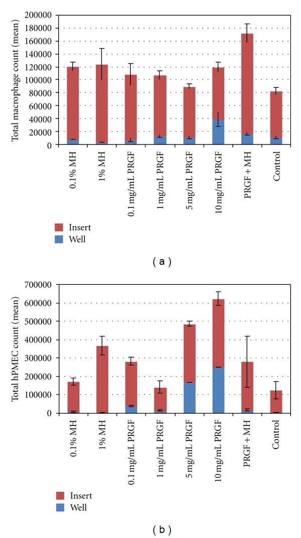
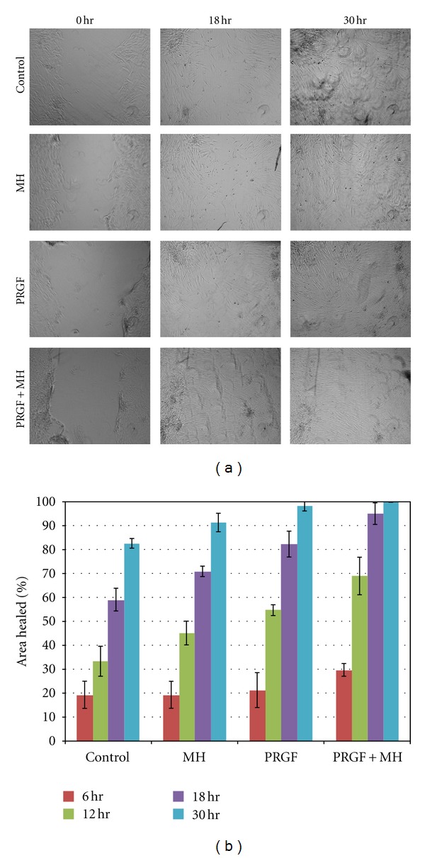
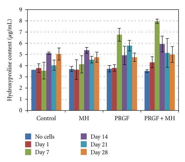
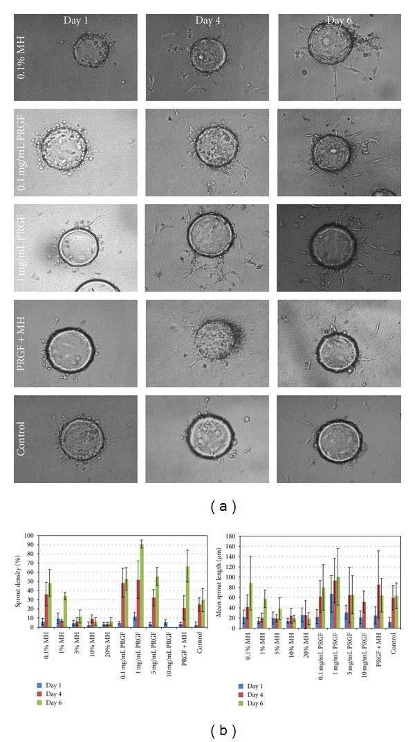
Aim. The purpose of this study was to determine the in vitro response of cells critical to the wound healing process in culture media supplemented with a lyophilized preparation rich in growth factors (PRGF) and Manuka honey. Materials and Methods. This study utilized cell culture media supplemented with PRGF, as well as whole Manuka honey and the medical-grade Medihoney (MH), a Manuka honey product. The response of human fibroblasts (hDF), macrophages, and endothelial cells (hPMEC) was evaluated, with respect to cell proliferation, chemotaxis, collagen matrix production, and angiogenic potential, when subjected to culture with media containing PRGF, MH, Manuka honey, and a combination of PRGF and MH. Results. All three cell types demonstrated increases in cellular activity in the presence of PRGF, with further increases in activity seen in the presence of PRGF+MH. hDFs proved to be the most positively responsive cells, as they experienced enhanced proliferation, collagen matrix production, and migration into an in vitro wound healing model with the PRGF+MH-supplemented media. Conclusion. This preliminary in vitro study is the first to evaluate the combination of PRGF and Manuka honey, two products with the potential to increase regeneration individually, as a combined product to enhance dermal regeneration.
Figures

References
-
- Simpson DG. Dermal templates and the wound-healing paradigm: the promise of tissue regeneration. Expert Review of Medical Devices. 2006;3(4):471–484. - PubMed
-
- Percival S, Cutting K. Microbiology of Wounds. Boca Raton, Fla, USA: CRC Press; 2010.
-
- Langemo DK, Hanson D, Anderson J, Thompson P, Hunter S. Use of honey for wound healing. Advances in Skin & Wound Care. 2009;22(3):113–118. - PubMed
-
- Rossiter K, Cooper AJ, Voegeli D, Lwaleed BA. Honey promotes angiogeneic activity in the rat aortic ring assay. Journal of Wound Care. 2010;19(10):440–446. - PubMed
-
- Lusby PE, Coombes A, Wilkinson JM. Honey: a potent agent for wound healing? Journal of Wound, Ostomy, and Continence Nursing. 2002;29(6):295–300. - PubMed
LinkOut - more resources
Full Text Sources
Other Literature Sources
