. 2012:2012:846171.
doi: 10.5402/2012/846171. Epub 2012 Dec 5.

Cardiac ion channelopathies and the sudden infant death syndrome

Affiliations

Cardiac ion channelopathies and the sudden infant death syndrome

Ronald Wilders. ISRN Cardiol. 2012.

Abstract

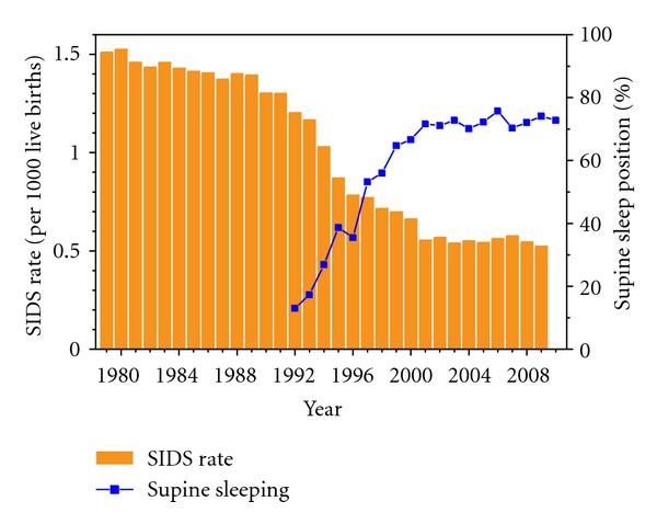
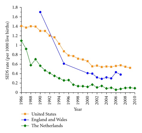
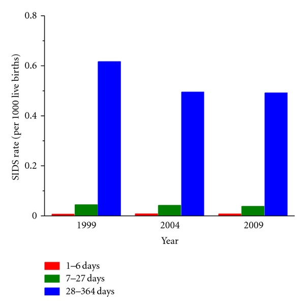
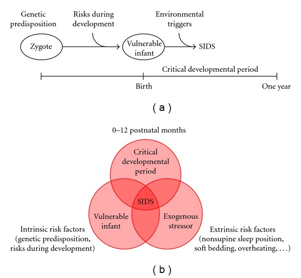
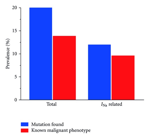
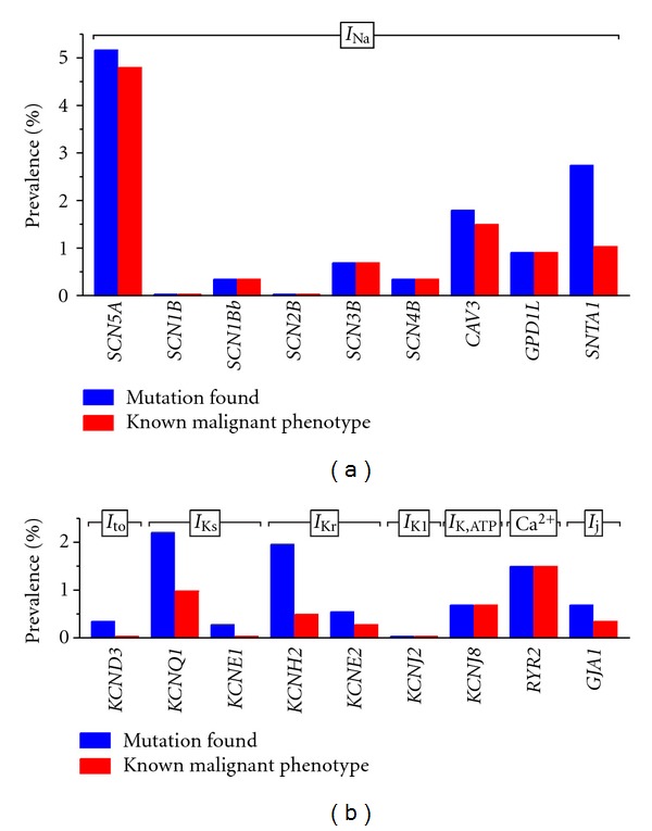
The sudden infant death syndrome (SIDS) causes the sudden death of an apparently healthy infant, which remains unexplained despite a thorough investigation, including the performance of a complete autopsy. The triple risk model for the pathogenesis of SIDS points to the coincidence of a vulnerable infant, a critical developmental period, and an exogenous stressor. Primary electrical diseases of the heart, which may cause lethal arrhythmias as a result of dysfunctioning cardiac ion channels ("cardiac ion channelopathies") and are not detectable during a standard postmortem examination, may create the vulnerable infant and thus contribute to SIDS. Evidence comes from clinical correlations between the long QT syndrome and SIDS as well as genetic analyses in cohorts of SIDS victims ("molecular autopsy"), which have revealed a large number of mutations in ion channel-related genes linked to inheritable arrhythmogenic syndromes, in particular the long QT syndrome, the short QT syndrome, the Brugada syndrome, and catecholaminergic polymorphic ventricular tachycardia. Combining data from population-based cohort studies, it can be concluded that at least one out of five SIDS victims carries a mutation in a cardiac ion channel-related gene and that the majority of these mutations are of a known malignant phenotype.


Figures

Figure 1
Figure 1
Rate of SIDS (orange bars), expressed as the number of SIDS cases per 1,000 live births, and the percent of infants put to sleep in the supine position (blue line with squares) in the United States over the period from 1979 to 2010. Data on SIDS rate (1979–2009) from the Centers for Disease Control and Prevention (CDC), as provided on the CDC Wide-ranging Online Data for Epidemiologic Research (WONDER) website (http://wonder.cdc.gov/mortSQL.html). For the years 1979–1999, the data refer to all deaths labeled with code 798.0 (“sudden infant death syndrome”) of the Ninth Revision of the International Classification of Diseases (ICD-9) of the World Health Organization. For the years 2000–2009, the data refer to all deaths specified with code R95 (“sudden infant death syndrome”) of the Tenth Revision (ICD-10). Data on supine sleep position (1992–2010) from the National Infant Sleep Position Study (NISP) of the National Institute of Child Health and Human Development (NICHD), as provided on the NISP public access website hosted by Boston University (http://slone-web2.bu.edu/ChimeNisp/Main_Nisp.asp).
Figure 2
Figure 2
Rate of SIDS, expressed as the number of SIDS cases per 1,000 live births, over the period from 1986 to 2010 in the United States (orange solid line with squares), England and Wales (blue solid line with circles), and The Netherlands (green solid line with diamonds). Data on SIDS rate for the United States as in Figure 1. Data for England and Wales from Hauck and Tanabe [13] (1990–2005), completed with data for 2006 and 2007 from the International Society for the Study and Prevention of Perinatal and Infant Death (ISPID), as provided on the ISPID public access website (http://www.ispid.org/statistics.html). Data for The Netherlands from Statistics Netherlands (Centraal Bureau voor de Statistiek, CBS), as provided on the CBS public access website (http://www.cbs.nl/).
Figure 3
Figure 3
Rate of SIDS, expressed as the number of SIDS cases per 1,000 live births, occurring during week 1 of the infant's life (days 1–6, red bars), during weeks 2–4 (days 7–27, green bars), and during week 5 and later (days 28–364, blue bars) in the United States in 1999, 2004, and 2009. Data from the Compressed Mortality database as provided on the Wide-ranging Online Data for Epidemiologic Research (WONDER) website of the Centers for Disease Control and Prevention (CDC, http://wonder.cdc.gov/mortSQL.html).
Figure 4
Figure 4
Triple risk model in the sudden infant death syndrome (SIDS). (a) Visualization of the triple risk model after Courts and Madea [50]. Genetic predisposition and risks during development create a vulnerable infant. If this vulnerable infant encounters environmental triggers during a critical developmental period, it may become an SIDS victim. (b) Visualization of the triple risk model after Filiano and Kinney [48] and Trachtenberg et al. [34]. A combination of intrinsic risk factors (genetic predisposition, risks during development) and extrinsic risk factors during a critical developmental period may create an SIDS victim.
Figure 5
Figure 5
Electrocardiographic data in 42 sets of parents who had infants with SIDS [80]. In 27 sets of parents, both parents had a normal ECG (left bar). In 15 sets (36%, middle bar), at least one parent had an abnormal ECG (middle bar), including a long QT interval in 11 sets (26%, right bar).
Figure 6
Figure 6
Action potential formation and propagation in ventricular myocytes. (a) Diagram of an action potential in an isolated ventricular myocyte with individual ion currents that “push up” (inward currents, upward arrows) or “pull down” (outward currents, downward arrows) the action potential. I Ca,L: L-type calcium current; I K,ACh: acetylcholine-sensitive potassium current; I K,ATP: ATP-sensitive potassium current; I K1: inward rectifier potassium current; I Kr: rapid delayed rectifier potassium current; I Ks: slow delayed rectifier potassium current; I Na: fast sodium current; I NaCa: sodium-calcium exchange current; I to: transient outward potassium current. Grey symbols refer to currents that become important under special conditions (release of ACh, shortage of ATP and calcium overload for I K,ACh, I K,ATP, and I NaCa, resp.). Loss of function of “pull down” currents may result in action potential lengthening (inset, red dashed trace labeled “1”), whereas loss of function of “push up” currents may result in action potential shortening (inset, red dashed trace labeled “2”) and loss of the action potential dome (inset, red dashed trace labeled “3”). (b) Diagram of action potential propagation. The propagating action potential (horizontal arrow) brings neighboring cells to threshold through the I Na driven gap junctional current I j.
Figure 7
Figure 7
Prevalence of cardiac ion channelopathies in SIDS victims, based on mutations found in population-based cohort studies (Tables 5 and 6). Left: overall prevalence (blue bar) and prevalence of mutations with a known malignant phenotype (red bar). Right: prevalence of fast sodium current (I Na) related mutations (blue bar) and those of a known malignant phenotype (red bar).
Figure 8
Figure 8
Prevalence of cardiac ion channelopathies in SIDS victims, based on data from population-based cohort studies (Tables 5 and 6). Blue bars indicate the prevalence of mutations and red bars that of mutations with a known malignant phenotype. (a) Mutations in genes affecting the fast sodium current I Na. (b) Mutations in genes affecting the transient outward potassium current I to (KCND3), the slow delayed rectifier potassium current I Ks(KCNQ1 and KCNE1), the rapid delayed rectifier potassium current I Kr (KCNH2 and KCNE2), the inward rectifier potassium current I K1 (KCNJ2), the ATP-sensitive potassium current I K,ATP (KCNJ8), the RyR2 calcium release channel (RYR2), and the gap junctional current I j (GJA1).

Similar articles

Cited by

References

    1. Beckwith JB. Discussion of terminology and definition of sudden infant death syndrome. In: Bergman AB, Beckwith JB, Ray CG, editors. Proceedings of the 2nd International Conference on Causes of Sudden Death in Infants; Seattle, Wash, USA. University of Washington Press; 1970. pp. 14–22.
    1. Willinger M, James LS, Catz C. Defining the sudden infant death syndrome (SIDS): deliberations of an expert panel convened by the National Institute of Child Health and Human Development. Pediatric Pathology. 1991;11(5):677–684. - PubMed
    1. Krous HF, Beckwith JB, Byard RW, et al. Sudden infant death syndrome and unclassified sudden infant deaths: a definitional and diagnostic approach. Pediatrics. 2004;114(1):234–238. - PubMed
    1. Krous HF. Sudden unexpected death in infancy and the dilemma of defining the sudden infant death syndrome. Current Pediatric Reviews. 2010;6(1):5–12.
    1. Byard RW, Krous HF. Sudden infant death syndrome: overview and update. Pediatric and Developmental Pathology. 2003;6(2):112–127. - PubMed

LinkOut - more resources