Spontaneous hemocholecyst in an end-stage renal failure patient on low molecular weight heparin hemodialysis
- PMID: 23304616
- PMCID: PMC3529897
- DOI: 10.1155/2012/363924
Spontaneous hemocholecyst in an end-stage renal failure patient on low molecular weight heparin hemodialysis
Abstract
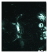
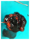
The present paper describes a case of spontaneous hemocholecyst in a patient with end-stage renal failure on low molecular weight heparin hemodialysis. The patient presented with acute right upper quadrant pain. An initial ultrasound scan demonstrated a distended gallbladder containing echogenic bile without stones. During hospitalization the patient became febrile, and jaundiced, developed leukocytosis, and had an elevation in serum bilirubin, transaminases, and alkaline phosphatase. A new ultrasound demonstrated a thick-walled gallbladder containing echogenic bile and pericholecystic fluid. MRI depicted a distended gallbladder containing material of mixed signal intensity and a normal biliary tract. Open cholecystectomy revealed a gallbladder filled with blood and clots, and transcystic common bile duct exploration flushed blood clots out of the bile duct. To our knowledge this is the second case of spontaneous hemocholecyst reported in the literature as a consequence of uremic bleeding and LMWH hemodialysis in the absence of other pathology.
Figures


Similar articles
-
Palliative percutaneous transhepatic gallbladder drainage of gallbladder empyema before laparoscopic cholecystectomy.Hepatogastroenterology. 2000 Jul-Aug;47(34):932-6. Hepatogastroenterology. 2000. PMID: 11020851
-
[Hemocholecyst complicated by rupture of the gallbladder].Pan Afr Med J. 2019 Sep 24;34:45. doi: 10.11604/pamj.2019.34.45.18682. eCollection 2019. Pan Afr Med J. 2019. PMID: 31762912 Free PMC article. French.
-
Acute cholecystitis caused by hemocholecyst: unusual clinical manifestation of gallbladder cancer.Acta Gastroenterol Belg. 2013 Mar;76(1):57-8. Acta Gastroenterol Belg. 2013. PMID: 23650784
-
Secondary hemocholecyst after radiofrequency ablation therapy for hepatocellular carcinoma.J Gastroenterol. 2003;38(4):399-403. doi: 10.1007/s005350300071. J Gastroenterol. 2003. PMID: 12743783 Review.
-
Biliary tract injuries during laparoscopic cholecystectomy: three case reports and literature review.G Chir. 2010 Jan-Feb;31(1-2):16-9. G Chir. 2010. PMID: 20298660 Review.
Cited by
-
Gallbladder bleeding-related severe gastrointestinal bleeding and shock in a case with end-stage renal disease: A case report.Medicine (Baltimore). 2016 Jun;95(23):e3870. doi: 10.1097/MD.0000000000003870. Medicine (Baltimore). 2016. PMID: 27281100 Free PMC article.
References
-
- Herrero-Calvo JA, González-Parra E, Pérez-García R, Tornero-Molina F. Spanish study of anticoagulation in haemodialysis. Nefrologia. 2012;32:143–152. - PubMed
-
- Chin MW, Enns R. Hemobilia. Current Gastroenterology Reports. 2010;12(2):121–129. - PubMed
-
- Laing FC, Frates MC, Feldstein VA, Goldstein RB, Mondro S. Sonographic appearances in the gallbladder and biliary tree with emphasis on intracholecystic blood. Journal of Ultrasound in Medicine. 1997;16(8):537–543. - PubMed
-
- Siu WT, Chau CH, Law BKB, Yau KK, Luk YW, Li MK. Non-operative management of endoscopic iatrogenic haemobilia: case report and review of literature. Acta Gastro-Enterologica Belgica. 2005;68(4):428–431. - PubMed
-
- Gunay Y, Bircan HY, Emek E, Cevik H, Altaca G, Moray G. The management of acute cholecystitis in chronic hemodialysis patients: percutaneous cholecystostomy versus cholecystectomy. Journal of Gastrointestinal Surgery. In press. - PubMed
LinkOut - more resources
Full Text Sources

