. 2013 Feb 5;85(3):1880-8.
doi: 10.1021/ac3031527. Epub 2013 Jan 23.

Top down proteomics of human membrane proteins from enriched mitochondrial fractions

Affiliations

Top down proteomics of human membrane proteins from enriched mitochondrial fractions

Adam D Catherman et al. Anal Chem. .

Abstract

The interrogation of intact integral membrane proteins has long been a challenge for biological mass spectrometry. Here, we demonstrate the application of top down mass spectrometry to whole membrane proteins below 60 kDa with up to 8 transmembrane helices. Analysis of enriched mitochondrial membrane preparations from human cells yielded identification of 83 integral membrane proteins, along with 163 membrane-associated or soluble proteins, with a median q value of 3 × 10(-10). An analysis of matching fragment ions demonstrated that significantly more fragment ions were found within transmembrane domains than would be expected based upon the observed protein sequence. In total, 46 proteins from the complexes of oxidative phosphorylation were identified which exemplifies the increasing ability of top down proteomics to provide extensive coverage in a biological network.


Figures

Figure 1
Figure 1. Platform for high-throughput Top Down Proteomics of mitochondrial membrane proteins
Cultured HeLa cells are subjected to subcellular fractionation to yield enriched mitochondria which are further fractionated to yield mitochondrial membrane proteins. These proteins are then separated according to molecular weight by GELFrEE, yielding 12 fractions. Each fraction is injected onto a nanoLC column and intact mass measurement and fragmentation spectra are collected with a 12 Tesla LTQ Velos FT Ultra. ProSightPC is used for identification prior to analysis of transmembrane domain content.
Figure 2
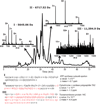
Figure 2. Representative data for the LC-MS/MS analysis of a single GELFrEE fraction containing <15 kDa proteins
The total ion chromatogram (TIC) shows the separation of proteins according to hydrophobicity, demonstrating the orthogonal nature of GELFrEE and RPLC. Intact spectra for three proteins are shown which allow for monoisotopic intact masses to be determined within 5 ppm of the expected molecular weight. Collisionally-induced fragmentation of each of the three proteins resulted in highly confident identifications, each demonstrating 100% sequence coverage using 10 ppm mass accuracy on the fragment ions. ATP synthase subunit epsilon was identified as the Met off form, cytochrome c oxidase polypeptide 7A2 with a 23 residue transit peptide cleavage, and transmembrane protein 14C with N-terminal acetylation. The TMHMM predicted transmembrane domain helices are displayed in red.
Figure 3
Figure 3. Identification of post-translational modifications and a mis-annotated start site on a newly observed human protein
A, Myristoylation identified on the N-terminus of the methionine cleaved form of NADH dehydrogenase 1 beta subcomplex subunit 7. B, Lysine 43 trimethylation was identified on ATP synthase subunit c following signal sequence cleavage. C, Signal sequence cleavage also occurs on transmembrane protein 109, leaving an N-terminal glutamine which cyclizes to a pyroglutamate. D, First evidence of a protein from a gene expected to be non-coding was identified as a 55 amino acid, single TMD containing product.
Figure 4
Figure 4. A selected example of gene-specific identifications illustrate that this is routine in Top Down Proteomics
ER lumen protein retaining receptor 1 and 2 were unambiguously differentiated from one another. Although many of the matching fragments were identical or isobaric between the two species, several unique ions reveal each individual identification.
Figure 5
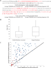
Figure 5. Examination of the propensity for transmembrane helices to fragment in the gas phase
A) Graphical fragment maps for two identified proteins demonstrating the propensity of transmembrane domain helices (shown in red) to fragment relative to the soluble regions. Also displayed is the average TMHMM value for all of the corresponding backbone positions as well as the average value for the entire protein. B) Distribution of lowest obtained q values for each of the identified proteins separated as predicted integral membrane and soluble/membrane associated proteins. The whiskers represent the 10th/90th percentiles. The distribution demonstrates that integral membrane proteins were, on average, identified with a greater confidence than the soluble proteins. C) Scatter plot displaying the TMHMM scores for fragment ions and entire proteins (as in A) where each points represents a unique protein. Integral membrane proteins are shown as blue circles and soluble/membrane associated proteins in red circles. The y=x line is also plotted to demonstrate where the data points would lie if the fragmentation was truly random. Points above this line indicate that the fragments were more likely to be derived from TMDs.

References

    1. Siuti N, Roth MJ, Mizzen CA, Kelleher NL, Pesavento JJ. J. Proteome Res. 2006;5:233–239. - PubMed
    1. Pesavento JJ, Bullock CR, Leduc RD, Mizzen CA, Kelleher NL. J. Biol. Chem. 2008;283:14927–14937. - PMC - PubMed
    1. Resemann A, Wunderlich D, Rothbauer U, Warscheid B, Leonhardt H, Fuchser J, Kuhlmann K, Suckau D. Anal. Chem. 2010;82:3283–3292. - PubMed
    1. Meng FY, Cargile BJ, Patrie SM, Johnson JR, McLoughlin SM, Kelleher NL. Anal. Chem. 2002;74:2923–2929. - PubMed
    1. Roth MJ, Forbes AJ, Boyne MT, Kim YB, Robinson DE, Kelleher NL. Mol. Cell. Proteomics. 2005;4:1002–1008. - PMC - PubMed

Publication types

Substances

LinkOut - more resources