. 2013 Jun;161(6):495-504.
doi: 10.1016/j.trsl.2012.12.008. Epub 2013 Jan 8.

Fragmented oxidation products define barrier disruptive endothelial cell response to OxPAPC

Affiliations

Fragmented oxidation products define barrier disruptive endothelial cell response to OxPAPC

Anna A Birukova et al. Transl Res. 2013 Jun.

Abstract

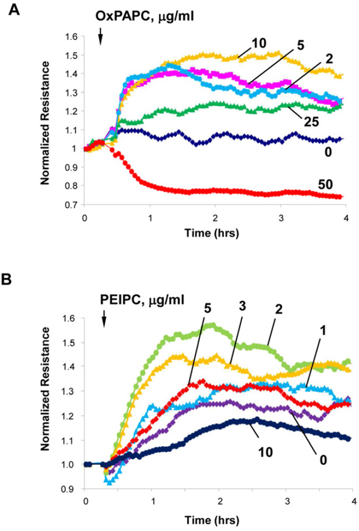
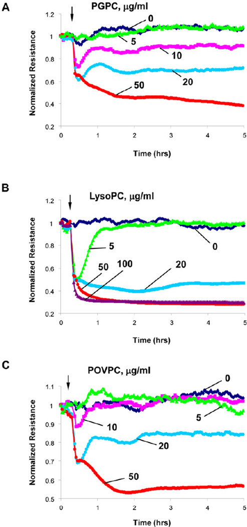
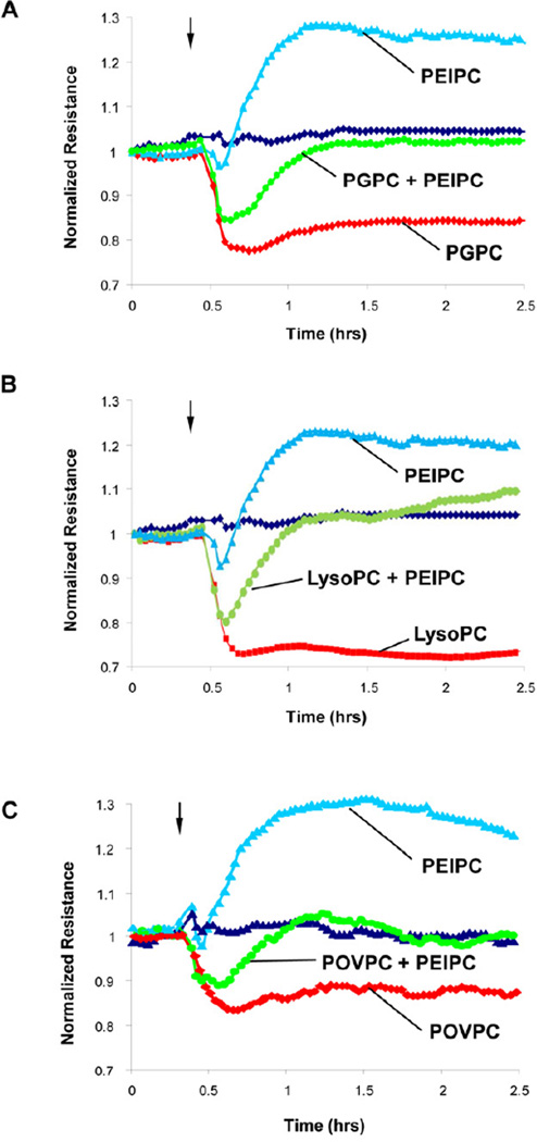
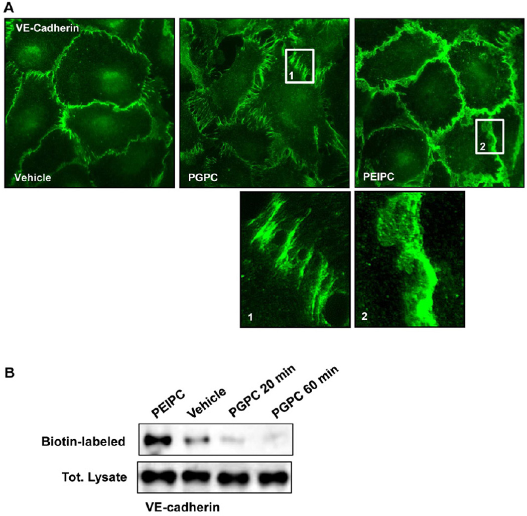
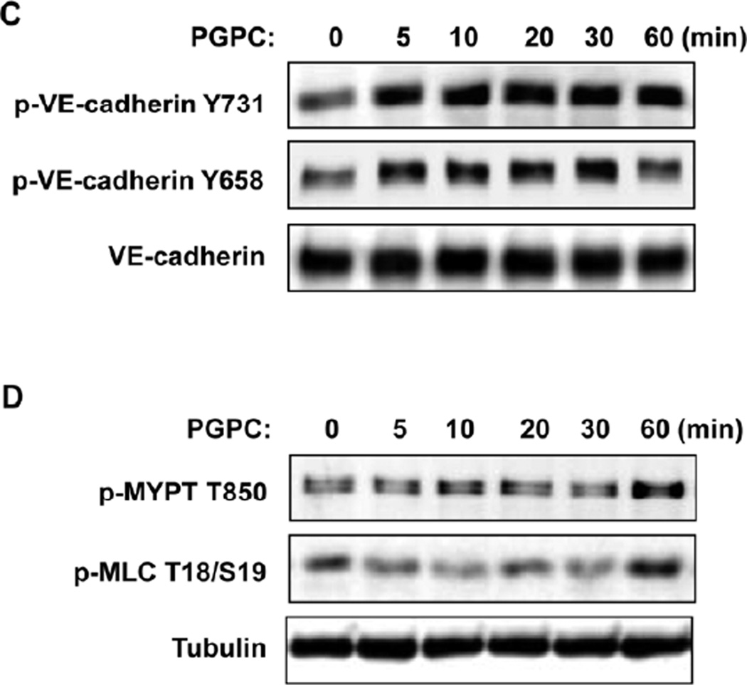
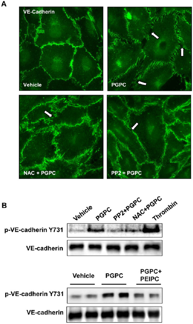
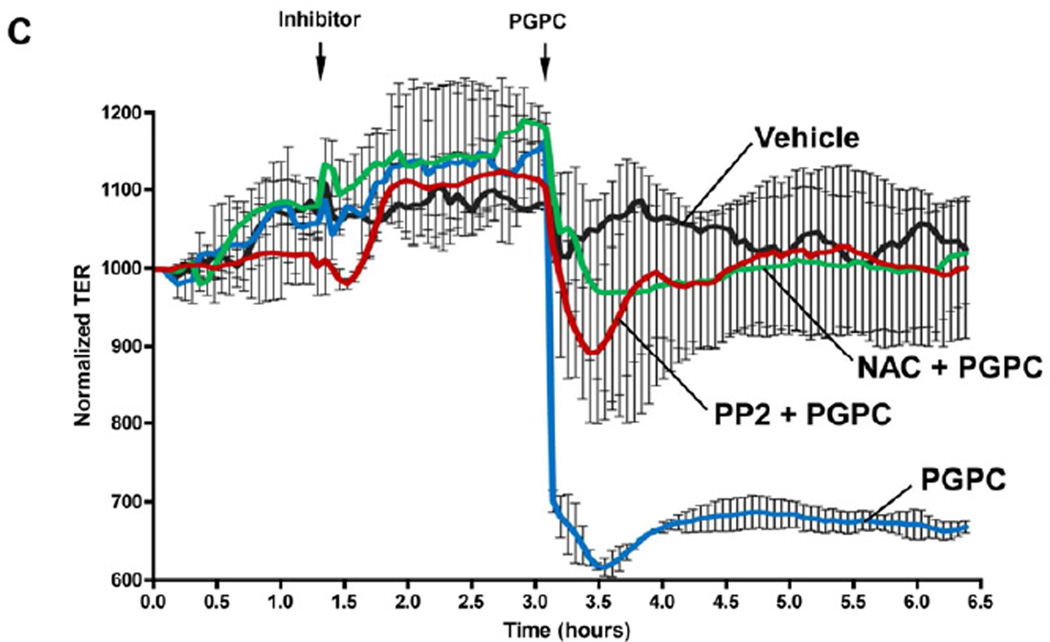
Excessive concentrations of oxidized phospholipids (OxPL), the products of 1-palmitoyl-2-arachidonoyl-sn-glycero-3-phosphatidylcholine (PAPC) oxidation have been detected in atherosclerosis, septic inflammation, and acute lung injury (ALI) and have been shown to induce vascular barrier dysfunction. In contrast, oxidized PAPC (OxPAPC) at low concentrations exhibit potent barrier protective effects. The nature of such biphasic effects remains unclear. We tested the hypothesis that barrier-disruptive effects of high OxPAPC doses on endothelial cell (EC) monolayer are defined by fragmented products of PAPC oxidation (lysophosphatidyl choline [lyso-PC], 1-palmitoyl-2-(5-oxovaleroyl)-sn-glycero-phosphatidylcholine [POVPC], 1-palmitoyl-2-glutaroyl-sn-glycero-phosphatidylcholine [PGPC]), whereas barrier enhancing effects are mediated by full length oxidated PAPC products and may be reproduced by single compounds contained in the OxPAPC such as 1-palmitoyl-2-(5,6-epoxyisoprostane E2)-sn-glycero-3-phosphatidyl choline (PEIPC). All 3 fragmented OxPAPC products increased EC permeability in a dose-dependent manner, whereas PEIPC decreased it and reversed barrier disruptive effects of lyso-PC, POVPC, and PGPC monitored by measurements of transendothelial electrical resistance. Immunofluorescence staining and western blot analysis showed that PGPC mimicked the cytoskeletal remodeling and tyrosine phosphorylation of adherens junction (AJ) protein vascular endothelial (VE)-cadherin leading to EC barrier dysfunction induced by high OxPAPC concentrations. Barrier-disruptive effects of PGPC were abrogated by reactive oxygen species (ROS) inhibitor, N-acetyl cysteine, or Src kinase inhibitor, PP-2. The results of this study show that barrier disruptive effects of fragmented OxPAPC constituents (lyso-PC, POVPC, PGPC) are balanced by barrier enhancing effects of full length oxygenated products (PEIPC). These data strongly suggest that barrier disruptive effects of OxPAPC at higher concentrations are dictated by predominant effects of fragmented phospholipids such as PGPC, which promote ROS-dependent activation of Src kinase and VE-cadherin phosphorylation at Tyr(658) and Tyr(731) leading to disruption of endothelial cell AJs.


Conflict of interest statement

The authors have no conflicts of interest to declare. All authors have read the journal's policy on disclosure of potential conflicts of interest.

Figures

Figure 1
Figure 1. Dose-dependent effects of OxPAPC and PEIPC on transendothelial electrical resistance
Human pulmonary artery endothelial cells (HPAECs) were seeded in polycarbonate wells with gold microelectrodes. After 24 hr of culture, HPAEC were stimulated with various concentrations of: A - OxPAPC (2, 5, 10, 25 and 50 µg/ml); or B - PEIPC (1, 2, 3, 5 and 10 µg/ml), and measurements of transendothelial electrical resistance (TER) were monitored over 4 hrs using an electrical cell-substrate impedance sensing system (ECIS). Results are representative of five independent experiments.
Figure 2
Figure 2. Dose-dependent effects of PGPC, lyso-PC and POVPC on transendothelial electrical resistance
HPAECs seeded in polycarbonate wells with gold microelectrodes were stimulated with various concentrations of: A – PGPC; B – lyso-PC; and B – POVPC, and measurements of transendothelial electrical resistance (TER) were monitored over 5 hrs using an electrical cell-substrate impedance sensing system (ECIS). Results are representative of five independent experiments.
Figure 3
Figure 3. Effects of PEIPC cotreatment with PGPC, lyso-PC and POVPC on transendothelial electrical resistance
HPAECs seeded in polycarbonate wells with gold microelectrodes were stimulated with PEIPC (2 µg/ml) alone or were co-stimulated with PEIPC and: A – PGPC (20 µg/ml); B – lyso-PC (20 µg/ml); and B – POVPC (20 µg/ml), and measurements of transendothelial electrical resistance (TER) were monitored over 5 hrs using an electrical cell-substrate impedance sensing system (ECIS). Results are representative of three independent experiments.
Figure 4
Figure 4. Effects of PGPC and PEIPC on intracellular distribution and phosphorylation of VE-cadherin and downstream Rho targets
A – Endothelial monolayers grown on glass coverslips were stimulated with PGPC (30 µg/ml, 30 min) or PEIPC (2 µg/ml, 30 min) followed by immunofluorescence staining for VE-cadherin. Insets depict higher magnification areas of VE-cadherin immunoreactivity at cell-cell junctions of PGPC and PEIPC treated endothelial monolayers. Shown are representative results of three independent experiments. B - HPAECs were stimulated with PEIPC or PGPC for indicated periods of time, washed, and cell surface proteins were labeled with Sulfo-NHS-SS-Biotin as described in Methods. Cells were lysed, and biotinylated proteins precipitated with streptavidin-agarose. Presence of biotinylated VE-cadherin was evaluated by Western blot analysis. C - EC were stimulated with PGPC (30 µg/ml) for indicated periods of time, and VE-cadherin phosphorylation at Tyr731 and Tyr658 was examined by western blot. Staining with VE-cadherin antibody was used as normalization control. D - Time course of myosin light chain phosphatase (MYPT) and myosin light chain (MLC) phosphorylation induced by PGPC was examined by western blot with corresponding phospho-specific antibodies. µ-Tubulin antibody was used as normalization control.
Figure 5
Figure 5. Effects of SRC and ROS inhibitors on remodeling of VE-cadherin-positive adherens junctions, VE-cadherin phosphorylation and EC permeability induced by PGPC
HPAECs were pretreated with vehicle, PP2 (1µM), or NAC (1mM) for 30 min followed by stimulation with 30 µg/ml PGPC. A – Immunofluorescence staining for VE-cadherin was performed after 15 min of PGPC stimulation. Arrows depict areas of intercellular gaps. Shown are representative results of three independent experiments. B – The levels of VE-cadherin phosphorylated at Tyr731 and Y658 were detected by Western blot analysis and normalized to the total VE-cadherin content. Lower panels depict effect of co-treatment with PEIPC (2 µg/ml) on PGPC-induced VE-cadherin phosphorylation at Y731. C – TER measurements were monitored over 6.5 hrs. Time points of inhibitor and PGPC addition are indicated by vertical arrows. Shown are pooled data from three independent experiments expressed as mean ± SD.

References

    1. Bochkov VN, Oskolkova OV, Birukov KG, et al. Generation and biological activities of oxidized phospholipids. Antioxid Redox Signal. 2010;12(8):1009–1059. - PMC - PubMed
    1. Quinlan GJ, Lamb NJ, Evans TW, Gutteridge JM. Plasma fatty acid changes and increased lipid peroxidation in patients with adult respiratory distress syndrome. Crit Care Med. 1996;24(2):241–246. - PubMed
    1. Montuschi P, Collins JV, Ciabattoni G, et al. Exhaled 8-isoprostane as an in vivo biomarker of lung oxidative stress in patients with copd and healthy smokers. Am J Respir Crit Care Med. 2000;162(3 Pt 1):1175–1177. - PubMed
    1. Carpenter CT, Price PV, Christman BW. Exhaled breath condensate isoprostanes are elevated in patients with acute lung injury or ards. Chest. 1998;114(6):1653–1659. - PubMed
    1. Jerlich A, Schaur RJ, Pitt AR, Spickett CM. The formation of phosphatidylcholine oxidation products by stimulated phagocytes. Free Radic Res. 2003;37(6):645–653. - PubMed

Publication types

MeSH terms

Substances