. 2013 Mar 7;121(10):1868-74.
doi: 10.1182/blood-2012-09-455436. Epub 2013 Jan 10.

Hepatocyte tissue factor activates the coagulation cascade in mice

Affiliations

Hepatocyte tissue factor activates the coagulation cascade in mice

Bradley P Sullivan et al. Blood. .

Abstract

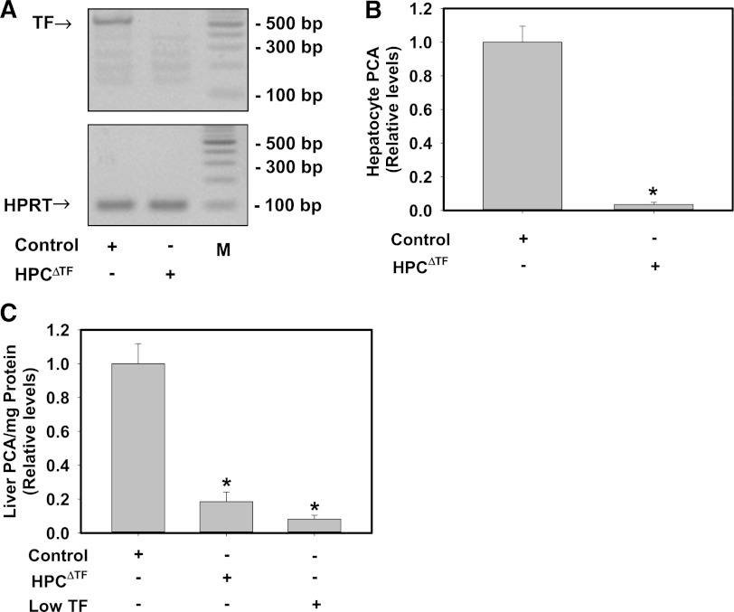
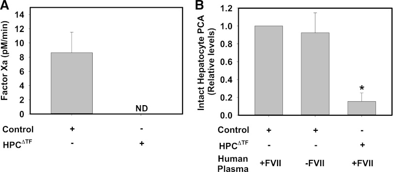
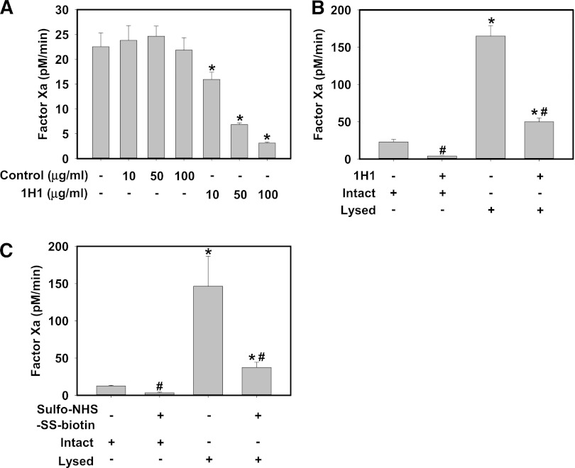
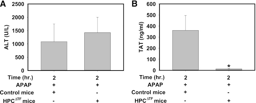
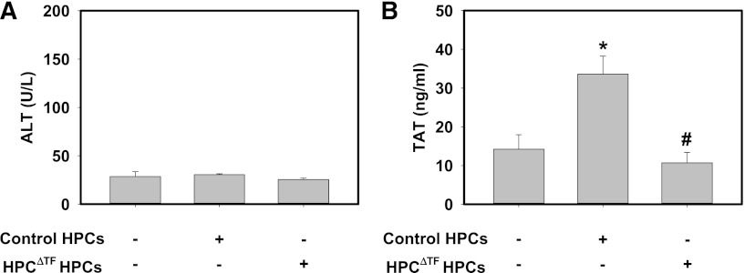
In this study, we characterized tissue factor (TF) expression in mouse hepatocytes (HPCs) and evaluated its role in mouse models of HPC transplantation and acetaminophen (APAP) overdose. TF expression was significantly reduced in isolated HPCs and liver homogenates from TF(flox/flox)/albumin-Cre mice (HPC(ΔTF) mice) compared with TF(flox/flox) mice (control mice). Isolated mouse HPCs expressed low levels of TF that clotted factor VII-deficient human plasma. In addition, HPC TF initiated factor Xa generation without exogenous factor VIIa, and TF activity was increased dramatically after cell lysis. Treatment of HPCs with an inhibitory TF antibody or a cell-impermeable lysine-conjugating reagent prior to lysis substantially reduced TF activity, suggesting that TF was mainly present on the cell surface. Thrombin generation was dramatically reduced in APAP-treated HPC(ΔTF) mice compared with APAP-treated control mice. In addition, thrombin generation was dependent on donor HPC TF expression in a model of HPC transplantation. These results suggest that mouse HPCs constitutively express cell surface TF that mediates activation of coagulation during hepatocellular injury.


Figures

Figure 1
Figure 1
HPCs express TF and contribute the majority of TF-dependent PCA in liver. (A) Representative agarose gel visualization of a TF PCR amplicon from primary HPCs isolated from control mice (TFflox/flox mice) or HPCΔTF mice. HPRT is shown for loading. M: DNA ladder. (B) TF-dependent PCA from lysates of control or HPCΔTF HPCs. Data are expressed as mean ± SEM relative to control HPCs (n = 3 independent experiments). *Significantly different from control HPCs (P < .05). (C) PCA of liver homogenates from control mice, HPCΔTF mice, and low TF mice. Data are expressed as mean +SEM relative to control mice (n = 3-5 mice/group). *Significantly different from control liver homogenates (P < .05).
Figure 2
Figure 2
HPC TF PCA does not require exogenous FVIIa. (A) Assessment of FXa generation by intact primary HPCs isolated from control mice (TFflox/flox mice) or HPCΔTF mice. FXa generation by HPCs was assessed in the absence of exogenous FVIIa (see Methods). FXa levels are expressed as mean + SEM (pM/min) from 3 independent experiments. FXa generation was not detectable (ND) in cells from HPCΔTF mice. (B) PCA of intact control or HPCΔTF primary HPCs determined using a single-stage clotting assay using normal human pooled plasma or FVII-deficient human plasma. Data are expressed as mean fold change + SEM of control HPCs clotted with normal human pooled plasma from 3 independent experiments. *Significantly different from control HPCs clotted with normal human pooled plasma (P < .05).
Figure 3
Figure 3
Isolated primary mouse HPCs express encrypted TF on the cell surface. Primary HPCs were isolated from control mice (TFflox/flox mice). (A) Intact HPCs were incubated with various concentrations of inhibitory rat anti-mouse TF antibody (clone 1H1, 10-100 μg/mL) or isotype control antibody (clone 54447) for 15 min prior to determination of FXa generation. (B) Intact HPCs were incubated with rat anti-mouse TF antibody (clone 1H1, 100 μg/mL) for 15 min, the unbound antibody was removed by washing, and FXa generation by intact and detergent-lysed (see Methods) HPCs was determined. (C) Intact HPCs were incubated with 15 mM sulfo-NHS-SS-biotin for 15 min, excess reagent removed by washing, and FXa generation by intact and detergent-lysed (see Methods) HPCs was determined. FXa generation by HPCs was assessed in the absence of exogenous FVIIa (see Methods). FXa levels are expressed as mean + SEM (pM/min) from 3 independent experiments. For panel A, *significantly different from respective isotype control group. For panels B-C,* significantly different from respective treatment without lysis. #Significantly different from respective group without TF inhibitor (P < .05).
Figure 4
Figure 4
HPC TF is required for early thrombin generation in mice given a hepatotoxic dose of APAP. Fasted control mice (TFflox/flox mice) and HPCΔTF mice were treated with 300 mg/kg APAP. (A) Serum ALT activity and (B) plasma TAT levels were determined 2 h after APAP administration. Data are expressed as mean + SEM. *Significantly different from control mice (P < .05). n = 5 to 10 mice/group.
Figure 5
Figure 5
Donor HPC TF triggers systemic thrombin generation in a model of HPC transplantation. Anesthetized mice were given 2 × 10⁵ primary control HPCs, HPCΔTF HPCs, or vehicle (100 µL sterile HBSS) via a portal vein injection. (A) Serum levels of ALT activity and (B) plasma TAT were determined 15 min after HPC injection. Data are expressed as mean +SEM. *Significantly different from control mice injected with vehicle (P < .05). #Significantly different from control mice injected with control primary HPCs (P < .05). n = 6 to 7 mice/group.

Similar articles

Cited by

References

    1. Mackman N. Role of tissue factor in hemostasis, thrombosis, and vascular development. Arterioscler Thromb Vasc Biol. 2004;24(6):1015–1022. - PubMed
    1. Drake TA, Morrissey JH, Edgington TS. Selective cellular expression of tissue factor in human tissues. Implications for disorders of hemostasis and thrombosis. Am J Pathol. 1989;134(5):1087–1097. - PMC - PubMed
    1. Rao LV, Pendurthi UR. Regulation of tissue factor coagulant activity on cell surfaces[published online ahead of print Sep 24, 2012]. J Thromb Haemost - PMC - PubMed
    1. Morrissey JH, Davis-Harrison RL, Tavoosi N, et al. Protein-phospholipid interactions in blood clotting. Thromb Res. 2010;125(Suppl 1):S23–S25. - PMC - PubMed
    1. Ahamed J, Versteeg HH, Kerver M, et al. Disulfide isomerization switches tissue factor from coagulation to cell signaling. Proc Natl Acad Sci USA. 2006;103(38):13932–13937. - PMC - PubMed

Publication types

MeSH terms