

Sex differences in activated corticotropin-releasing factor neurons within stress-related neurocircuitry and hypothalamic-pituitary-adrenocortical axis hormones following restraint in rats
- PMID: 23305762
- PMCID: PMC3594441
- DOI: 10.1016/j.neuroscience.2012.12.051
Sex differences in activated corticotropin-releasing factor neurons within stress-related neurocircuitry and hypothalamic-pituitary-adrenocortical axis hormones following restraint in rats
Abstract
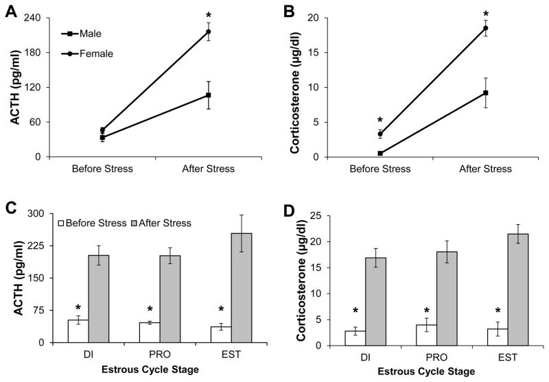
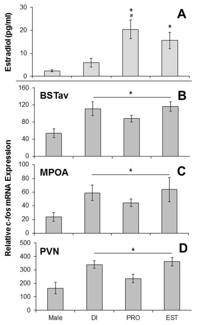
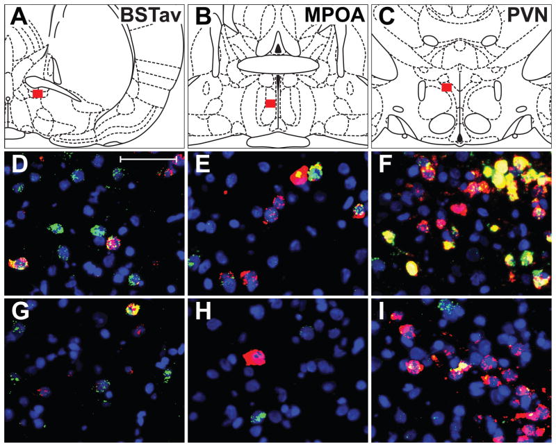
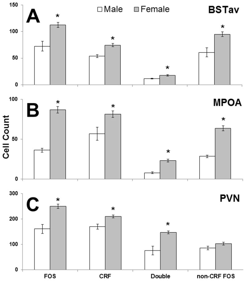
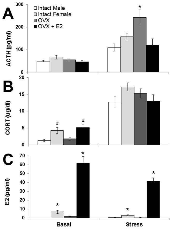
Women may be more vulnerable to certain stress-related psychiatric illnesses than men due to differences in hypothalamic-pituitary-adrenocortical (HPA) axis function. To investigate potential sex differences in forebrain regions associated with HPA axis activation in rats, these experiments utilized acute exposure to a psychological stressor. Male and female rats in various stages of the estrous cycle were exposed to 30min of restraint, producing a robust HPA axis hormonal response in all animals, the magnitude of which was significantly higher in female rats. Although both male and female animals displayed equivalent c-fos expression in many brain regions known to be involved in the detection of threatening stimuli, three regions had significantly higher expression in females: the paraventricular nucleus of the hypothalamus (PVN), the anteroventral division of the bed nucleus of the stria terminalis (BSTav), and the medial preoptic area (MPOA). Dual fluorescence in situ hybridization analysis of neurons containing c-fos and corticotropin-releasing factor (CRF) mRNA in these regions revealed significantly more c-fos and CRF single-labeled neurons, as well as significantly more double-labeled neurons in females. Surprisingly, there was no effect of the estrous cycle on any measure analyzed, and an additional experiment revealed no demonstrable effect of estradiol replacement following ovariectomy on HPA axis hormone induction following stress. Taken together, these data suggest sex differences in HPA axis activation in response to perceived threat may be influenced by specific populations of CRF neurons in key stress-related brain regions, the BSTav, MPOA, and PVN, which may be independent of circulating sex steroids.
Copyright © 2013 IBRO. Published by Elsevier Ltd. All rights reserved.
Figures

References
-
- Aloisi AM, Albonetti ME, Carli G. Sex differences in the behavioural response to persistent pain in rats. Neurosci Letters. 1994;179:79–82. - PubMed
-
- Aloisi AM, Zimmermann M, Herdegen T. Sex-dependent effects of formalin and restraint on c-fos expression in the septum and hippocampus of the rat. Neurosci. 1997;81:951–958. - PubMed
-
- Aloisi AM, Ceccarelli I, Lupo C. Behavioural and hormonal effects of restraint stress and formalin test in male and female rats. Brain Res Bull. 1998;47:57–62. - PubMed
-
- Atkinson HC, Waddell BJ. Circadian variation in basal plasma corticosterone and adrenocorticotropin in the rat: sexual dimorphism and changes across the estrous cycle. Endocrinol. 1997;138:3842–3848. - PubMed
Publication types
MeSH terms
Substances
Grants and funding
LinkOut - more resources
Full Text Sources
Other Literature Sources
Medical
