. 2013 Mar 15;114(6):742-51.
doi: 10.1152/japplphysiol.01389.2012. Epub 2013 Jan 10.

Responses of vestibular nucleus neurons to inputs from the hindlimb are enhanced following a bilateral labyrinthectomy

Affiliations

Responses of vestibular nucleus neurons to inputs from the hindlimb are enhanced following a bilateral labyrinthectomy

Andrew A McCall et al. J Appl Physiol (1985). .

Abstract

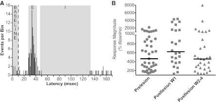
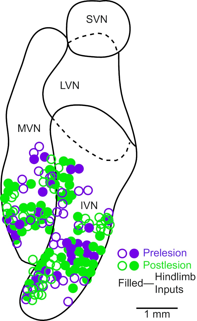
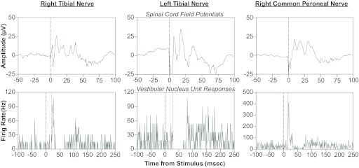
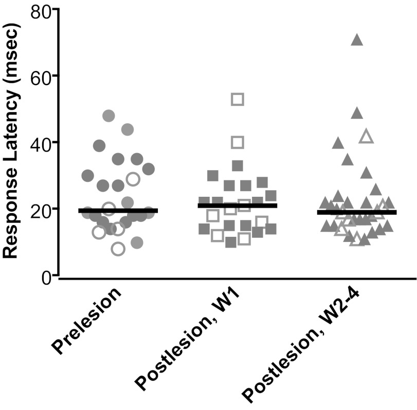
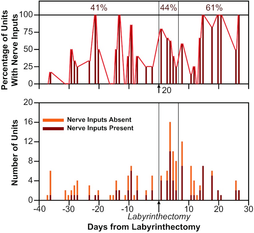
Vestibular nucleus neurons have been shown to respond to stimulation of afferents innervating the limbs. However, a limitation in the potential translation of these findings is that they were obtained from decerebrate or anesthetized animals. The goal of the present study was to determine whether stimulation of hindlimb nerves similarly affects vestibular nucleus neuronal activity in conscious cats, and whether the responsiveness of neurons to the stimuli is altered following a bilateral labyrinthectomy. In labyrinth-intact animals, the firing rate of 24/59 (41%) of the neurons in the caudal vestibular nucleus complex was affected by hindlimb nerve stimulation. Most responses were excitatory; the median response latency was 20 ms, but some units had response latencies as short as 10 ms. In the first week after a bilateral labyrinthectomy, the proportion of vestibular nucleus neurons that responded to hindlimb nerve stimulation increased slightly (to 24/55 or 44% of units). However, during the subsequent postlabyrinthectomy survival period, the proportion of vestibular nucleus neurons with hindlimb inputs increased significantly (to 30/49 or 61% of units). Stimuli to hindlimb nerves needed to elicit neuronal responses was consistently over three times the threshold for eliciting an afferent volley. These data show that inputs from hindlimb afferents smaller than those innervating muscle spindles and Golgi tendon organs affect the processing of information in the vestibular nuclei, and that these inputs are enhanced following a bilateral labyrinthectomy. These findings have implications for the development of a limb neuroprosthetics device for the management of bilateral vestibular loss.


Figures

Fig. 1.
Fig. 1.
Method to determine whether responses to nerve stimulation were present. A: poststimulus histograms were first inspected to determine which responses putatively contained excitatory (E) and/or inhibitory (I) components, where activity deviated from that during the baseline period 10 ms before nerve stimulation was provided. In this example, E and I responses, as well as baseline activity, are designated on a poststimulus histogram generated from data collected 15 days following the bilateral labyrinthectomy. A vertical dashed line indicates when the stimulus was delivered; data from 61 sequential stimulus presentations are represented in the trace. The average number of events per bin during the E and I responses were determined and compared with the average number of events per bin during the baseline period. In this example, the average number of events per bin in the E, I, and baseline periods were, respectively, 5.3, 0.1, and 0.6. The SD of bin counts during the baseline period was calculated to be 0.8. The maximal bin count during the E period was 16, and the minimal bin count during the I period was 0. Two criteria were used to designate a significant E response: an average response bin count over 1 SD larger that the average bin count during the baseline period, and a peak response over 3 SDs larger than the average bin count during the baseline period. In this case, the average bin count during the baseline period (0.6) plus 1 SD (0.8) was 1.4 counts/bin. Thus the average activity during the E period (5.3 counts/bin) was over 1 SD larger than the average baseline activity, and the peak response (16 events/bin) was over 3 SDs larger than average baseline activity. Hence, the response was deemed to be significant. Although the I response is evident in the trace, we could not use the same criteria to demonstrate that it was significant, since mean baseline activity (0.6) minus 1 SD (0.8) was less than zero. B: magnitudes of responses relative to baseline activity. For E responses, response magnitudes represent: 100 × average bin count during response/average bin count during baseline period. For I responses, response magnitudes represent 100 × 1/(average bin count during response/average bin count during baseline period). When both I and E responses were present, the response magnitudes for each component were averaged to generate the value that was plotted. Horizontal lines show median values. Abbreviations: W1, first week after bilateral labyrinthectomy; W2–4, the subsequent 3 wk (weeks 2–4) after bilateral labyrinthectomy.
Fig. 2.
Fig. 2.
Locations of neurons tested for responses to hindlimb nerve stimulation. The locations are plotted on a horizontal section through the vestibular nuclei. Open symbols indicate neurons that failed to respond to nerve stimulation, whereas solid symbols denote units with hindlimb inputs. IVN, inferior vestibular nucleus; LVN, lateral vestibular nucleus; MVN, medial vestibular nucleus; SVN, superior vestibular nucleus.
Fig. 3.
Fig. 3.
Averaged responses of a neuron to rotations in vertical planes. To facilitate comparisons of responses, firing rates are indicated as Hz/° of tilt, as smaller stimulus amplitudes were delivered at higher stimulus frequencies. A and B: responses to 0.5 Hz, 5° clockwise (CW) and counterclockwise (CCW) wobble stimuli. Solid curves superimposed on traces are sine waves fit to the responses. C: responses to 0.5 Hz, 5° sinusoidal rotations in the roll plane. A dashed line indicates table position. Roll tilt did not produce a significant modulation of the unit's activity. D–F: responses to sinusoidal rotations in the pitch plane at 0.1 Hz (10°), 0.5 Hz (5°), and 1.0 Hz (2.5°). The gain of the response to 1-Hz rotations was 14 times the gain of the response to 0.1-Hz tilts. LED, left ear-down roll; ND, nose-down pitch; NU, nose-up pitch; RED, right ear-down roll. The number of sweeps averaged to generate each trace were as follows: 31 (A), 60 (B), 13 (C), 10 (D), 29 (E), and 47 (F).
Fig. 4.
Fig. 4.
Characteristics of responses of neurons to tilts in vertical planes. Data are segregated based on whether the response gain for a particular unit increased more (advancing gain) or less (flat gain) than fivefold per stimulus decade. A and B: polar plots showing response vector orientations and gains of units. Response vector orientations were determined using wobble stimuli delivered at 0.5 Hz. The maximal radius of each plot designates a response gain of 15 spikes·s−1·°−1. C and D: Bode plots illustrating the dynamic properties of responses of neurons to rotations in a fixed plane near the response vector orientation at multiple frequencies. Response gain and phase were plotted with respect to stimulus position. Thin gray lines show data for individual neurons; thick solid lines indicate average values. Error bars designate 1 SE. CED, contralateral ear-down roll; IED, ipsilateral ear-down roll tilt.
Fig. 5.
Fig. 5.
Effects of hindlimb nerve stimulation on the activity of a vestibular nucleus neuron. In each column, the bottom panel is a poststimulus histogram indicating the responses of the unit to a single shock of 250-μA magnitude, which was 10 times the threshold for producing a spinal cord field potential (shown in the top panel). A gray vertical line at time zero indicates when the stimulus was delivered, whereas a red vertical line designates the onset of each response. The poststimulus histograms were generated from the following number of sweeps: right tibial nerve, 65; left tibial nerve, 56; right common peroneal nerve, 63.
Fig. 6.
Fig. 6.
Latencies of responses of vestibular nucleus neurons to hindlimb nerve stimulation (indicated by shaded symbols). If a neuron's activity was affected by stimulation of more than one nerve, the shortest response latency was plotted. Solid symbols indicate latencies of responses that were exclusively excitatory, whereas open signals show latencies of responses that included inhibition (such as excitation followed by inhibition). Different columns show response latencies determined before vestibular lesions (prelesion), in W1 (postlesion), and during W2–4 (postlesion). Solid lines designate median values.
Fig. 7.
Fig. 7.
The bottom diagram shows the number of vestibular nucleus neurons tested on each day (in all animals combined) that did and did not respond to hindlimb nerve stimulation. The top diagram shows the same data, designated as percentage of units recorded on each day responsive to hindlimb inputs. Testing days are indicated relative to the date of the bilateral labyrinthectomy, which was performed on day 0, as indicated on the graph. Vertical lines separate the three time periods considered during the study: prelesion (left side of diagrams), W1 (middle section of diagrams), and W2–4 (right side of diagram). Numbers above the top panel indicate the fraction of neurons recorded during each time period that responded to hindlimb nerve stimulation.

Similar articles

Cited by

References

    1. Anastasopoulos D, Mergner T. Canal-neck interaction in vestibular nuclear neurons of the cat. Exp Brain Res 46: 269–280, 1982 - PubMed
    1. Andersson G, Oscarsson O. Climbing fiber microzones in cerebellar vermis and their projection to different groups of cells in the lateral vestibular nucleus. Exp Brain Res 32: 565–579, 1978 - PubMed
    1. Armstrong DM, Harvey RJ. Responses to a spino-olivo-cerebellar pathway in the cat. J Physiol 194: 147–168, 1968 - PMC - PubMed
    1. Baker J, Goldberg J, Hermann G, Peterson B. Spatial and temporal response properties of secondary neurons that receive convergent input in vestibular nuclei of alert cats. Brain Res 294: 138–143, 1984 - PubMed
    1. Barman SM, Sugiyama Y, Suzuki T, Cotter LA, DeStefino VJ, Reighard DA, Cass SP, Yates BJ. Rhythmic activity of neurons in the rostral ventrolateral medulla of conscious cats: effect of removal of vestibular inputs. Am J Physiol Regul Integr Comp Physiol 301: R937–R946, 2011 - PMC - PubMed

Publication types

LinkOut - more resources