

A mechanism of rapidly reversible cerebral ventricular enlargement independent of tissue atrophy
- PMID: 23306181
- PMCID: PMC3629396
- DOI: 10.1038/npp.2013.11
A mechanism of rapidly reversible cerebral ventricular enlargement independent of tissue atrophy
Abstract
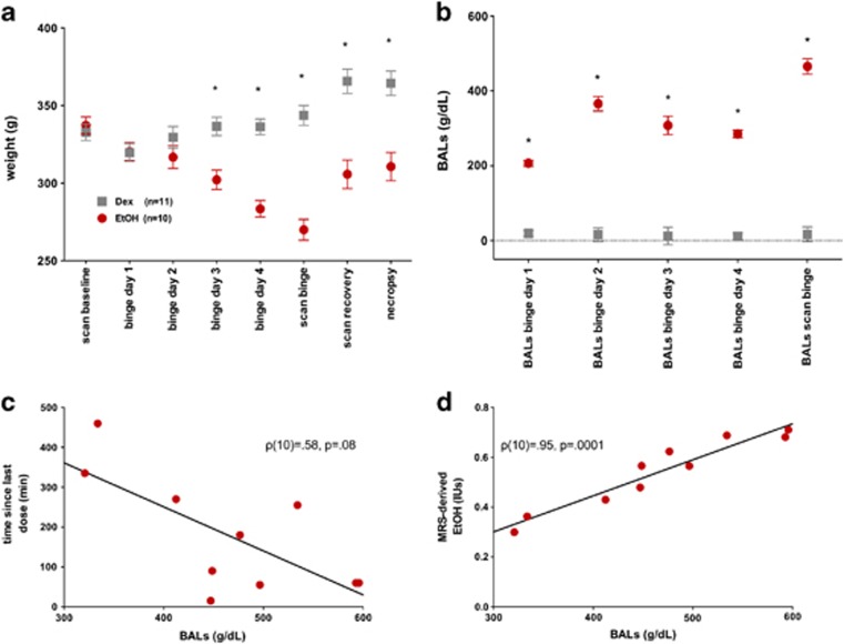
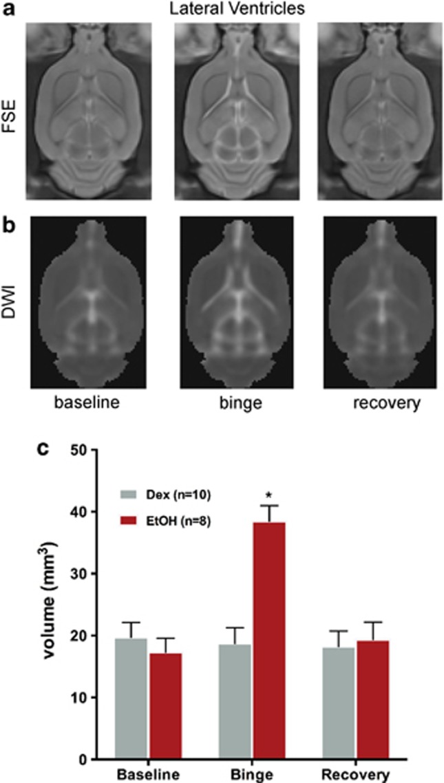
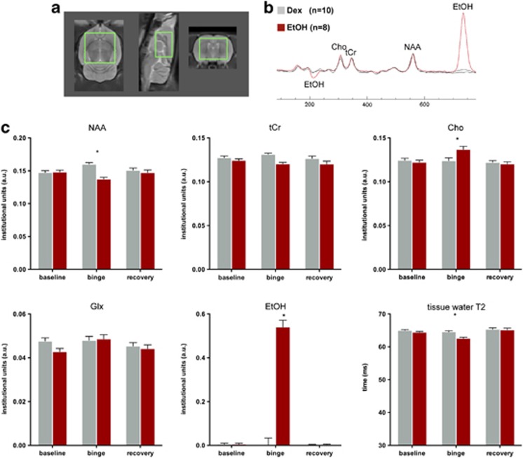
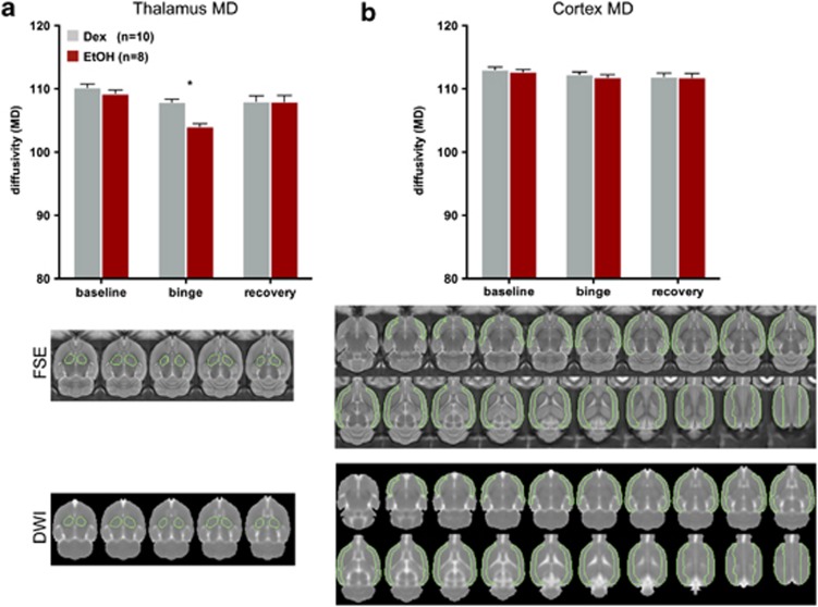
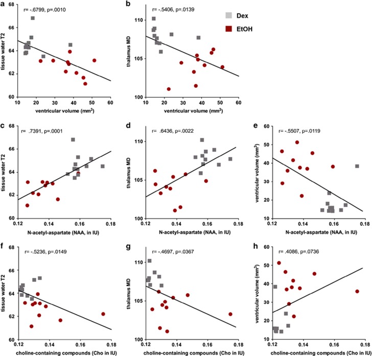
Ventricular enlargement, a common in vivo marker of aging, disease, and insult, is presumed to reflect atrophy of surrounding brain regions. Pathological mechanisms underlying ventricular enlargement, however, are likely specific to the condition under investigation. Here, multimodal imaging, incorporating structural magnetic resonance imaging (MRI), MR spectroscopy (MRS), and diffusion weighted imaging (DWI), was used in rats exposed to binge ethanol (EtOH) to provide insight into a mechanism of reversible ventricular enlargement. During intoxication, MRI revealed expansion of ventricles, but volume changes in dorsal or ventral hippocampi, caudate-putamen, or thalamus were not detectible. MRS of whole-brain parenchyma showed decreases in N-acetylasparate (NAA) and tissue water T2, and increases in choline-containing compounds (Cho). DWI showed decreased diffusivity selective to the thalamus. All MR parameters returned to baseline with 7 days of recovery. Rapid recovery of ventricular volume and the absence of detectable tissue volume reductions in brain regions adjacent to ventricles argue against atrophy as a mechanism of ventricular expansion. Decreased tissue water T2 and decreased thalamic diffusivity suggest lower tissue water content and a role for both NAA and Cho, as osmolytes is proposed. Together, these data support a model of fluid redistribution during acute EtOH intoxication and recovery to account for rapid ventricular volume changes.
Figures
Similar articles
-
Rat strain differences in brain structure and neurochemistry in response to binge alcohol.Psychopharmacology (Berl). 2014 Jan;231(2):429-45. doi: 10.1007/s00213-013-3253-z. Epub 2013 Sep 13. Psychopharmacology (Berl). 2014. PMID: 24030467 Free PMC article.
-
Brain injury and recovery following binge ethanol: evidence from in vivo magnetic resonance spectroscopy.Biol Psychiatry. 2010 May 1;67(9):846-54. doi: 10.1016/j.biopsych.2009.10.028. Epub 2009 Dec 30. Biol Psychiatry. 2010. PMID: 20044076 Free PMC article.
-
Transient CNS responses to repeated binge ethanol treatment.Addict Biol. 2016 Nov;21(6):1199-1216. doi: 10.1111/adb.12290. Epub 2015 Aug 18. Addict Biol. 2016. PMID: 26283309 Free PMC article.
-
Development and resolution of brain lesions caused by pyrithiamine- and dietary-induced thiamine deficiency and alcohol exposure in the alcohol-preferring rat: a longitudinal magnetic resonance imaging and spectroscopy study.Neuropsychopharmacology. 2007 May;32(5):1159-77. doi: 10.1038/sj.npp.1301107. Epub 2006 May 24. Neuropsychopharmacology. 2007. PMID: 16723995
-
New and enlarging white matter lesions adjacent to the ventricle system and thalamic atrophy are independently associated with lateral ventricular enlargement in multiple sclerosis.J Neurol. 2020 Jan;267(1):192-202. doi: 10.1007/s00415-019-09565-w. Epub 2019 Oct 14. J Neurol. 2020. PMID: 31612322
Cited by
-
Alcohol Withdrawal and the Associated Mood Disorders-A Review.Int J Mol Sci. 2022 Nov 29;23(23):14912. doi: 10.3390/ijms232314912. Int J Mol Sci. 2022. PMID: 36499240 Free PMC article. Review.
-
Alcohol's effects on the mouse brain are modulated by age and sex.Addict Biol. 2022 Sep;27(5):e13209. doi: 10.1111/adb.13209. Addict Biol. 2022. PMID: 36001428 Free PMC article.
-
Rat strain differences in brain structure and neurochemistry in response to binge alcohol.Psychopharmacology (Berl). 2014 Jan;231(2):429-45. doi: 10.1007/s00213-013-3253-z. Epub 2013 Sep 13. Psychopharmacology (Berl). 2014. PMID: 24030467 Free PMC article.
-
The Aging Brain With HIV Infection: Effects of Alcoholism or Hepatitis C Comorbidity.Front Aging Neurosci. 2018 Mar 22;10:56. doi: 10.3389/fnagi.2018.00056. eCollection 2018. Front Aging Neurosci. 2018. PMID: 29623036 Free PMC article. Review.
-
Systemic Administration of the TLR7/8 Agonist Resiquimod (R848) to Mice Is Associated with Transient, In Vivo-Detectable Brain Swelling.Biology (Basel). 2022 Feb 10;11(2):274. doi: 10.3390/biology11020274. Biology (Basel). 2022. PMID: 35205140 Free PMC article.
References
-
- Adalsteinsson E, Hurd RE, Mayer D, Sailasuta N, Sullivan EV, Pfefferbaum A. In vivo 2D J-resolved magnetic resonance spectroscopy of rat brain with a 3-T clinical human scanner. NeuroImage. 2004;22:381–386. - PubMed
-
- Alling C, Bostrom K. Demyelination of the mamillary bodies in alcoholism. A combined morphological and biochemical study. Acta Neuropathologica (Berl) 1980;50:77–80. - PubMed
-
- Altura BM, Altura BT. Alcohol, the cerebral circulation and strokes. Alcohol (Fayetteville, NY) 1984;1:325–331. - PubMed
-
- Baslow MH, Suckow RF, Hungund BL. Effects of ethanol and of alcohol dehydrogenase inhibitors on the reduction of N-acetylaspartate levels of brain in mice in vivo: a search for substances that may have therapeutic value in the treatment of Canavan disease. J Inherit Metab Dis. 2000;23:684–692. - PubMed
-
- Baslow MH, Suckow RF, Sapirstein V, Hungund BL. Expression of aspartoacylase activity in cultured rat macroglial cells is limited to oligodendrocytes. J Mol Neurosci. 1999;13:47–53. - PubMed
Publication types
MeSH terms
Substances
Grants and funding
LinkOut - more resources
Full Text Sources
Other Literature Sources
