. 2013 Mar 8;288(10):6788-800.
doi: 10.1074/jbc.M112.445056. Epub 2013 Jan 10.

Intracellular Mycobacterium tuberculosis exploits host-derived fatty acids to limit metabolic stress

Affiliations

Intracellular Mycobacterium tuberculosis exploits host-derived fatty acids to limit metabolic stress

Wonsik Lee et al. J Biol Chem. .

Abstract

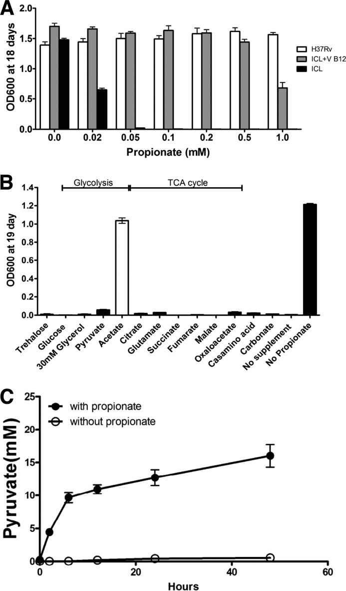
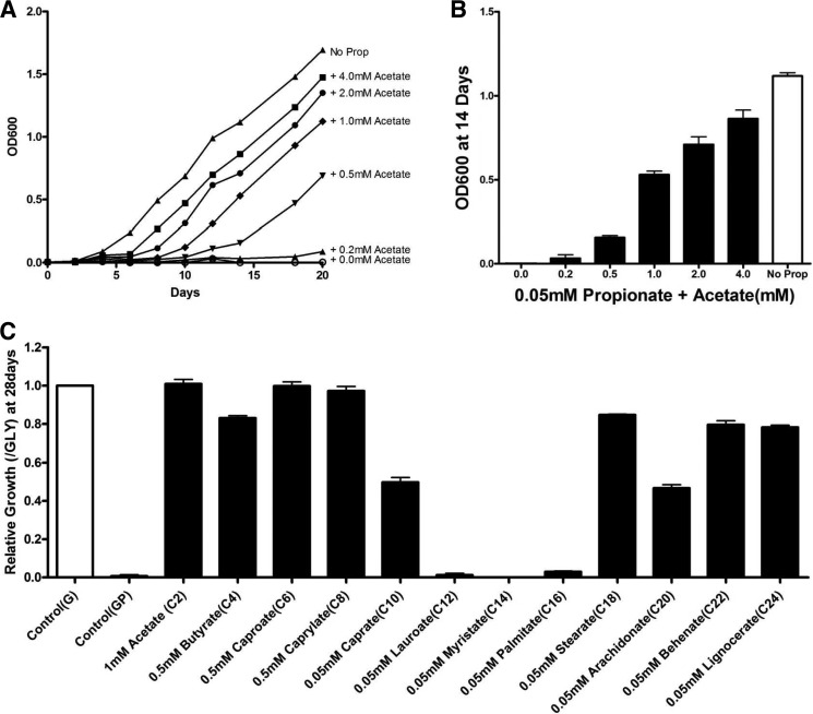
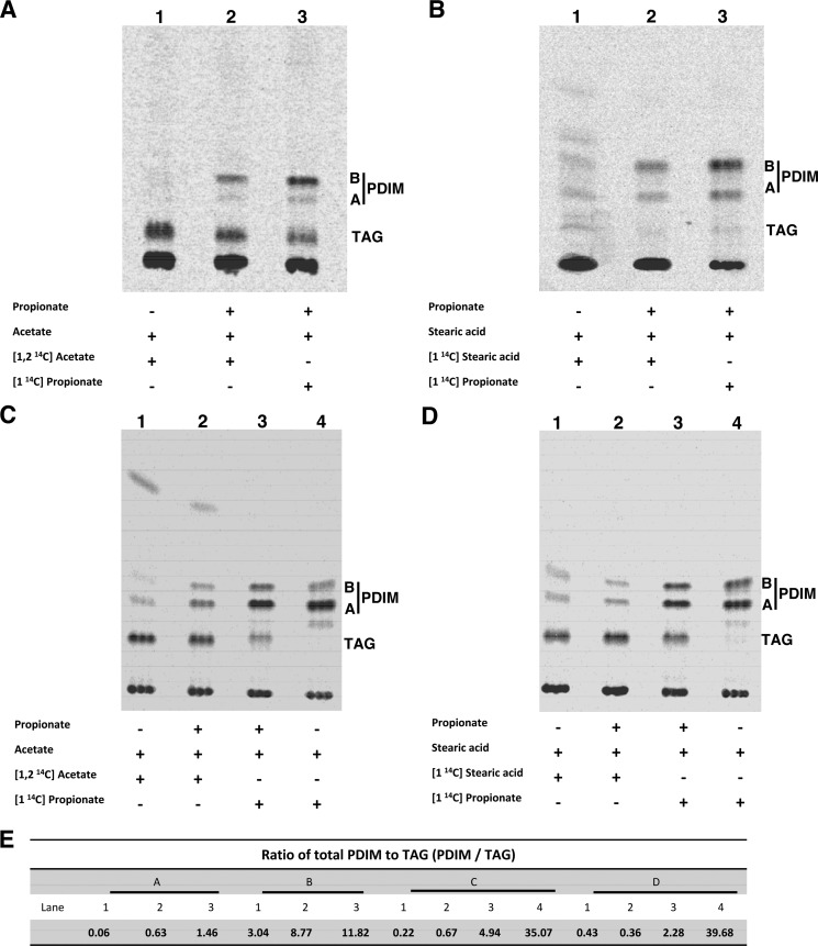
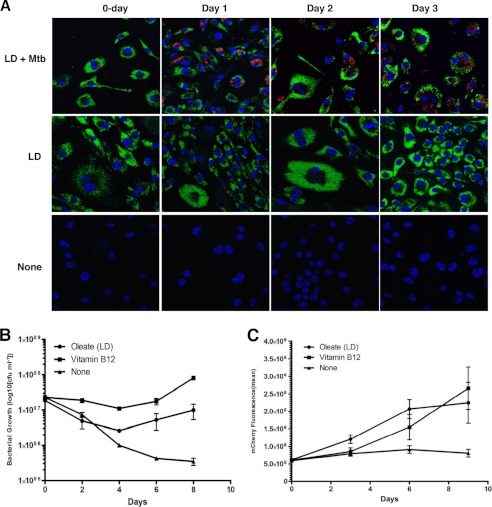
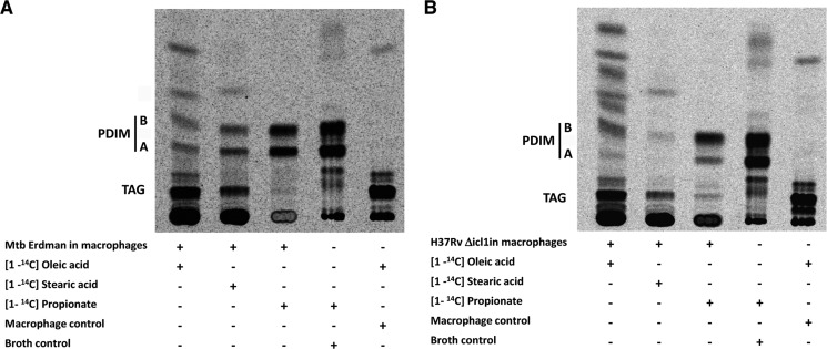
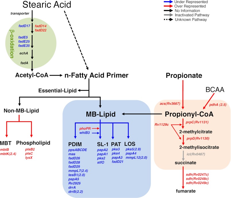
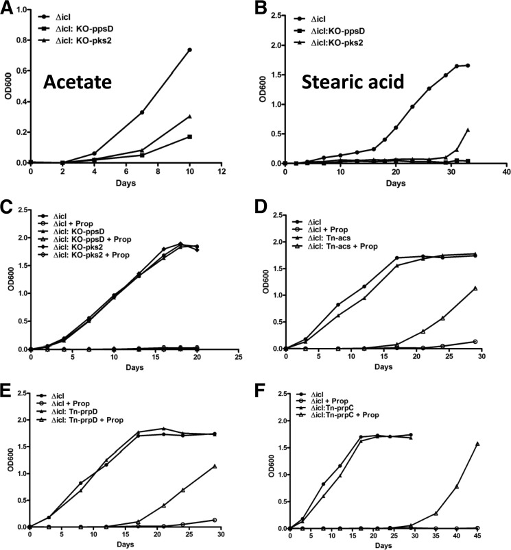
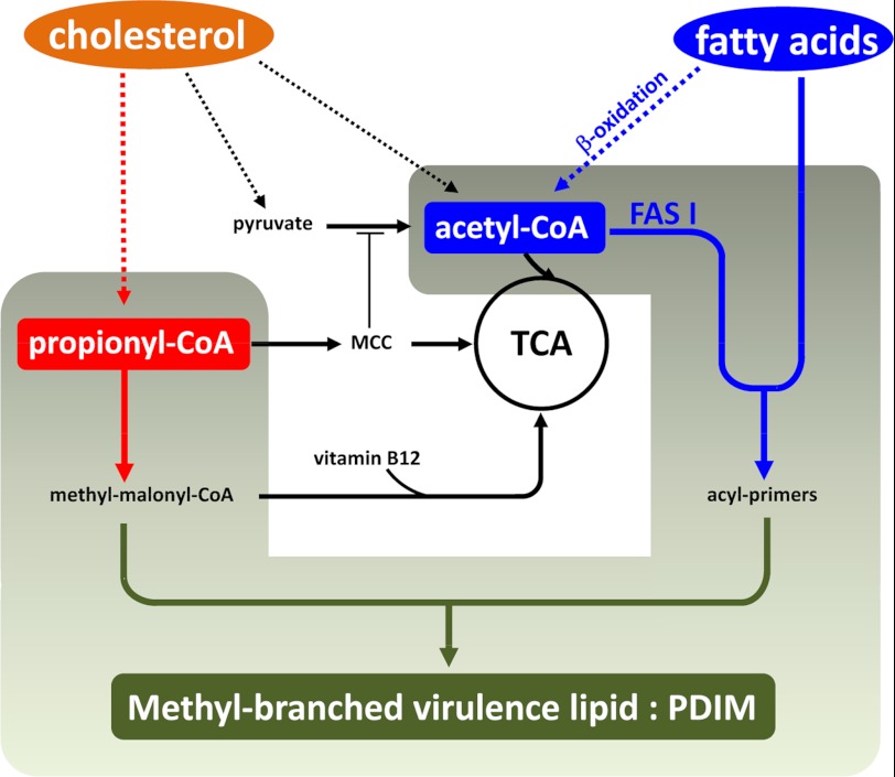
Recent data indicate that the nutrients available to Mycobacterium tuberculosis (Mtb) inside its host cell are restricted in their diversity. Fatty acids and cholesterol appear more favored; however, their degradation can result in certain metabolic stresses. Their breakdown can generate propionyl-CoA, which gives rise to potentially toxic intermediates. Detoxification of propionyl-CoA relies on the activity of the methylcitrate cycle, the methylmalonyl pathway, or incorporation of the propionyl-CoA into methyl-branched lipids in the cell wall. The current work explores carbon flux through these pathways, focusing primarily on those pathways responsible for the incorporation of propionyl-CoA into virulence-associated cell wall lipids. Exploiting both genetic and biochemical rescue, we demonstrate that these metabolic pressures are experienced by Mtb inside its host macrophage and that the bacterium accesses host fatty acid stores. The metabolism of these host lipids expands the acetyl-CoA pool and alleviates the pressure from propionyl-CoA. These data have major implications for our appreciation of central metabolism of Mtb during the course of infection.


Figures

FIGURE 1.
FIGURE 1.
Mitigation of propionate toxicity in a Δicl1 mutant strain by acetate and VitB12. The Δicl1 mutant strain of Mtb grows poorly in the presence of propionate, but growth can be rescued by the addition of either VitB12 or acetate. A, bacterial growth, measured by absorbance at 600 nm, was determined for Mtb H37Rv and Δicl strain grown for 18 days in minimal medium containing 10 mm glycerol as the primary carbon source and propionate across the range from 0.02 to 1 mm. Vitamin B12 at a concentration of 10 μg/ml reversed the propionate toxicity observed in the Δicl1 mutant strain. B, propionate toxicity is rescued in the Δicl1 mutant strain with acetate. Of the central metabolic intermediates tested, only acetate (4 mm) was able to rescue growth of the Δicl1 mutant strain in the presence of propionate (0.05 mm). C, pyruvate accumulates and is released from the Δicl1 mutant when grown in the presence of propionate over the course of 48 h. Data are representative of three experiments. Error bars, Standard Deviation.
FIGURE 2.
FIGURE 2.
Fatty acids of differing chain lengths rescue the Δicl1 strain from propionate toxicity. Acetate, short chain fatty acids, and long chain fatty acids similar to those found in the host macrophage can rescue propionate toxicity in Δicl1 Mtb. A and B, the C2 compound, acetate, rescues propionate toxicity in Δicl1 Mtb in a dose-dependent manner. Bacteria were grown for 18 days in medium containing 10 mm glycerol (G) and 0.05 mm propionate supplemented with acetate across the range from 0.05 to 4 mm. C, short to mid-length and long chain fatty acids also rescue propionate toxicity in Δicl1 Mtb. Bacteria were grown for 18 days in medium containing 10 mm glycerol and 0.05 mm propionate (GP) supplemented with saturated even chain length fatty acids (C2–C24) at the following concentrations. Acetate (C2) was added at 1 mm, short chain fatty acids (C4–C8) at 0.5 mm, and intermediate and long chain fatty acids (C10–C24) at 0.05 mm. Bacterial growth was measured by absorbance at 600 nm, and results are representative of three replicates. Error bars, Standard Deviation.
FIGURE 3.
FIGURE 3.
Fatty acids rescue propionate toxicity in Mtb through incorporation into methyl-branched lipids, such as PDIM, in both the Δicl1 mutant strain and wild-type Mtb. TLC analysis of the cell lipids indicates that radiolabel derived from [1-14C]propionate, [1,2-14C]acetate, or [1-14C]stearic acid is incorporated into the cell wall lipid PDIM. 14C incorporation into PDIM is enhanced by 0.05 mm propionate but is a constitutive process in both the Δicl1 mutant strain and in the high PDIM-producing wild-type Erdman strain. Bacteria were grown in glycerol medium containing propionate, acetate, and stearic acid as indicated. A, lipids from the Δicl1 mutant incubated with [1,2-14C]acetate in the absence of propionate (lane 1), incubated with [1,2-14C] acetate in the presence of 0.05 mm unlabeled propionate (lane 2), or incubated with [1-14C]propionate in the presence of 0.05 mm unlabeled propionate (lane 3). C, lipids from Erdman. B, the experiment was repeated using the long chain fatty acid, stearic acid (C18), instead of acetate (C2). Shown are lipids from the Δicl1 mutant incubated with [1-14C] stearic acid in the absence of propionate (lane 1), with [1-14C]stearic acid in the presence of 0.05 mm unlabeled propionate (lane 2), or with [1-14C]propionate in the presence of 0.05 mm unlabeled propionate (lane 3). D, lipids from Erdman. E, ratio of PDIM to TAG. The identity of the PDIM and TAG bands had been established previously by mass spectrometry (32). PDIM A, phthiocerol dimycocerosate A; PDIM B, phthiodiolone dimycocerosate.
FIGURE 4.
FIGURE 4.
Intracellular growth is restored to the Δicl1 Mtb through the induction of oleate-containing lipid droplets in the infected cell. The survival of the Δicl1 mutant is impaired in macrophages; however, growth could be restored by preloading the host cells with exogenous oleate or by adding VitB12 to the cell culture medium to facilitate operation of the MMP. A, induction of macrophage lipids droplets, detected with BODIPY 493/503 (green), Δicl1 strain expressing pVV16-mCherry (smyc′::mCherry) (red), and nuclei (blue) in oleate-loaded macrophages versus untreated macrophages over the duration of the experimental period. B, bacterial cfu counts were determined at 2-day intervals across an 8-day period for the Δicl1 Mtb in untreated, control macrophages (triangle), in lipid droplet-containing, oleate-loaded macrophages (circle), and in macrophage supplemented by the addition of VitB12 to the medium (square). C, the experiment was repeated with a Δicl1 strain expressing pVV16-mCherry (smyc′::mCherry), and bacterial proliferation was quantified by measuring bacterial mCherry fluorescence across an 8-day infection period. Both methods demonstrated the enhanced growth and survival of the Δicl1 mutant in the presence of either oleate-induced droplets or exogenous VitB12. Error bars, Standard Deviation values from three representative replicates.
FIGURE 5.
FIGURE 5.
Mtb inside lipid droplet-loaded macrophage incorporates the host-derived fatty acids into PDIM. Radiolabeled fatty acid precursors are incorporated into PDIM of both the WT Mtb (A) and the Δicl1 Mtb (B) when grown in lipid droplet-containing macrophages. Lipid droplets were induced in macrophages with excess oleic acid containing tracer amounts of [1-14C]oleic acid (lane 1), [1-14C]stearic acid (lane 2), or [1-14C]propionate (lane 3) for 24 h. The lipid-loaded macrophages were infected with either WT Mtb or Δicl1 Mtb at an MOI of 5:1 for 4 h. At 5 days postinfection, Mtb cell wall lipids were extracted from macrophage and analyzed by TLC. Control labeling experiments were run with WT Mtb grown in broth culture labeled with [1-14C]propionate (lane 4) and macrophages alone labeled with [1-14C]oleic acid (lane 5). The identity of the PDIM species has been established previously by mass spectrometry (32). PDIM A, phthiocerol dimycocerosate A; PDIM B, phthiodiolone dimycocerosate.
FIGURE 6.
FIGURE 6.
Phenotypic TraSH screen identifies genes involved in propionate utilization and toxicification. Graphic illustration of the differentially represented genes in the context of the metabolic pathways most relevant to propionate utilization and detoxification. Genes required for Δicl1 Mtb growth in the presence propionate and long chain fatty acids as a means of propionate toxicity rescue are indicated in blue type. Genes that, when mutated, enhance Δicl1 Mtb growth in the presence of propionate and long chain fatty acids are indicated in red type. The differentially represented genes are shown in relation to the relevant carbon metabolic pathways: synthesis of non-essential MB lipids, synthesis of non-MB lipids, the MMC, and genes involved in the β-oxidation breakdown of long chain fatty acids. All genes selected were >3-fold overrepresented or >3-fold underrepresented with p < 0.05. The full list of overrepresented and underrepresented genes is provided in supplemental Tables S1 and S2. BCAA, branched chain amino acids; PAT, polyacyltrehalose; LOS, lipooligosaccharide.
FIGURE 7.
FIGURE 7.
Validation of the genes implicated in propionate utilization and detoxification in Δicl Mtb. Genes identified by the TraSH screen were validated through the generation of double mutants and characterized by growth phenotype. A–C, genes negatively selected for involved in methyl branch-containing lipid biosynthesis. Growth of the Δicl1 parental strain and Δicl1:KO-ppsD (PDIM biosynthesis) and Δicl1:KO-pks2 (SL-1 biosynthesis) double mutant strains was assessed in minimal media containing glycerol, propionate, and 1 mm acetate (A) or 0.05 mm stearic acid (B) or without any fatty acid supplement (C). In each instance, the mutants exhibited defective growth in fatty acid with propionate (A and B) yet grew normally in minimal medium with glycerol (C). D–F, genes positively selected as being involved in propionate utilization and the generation of toxic intermediates. Shown is growth of the Δicl1 parental strain Δicl1:Tn acs (Rv3667) (D), Δicl1:Tn prpD (Rv1130) (E), or Δicl1:Tn prpC (Rv1131) (F) in minimal media containing glycerol and 0.05 mm propionate (D and E) or 0.1 mm propionate (F). These mutants all grew normally in minimal medium, and each of the latter two mutations conferred partial resistance to propionate toxicity, consistent with their enrichment under propionate selection. Results are representative of three experiments.
FIGURE 8.
FIGURE 8.
Model of assimilation of host lipids and fatty acids into methyl-branched Mtb virulence lipids. Mtb has access to both cholesterol and fatty acids from the macrophage host cell. Degradation of cholesterol expands the pool of propionyl-CoA, which can be used to fuel central metabolism and biosynthetic pathways. Accumulation of propionyl-CoA and/or products of the MCC may limit the generation of acetyl-CoA from pyruvate by inhibiting PDH activity. Inhibition of PDH activity places more pressure on the acetyl-CoA pool, which is utilized as malonyl-CoA for the assembly of Mtb cell wall lipids. Additionally, MB cell wall lipids can be built using n-acyl primers generated either from de novo synthesis or from preformed long chain fatty acids imported by the bacterium. The integration of these two- and three-carbon metabolic pathways is clearly critical to the growth of Mtb and its success within its host cell environment.

References

    1. Bloch H., Segal W. (1956) Biochemical differentiation of Mycobacterium tuberculosis grown in vivo and in vitro. J. Bacteriol. 72, 132–141 - PMC - PubMed
    1. Cole S. T., Brosch R., Parkhill J., Garnier T., Churcher C., Harris D., Gordon S. V., Eiglmeier K., Gas S., Barry C. E., 3rd, Tekaia F., Badcock K., Basham D., Brown D., Chillingworth T., Connor R., Davies R., Devlin K., Feltwell T., Gentles S., Hamlin N., Holroyd S., Hornsby T., Jagels K., Krogh A., McLean J., Moule S., Murphy L., Oliver K., Osborne J., Quail M. A., Rajandream M. A., Rogers J., Rutter S., Seeger K., Skelton J., Squares R., Squares S., Sulston J. E., Taylor K., Whitehead S., Barrell B. G. (1998) Deciphering the biology of Mycobacterium tuberculosis from the complete genome sequence. Nature 393, 537–544 - PubMed
    1. Liu K., Yu J., Russell D. G. (2003) pckA-deficient Mycobacterium bovis BCG shows attenuated virulence in mice and in macrophages. Microbiology 149, 1829–1835 - PubMed
    1. Marrero J., Rhee K. Y., Schnappinger D., Pethe K., Ehrt S. (2010) Gluconeogenic carbon flow of tricarboxylic acid cycle intermediates is critical for Mycobacterium tuberculosis to establish and maintain infection. Proc. Natl. Acad. Sci. U.S.A. 107, 9819–9824 - PMC - PubMed
    1. McKinney J. D., Höner zu Bentrup K., Muñoz-Elías E. J., Miczak A., Chen B., Chan W. T., Swenson D., Sacchettini J. C., Jacobs W. R., Jr., Russell D. G. (2000) Persistence of Mycobacterium tuberculosis in macrophages and mice requires the glyoxylate shunt enzyme isocitrate lyase. Nature 406, 735–738 - PubMed

Publication types

MeSH terms