Spinal ventral epidural arteriovenous fistulas of the lumbar spine: angioarchitecture and endovascular treatment
- PMID: 23306215
- PMCID: PMC3582814
- DOI: 10.1007/s00234-012-1130-9
Spinal ventral epidural arteriovenous fistulas of the lumbar spine: angioarchitecture and endovascular treatment
Abstract
Introduction: Spinal ventral epidural arteriovenous fistulas (EDAVFs) are relatively rare spinal vascular lesions. We investigated the angioarchitecture of spinal ventral EDAVFs and show the results of endovascular treatment.
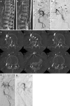
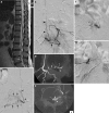
Methods: We reviewed six consecutive patients (four males and two females; mean age, 67.3 years) with spinal ventral EDAVFs treated at our institutions from May 2011 to October 2012. All patients presented with progressive myelopathy. The findings of angiography, including 3D/2D reformatted images, treatments, and outcomes, were investigated. A literature review focused on the angioarchitecture and treatment of spinal ventral EDAVFs is also presented.
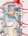
Results: The EDAVFs were located in the ventral epidural space at the L1-L5 levels. All EDAVFs were supplied by the dorsal somatic branches from multiple segmental arteries. The ventral somatic branches and the radiculomeningeal arteries also supplied the AVFs in two patients. The AVFs drained via an epidural venous pouch into the perimedullary vein in four patients and into both the perimedullary vein and paravertebral veins in two patients. Four cases without paravertebral drainage were treated by transarterial embolization with diluted glue, and two cases with perimedullary and paravertebral drainages were treated by transvenous embolization alone or in combination with transarterial embolization. An angiographic cure was obtained in all patients. Clinical symptoms resolved in two patients, markedly improved in three patients, and minimally improved in one patient.
Conclusion: In our limited experience, spinal ventral EDAVFs were primarily fed by somatic branches. EDAVFs can be successfully treated by endovascular techniques selected based on the drainage type of the AVF.
Figures


Similar articles
-
Three-dimensional angioarchitecture of spinal dural arteriovenous fistulas, with special reference to the intradural retrograde venous drainage system.J Neurosurg Spine. 2013 Apr;18(4):398-408. doi: 10.3171/2013.1.SPINE12305. Epub 2013 Feb 22. J Neurosurg Spine. 2013. PMID: 23432326
-
Lumbar spinal epidural arteriovenous fistula with perimedullary venous drainage after endoscopic lumbar surgery.Interv Neuroradiol. 2015 Apr;21(2):249-54. doi: 10.1177/1591019915583212. Epub 2015 May 6. Interv Neuroradiol. 2015. PMID: 25948114 Free PMC article.
-
Spinal extradural arteriovenous fistulas: a clinical and radiological description of different types and their novel treatment with Onyx.J Neurosurg Spine. 2011 Nov;15(5):541-9. doi: 10.3171/2011.6.SPINE10695. Epub 2011 Jul 29. J Neurosurg Spine. 2011. PMID: 21800954
-
Venous manifestations of spinal arteriovenous fistulas.Neuroimaging Clin N Am. 2003 Feb;13(1):73-93. doi: 10.1016/s1052-5149(02)00077-1. Neuroimaging Clin N Am. 2003. PMID: 12802942 Review.
-
Spinal cord arteriovenous shunts: from imaging to management.Eur J Radiol. 2003 Jun;46(3):221-32. doi: 10.1016/s0720-048x(03)00093-7. Eur J Radiol. 2003. PMID: 12758116 Review.
Cited by
-
Diagnostic accuracy of three-dimensional-rotational angiography and heavily T2-weighted volumetric magnetic resonance fusion imaging for the diagnosis of spinal arteriovenous shunts.J Neurointerv Surg. 2022 Jan;14(1):neurintsurg-2020-017252. doi: 10.1136/neurintsurg-2020-017252. Epub 2021 Mar 4. J Neurointerv Surg. 2022. PMID: 33674393 Free PMC article.
-
Endovascular treatment of epidural arteriovenous fistula associated with sacral arteriovenous malformation: case report.Front Neurol. 2024 Feb 12;15:1326182. doi: 10.3389/fneur.2024.1326182. eCollection 2024. Front Neurol. 2024. PMID: 38410195 Free PMC article.
-
Angiographic and Clinical Characteristics of Thoracolumbar Spinal Epidural and Dural Arteriovenous Fistulas.Stroke. 2017 Dec;48(12):3215-3222. doi: 10.1161/STROKEAHA.117.019131. Epub 2017 Nov 7. Stroke. 2017. PMID: 29114089 Free PMC article.
-
Dural arteriovenous fistulas at the craniocervical junction: a systematic review and meta-analysis.Neurosurg Rev. 2024 Oct 23;47(1):812. doi: 10.1007/s10143-024-03018-3. Neurosurg Rev. 2024. PMID: 39441455
-
Transvenous embolization through a cutaneous lumbar vein to treat a spinal epidural arteriovenous fistula: illustrative case.J Neurosurg Case Lessons. 2025 Jul 28;10(4):CASE25329. doi: 10.3171/CASE25329. Print 2025 Jul 28. J Neurosurg Case Lessons. 2025. PMID: 40720910 Free PMC article.
References
Publication types
MeSH terms
LinkOut - more resources
Full Text Sources
Other Literature Sources

