Case Reports
. 2013;49(4):185-91.
doi: 10.1159/000345766. Epub 2013 Jan 10.

Corneal nerve architecture in a donor with unilateral epithelial basement membrane dystrophy

Affiliations
Case Reports

Corneal nerve architecture in a donor with unilateral epithelial basement membrane dystrophy

Jiucheng He et al. Ophthalmic Res. 2013.

Abstract

Background: Epithelial basement membrane dystrophy (EBMD) is by far the most common corneal dystrophy. In this study, we used a newly developed method of immunofluorescence staining and imaging to study the entire corneal nerve architecture of a donor with unilateral EBMD.

Method: Two fresh eyes from a 56-year-old male donor were obtained; the right eye of the donor was diagnosed with EBMD and the left was normal. After slit lamp examination, the corneas were immunostained with anti-β-tubulin III antibody. Images were recorded by a fluorescent microscope equipped with a Photometrics digital camera using MetaVue imaging software.

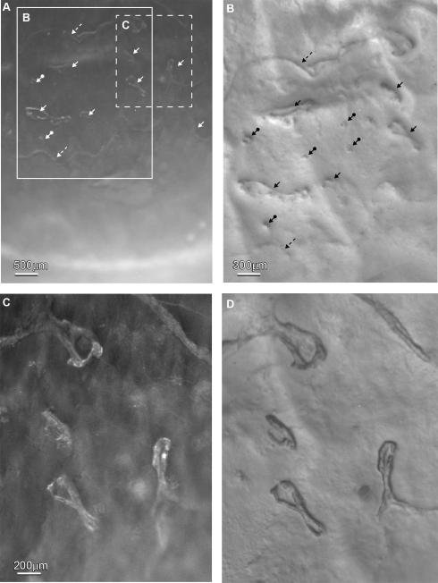
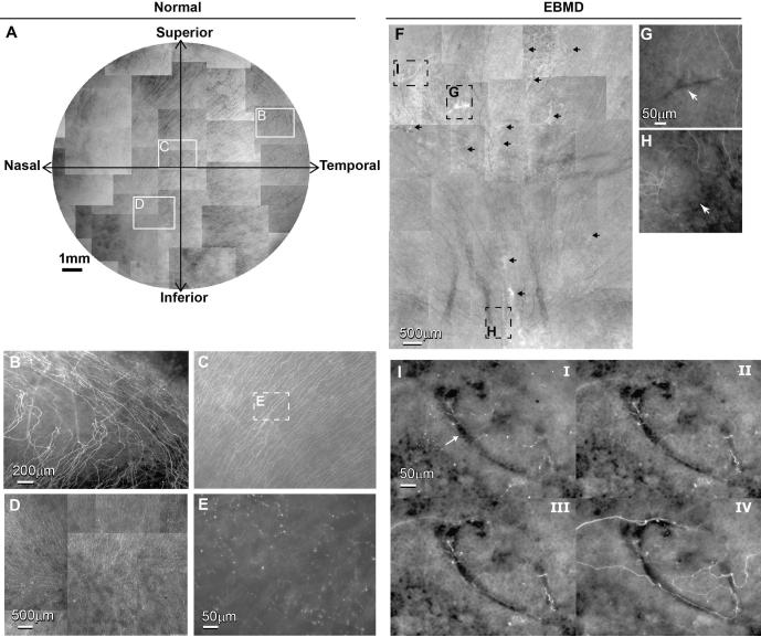
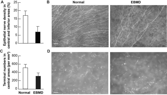
Results: The left cornea appeared normal as observed by slit lamp and stereomicroscope, but the right eye had numerous irregular geographic patches in the basement membrane. Immunofluorescence showed no difference in the stromal nerve distribution between the 2 eyes, but there were areas without innervations in the EBMD cornea. Subbasal nerve fibers also showed tortuous courses and fewer divisions. There was a significant decrease in the density of subbasal nerve fibers and the number of terminals in the right eye.

Conclusion: We show for the first time detailed nerve architecture in an EBMD cornea. Our results suggest that EBMD-induced abnormalities of basement membrane altered epithelial nerve architecture and decreased nerve density, contributing to the pathology of the disease.


Figures

Figure 1
Figure 1
Stereomicroscope images of the right cornea with diagnosed EBMD. A) Dark-field illumination at a low magnification showed that numerous irregular geographic shaped, faint gray-white patches were present in the central area of the inferior quadrants of the cornea. B) Highly oblique illumination of the large framed area in A showed that the pathologic lesions were present as finger-prints (arrows), dots (arrows with circles at the end) and ridge-like patterns (dashed arrows). C) Dark-field of the central area (dashed frame in A) in higher magnification and D) oblique illumination showed the details of four finger-print lesions.
Figure 2
Figure 2
A) Whole mount view of epithelial nerve distribution in the normal left cornea. Images were acquired in a time-lapse mode with a Nikon Eclipse TE200 and with a 5X lens in compliance with the natural shape of the cornea. The epithelial nerve bundles ran in a radial pattern from the periphery to merge in an area within the inferior-nasal quadrant beyond the corneal apex. Highlighted images showed the detailed architecture of the epithelial nerve distribution in the peripheral (B), central (C), and vortex areas (D). E) High magnification image recorded from the central area as marked in Figure 2C shows the nerve terminals at the superficial epithelia. F) Whole mount view of corneal epithelial nerve distribution in the central area of the inferior quadrant of the right cornea. The total area is about 45 mm2, consisting of 38 images recorded in a time-lapse mode with a 10X lens. Arrows indicate the pathological loci. G) and H) are highlighted images showing the detailed architecture of the epithelial nerve distribution in the central and vortex area as marked in F). I) Topographic images, recorded at the plane of superficial epithelia (i), wing cells (ii), basal cells (iii) and the basement membrane (iv), show detailed epithelial innervation in a finger-print lesion (arrow) at the central cornea.
Figure 3
Figure 3
Effect of EBMD on corneal subbasal nerve density and nerve terminals. (A) Subbasal nerve density in the central area of the inferior quadrant was calculated as percentage of total area in each image. A total of 36 images were taken at a magnification of 10 × lens from each eye. These images were changed to grayscale mode and placed against a white background to get better contrast using Photoshop imaging software. The nerve fibers of each image were carefully drawn with four-pixel lines following the course of each fiber. Percentage of nerve area was quantified for each image using the image analysis program. Data are expressed as average ± SD. (B) Representative images of EBMD and normal subbasal nerves. (C) Number of nerve terminals. 20 images for each eye from the central corneas were used. Nerve terminals in superficial epithelia in each image were counted. Because each image took up an area of 0.335 mm2, the terminal numbers per mm2 were calculated. Data are expressed as average ± SD. (D) Representative images of nerve terminals in both eyes.

References

    1. Vogt A. In: Lehrbuch und Atlas der Spalt lampen mikroskopie des lebenden Auges. 2. Vogt A, editor. Springer; 1930. pp. 264–265. part 1.
    1. Cogan DG, Donaldson DD, Kuwabara T, Marshall D. Microcystic dystrophy of the corneal epithelium. Trans Am Ophthalmol Soc. 1964;62:213–225. - PMC - PubMed
    1. Guerry D., III Observations on Cogan's microcystic dystrophy of the corneal epithelium. Trans Am Ophthalmol Soc. 1965;63:320–34. - PMC - PubMed
    1. Rodrigues MM, Fine BS, Laibson PR, Zimmermann LE. Disorders of the corneal epithelium: a clinic pathologic study of dot, geographic, and fingerprint patterns. Arch Ophthalmol. 1974;92:475–482. - PubMed
    1. Werblin TP, Hirst LW, Stark WJ, et al. Prevalence of map-dot-fingerprint changes in the cornea. Br J Ophthalmol. 1981;65(6):401–409. - PMC - PubMed

Publication types

Supplementary concepts

LinkOut - more resources