Comment
. 2013 Mar-Apr;7(2):219-25.
doi: 10.4161/cam.23184. Epub 2013 Jan 10.

Vinculin regulation of F-actin bundle formation: what does it mean for the cell?

Affiliations
Comment

Vinculin regulation of F-actin bundle formation: what does it mean for the cell?

Caitlin E Tolbert et al. Cell Adh Migr. 2013 Mar-Apr.

Abstract

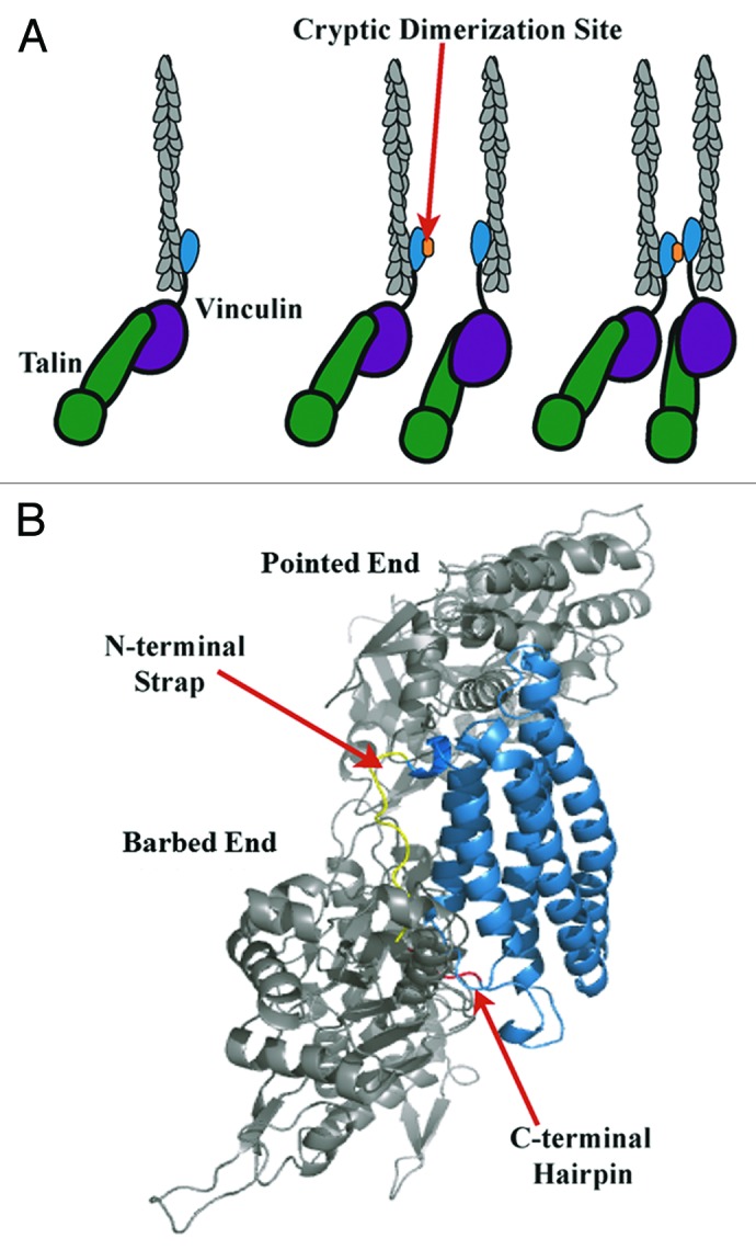
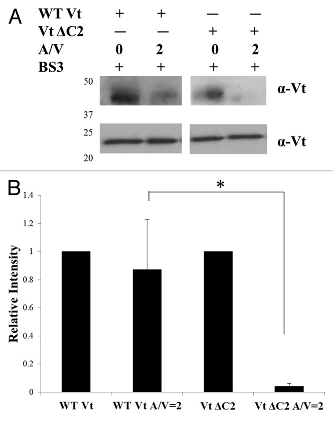
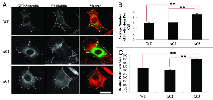
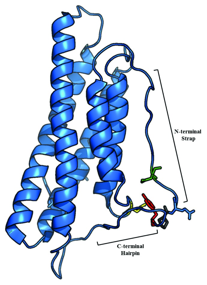
Vinculin is an essential cell adhesion protein, found at both focal adhesions and adherens junctions, where it couples transmembrane proteins to the actin cytoskeleton. Vinculin is involved in controlling cell shape, motility and cell survival, and has more recently been shown to play a role in force transduction. The tail domain of vinculin (Vt) has the ability to both bind and bundle actin filaments. Binding to actin induces a conformational change in Vt believed to promote formation of a Vt dimer that is able to crosslink actin filaments. We have recently provided additional evidence for the actin-induced Vt dimer and have shown that the vinculin carboxyl (C)-terminal hairpin is critical for both the formation of the Vt dimer and for bundling F-actin. We have also demonstrated the importance of the C-terminal hairpin in cells as deletion of this region impacts both adhesion properties and force transduction. Intriguingly, we have identified bundling deficient variants of vinculin that show different cellular phenotypes. These results suggest additional role(s) for the C-terminal hairpin, distinct from its bundling function. In this commentary, we will expand on our previous findings and further investigate these actin bundling deficient vinculin variants.

Keywords: F-actin bundling; dimerization; focal adhesion; scaffold; vinculin.


Figures

None
Figure 1. Model of vinculin activation, F-actin binding and bundling. (A) As modified from Janssen et al., release of autoinhibitory interactions within full length vinculin via binding of talin (green) to Vh (purple) and F-actin (gray) to Vt (blue), leads to vinculin activation. F-actin binding causes a conformational change in Vt that exposes a cryptic dimerization site (orange) that enables Vt self-association and F-actin bundling., (B) According to the Janssen model, the C-terminal hairpin (red) and N-terminal strap (yellow) point into the F-actin interface. However, the Janssen model may require further validation given the resolution of the micrograph, multiple conformational clashes between Vt and F-actin, and contrasting data that has arisen indicating that the C-terminal hairpin is necessary for bundle formation. Hence, given our findings, a refined or alternative model for this critical interaction should lead to more specific tools to study the function of the vinculin/F-actin interaction.
None
Figure 2. Detection of the actin-induced Vt dimer. Crosslinking experiments were conducted as previously described. (A) Western blots of WT Vt and Vt ΔC2 in the absence and presence of actin (A/V = 2) and probed against Vt. Crosslinked samples were run on SDS-PAGE gels (lower panel) to observe the Vt monomer band (21 kDa) or NuPAGE Bis-tris 10% gradient gels (Invitrogen) (upper panel) to observe dimer species and blotted with a rabbit anti-chicken Vt antibody [a gift from Dr. Susan Craig (John Hopkins University)]. (B) Quantification of crosslinked bands by densitometry. In the presence of actin, a significant decrease in the amount of the actin-induced dimer is observed for Vt ΔC2 relative to WT Vt. Similar results were previously obtained for the larger bundling deficient variant, Vt ΔC5. Densitometry is the average ± SEM combined from three independent experiments. Statistical significance was determined using the Student’s t-test. *p ≤ 0.05.
None
Figure 3. Vinculin C-terminal hairpin deletions alter cell morphology. (A) Vin−/− MEFs were transfected with GFP-tagged WT-, ΔC2- or ΔC5 vinculin and plated on FN for two hours as previously described. (B) The number of protrusions and (C) the relative protrusion area were measured with NIH ImageJ. Cells expressing ΔC5 vinculin show significantly more and larger protrusions per cell, relative to WT vinculin and ΔC2 vinculin. Results shown represent ~60 cells from three independent experiments. Statistical significance was determined by the Student’s t-test. **p ≤ 0.001 comparing WT vinculin and ΔC2 vinculin to ΔC5 vinculin. Scale bar is 30 µm.
None
Figure 4. Ribbon diagram of Vt (PDB ID 1ST6) highlighting interactions of the Y1065 side chain. The side chain of Y1065 (red) in the C-terminal hairpin forms hydrogen bonds with D882 (green) in the strap, K915 (yellow) in the helix 1/helix 2 loop and P878 (gray) in the proline-rich linker region.
None
Figure 5. Vinculin mutants deficient in F-actin bundling also alter vinculin’s scaffold function and prevent actin accumulation at adhesion complexes. Adhesion complexes were isolated from Vin−/− MEFs expressing either WT-, ΔC2-, ΔC5 vinculin or untransfected, after incubation with FN coated beads for 30 min.

Comment on

References

    1. Parsons JT, Horwitz AR, Schwartz MA. Cell adhesion: integrating cytoskeletal dynamics and cellular tension. Nat Rev Mol Cell Biol. 2010;11:633–43. doi: 10.1038/nrm2957. - DOI - PMC - PubMed
    1. Ziegler WH, Liddington RC, Critchley DR. The structure and regulation of vinculin. Trends Cell Biol. 2006;16:453–60. doi: 10.1016/j.tcb.2006.07.004. - DOI - PubMed
    1. Xu W, Baribault H, Adamson ED. Vinculin knockout results in heart and brain defects during embryonic development. Development. 1998;125:327–37. - PubMed
    1. Xu W, Coll JL, Adamson ED. Rescue of the mutant phenotype by reexpression of full-length vinculin in null F9 cells; effects on cell locomotion by domain deleted vinculin. J Cell Sci. 1998;111:1535–44. - PubMed
    1. Zemljic-Harpf AE, Ponrartana S, Avalos RT, Jordan MC, Roos KP, Dalton ND, et al. Heterozygous inactivation of the vinculin gene predisposes to stress-induced cardiomyopathy. Am J Pathol. 2004;165:1033–44. doi: 10.1016/S0002-9440(10)63364-0. - DOI - PMC - PubMed

Publication types