. 2013 Feb 1;41(4):2659-72.
doi: 10.1093/nar/gks1362. Epub 2013 Jan 9.

Rbm20 regulates titin alternative splicing as a splicing repressor

Affiliations

Rbm20 regulates titin alternative splicing as a splicing repressor

Shijun Li et al. Nucleic Acids Res. .

Abstract

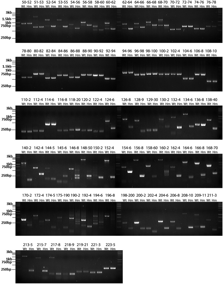
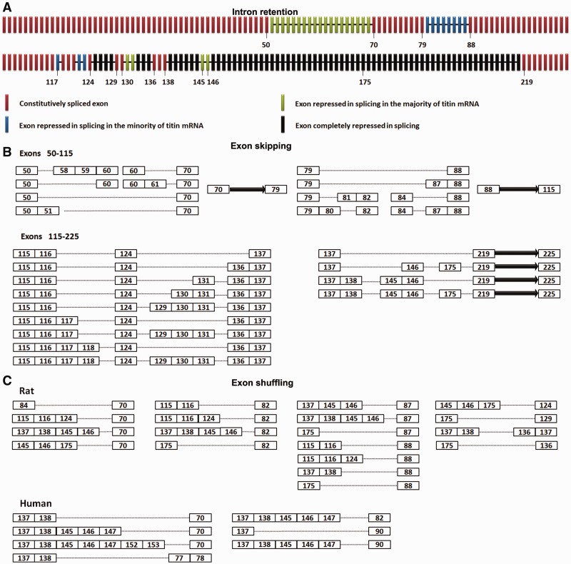
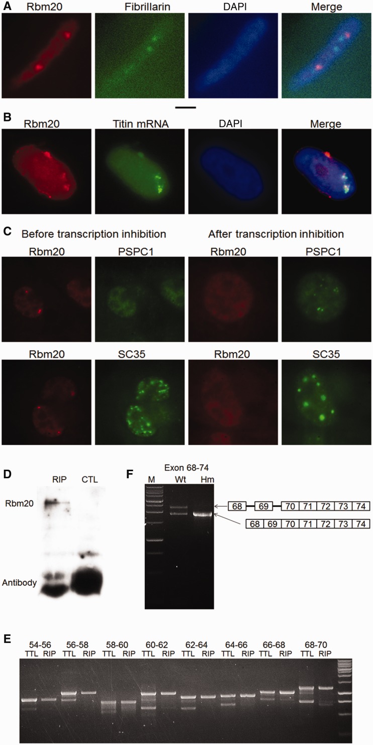
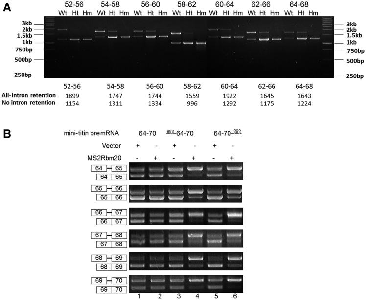
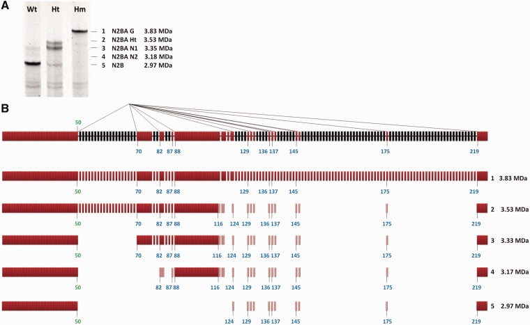
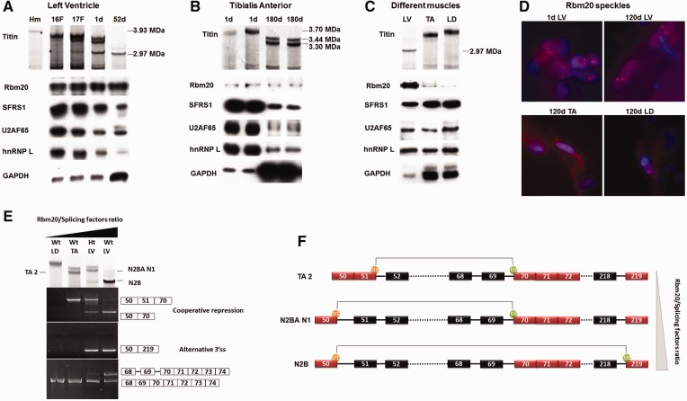
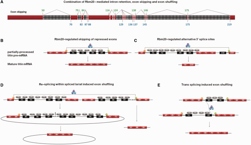
Titin, a sarcomeric protein expressed primarily in striated muscles, is responsible for maintaining the structure and biomechanical properties of muscle cells. Cardiac titin undergoes developmental size reduction from 3.7 megadaltons in neonates to primarily 2.97 megadaltons in the adult. This size reduction results from gradually increased exon skipping between exons 50 and 219 of titin mRNA. Our previous study reported that Rbm20 is the splicing factor responsible for this process. In this work, we investigated its molecular mechanism. We demonstrate that Rbm20 mediates exon skipping by binding to titin pre-mRNA to repress the splicing of some regions; the exons/introns in these Rbm20-repressed regions are ultimately skipped. Rbm20 was also found to mediate intron retention and exon shuffling. The two Rbm20 speckles found in nuclei from muscle tissues were identified as aggregates of Rbm20 protein on the partially processed titin pre-mRNAs. Cooperative repression and alternative 3' splice site selection were found to be used by Rbm20 to skip different subsets of titin exons, and the splicing pathway selected depended on the ratio of Rbm20 to other splicing factors that vary with tissue type and developmental age.


Figures

Figure 1.
Figure 1.
Splicing of titin mRNA is inhibited in the wild type (Wt, Rbm20+/+) but not in the homozygote mutant (Hm, Rbm20−/−). RT-PCR was used with primer pairs spanning two or three exons to amplify cardiac titin mRNA from Wt and Hm left ventricle (LV). Usually three bands were obtained in the Wt for the middle Ig region between exons 50 and 70 and exons 80 and 88, but only one band in the Hm. As exemplified by exons 68–70 in the top panel, the lower Wt band has the same size as the adjoining Hm band and is the product of constitutive splicing; the upper band contains product where two introns for the primer-spanned region are both retained; the middle band has one of the two spanned introns retained. The splicing in the PEVK region (exons 124–218) is repressed to a greater extent in the Wt. Under most circumstances (as exemplified by exons 124–126), PEVK introns cannot be removed from Wt titin mRNA so that only one band, representing the retained intron pattern with a larger size than the adjoining band in the Hm, can be detected for most primer pairs. No retained introns can be detected in the Hm. The size of marker is indicated on the left, and the expected size of the PCR product for each primer pairs are listed in the Supplementary Table S2. The PCR products match their predicted size for all the primer pairs.
Figure 2.
Figure 2.
Rbm20 mediates exon skipping and exon shuffling. (A) Schematic illustration of intron retention along cardiac titin mRNA in Wt rats. The titin exons and retained introns are shown with bars and lines, respectively. Colors denote the extent of Rbm20 repression on different regions. Black: complete repression, splicing repressed in all mRNAs; green: strong repression, splicing repressed in the majority of mRNAs; blue: mild repression, splicing repressed in the minority of mRNAs; red, no repression, normally spliced. The numbers of some exons are indicated. (B) Schematic illustration of exon skipping along cardiac titin mRNA in the Wt rat. The exons are displayed as numbered boxes; the solid arrow represents consecutive exons that result from constitutive splicing between the adjacent numbered exons. The dotted lines denote the exon skipping mediated by direct ligation between adjacent numbered exons. Results show that exon skipping only occurs in the Rbm20-repressed regions: exons 50–70, exons 79–88 and most of the PEVK region.(C) Diagram depicts the exon composition of exon-shuffling isoforms from the cardiac titin mRNA in the Wt rat and human. The exons are shown in numbered boxes, and the dotted lines denote direct attachment between the adjacent numbered exons.
Figure 3.
Figure 3.
Rbm20 aggregated on newly synthesized titin mRNAs forms Rbm20 speckles and represses the splicing of titin mRNA. (A) Immunofluorescence shows two Rbm20 speckles in the nucleus of cardiomyocytes from LV. The size of Rbm20 speckles was similar to nucleoli; Rbm20 is stained red, and nucleoli are stained green with antibody against fibrillarin. 4′,6 diamidino-2-phenylindole (DAPI)-stained nuclei are blue. Black scale bar: 2 microns. (B) Immunostaining of Rbm20 protein combined with titin mRNA FISH shows the co-localization of titin mRNA with Rbm20 speckles. Rbm20 is stained red; titin mRNA is stained green; and the overlap of red and green yields yellow. (C) Rbm20 speckles are transcription dependent. When the transcription of HL-1 cell is inhibited, Rbm20 speckles (red) dissociate. The behavior of paraspeckles (PSPC1) and splicing speckles (SC35) verified that transcription in the specific cell is inhibited. After transcription inhibition, crescent-shaped paraspeckles were formed, and the splicing speckles (green) become concentrated. Rbm20 is stained red; PSPC1 and SC35, marker proteins for paraspeckles and splicing speckles, respectively, are stained green. (D) Rbm20 can be specifically immunoprecipitated by anti-Rbm20 but not by control antibody. RIP: with anti-Rbm20, CTL: with control antibody (anti-spectrin α). (E) When using RT-PCR to amplify the total titin mRNA (TTL) from LV and the titin mRNA co-immunoprecipitated with Rbm20 (RIP), for the total titin mRNA, both retained introns (upper band in each primer pair) and constitutive splicing pattern (lower band) can be detected. However, for the Rbm20-immunoprecipitated titin mRNA, only the retained intron pattern can be clearly detected, meaning the constitutively spliced titin mRNA can barely be precipitated by Rbm20. (F) The co-existence of retained intron and constitutive splicing patterns on the same titin message were found in the Wt. The splicing on introns before exon 70 remains inhibited when the splicing on the downstream introns was finished. This situation does not occur in the Hm.
Figure 4.
Figure 4.
Rbm20 represses the splicing of titin mRNA in a cooperative manner for the region between exons 52 and 68. (A) All-intron retention and constitutive splicing are two major splicing patterns for all the primer pairs across the region from exon 52–68 in the Wt and Ht. Each primer pair used in this study spans five exons and four introns, with three exons and two introns overlapped with the spanned region of the adjacent primer pairs. The ratio of all-intron retention to constitutive splicing decreased from Wt to Ht; no intron retention can be found with Hm. The marker indication and the expected size of the PCR product for each primer pairs are shown. (B) The binding of Rbm20 to certain positions of titin mRNA can strengthen the repression of Rbm20 on distant exons. Lanes 1, 2, 3 and 5 show that neither MS2 structure ligated to titin mRNA nor Rbm20 fused with MS2 coat protein can repress the splicing of titin minigenes by themselves. Lane 4 and 6 show that when Rbm20 is recruited to titin mRNA at either upstream or downstream positions, all the titin exons on the titin mRNA are repressed, even for the exons that are far away from the Rbm20-binding site.
Figure 5.
Figure 5.
Rbm20-regulated alternative 3′ splice sites (ss). (A) The cardiac titin protein isoforms in the LV of the Wt, Ht and Hm. Based on their size from large to small, the titin isoforms are numbered from 1 to 5. The apparent molecular weights and the name of different titin isoforms are shown on the right. The 2.97-MDa titin isoform is the N2B titin isoform, which is dominant in the Wt rat heart. (B) Schematic illustration of the exon 50–involved alternative 3′ ss pathways in which the 5′ ss of exon 50 can splice with the 3′ ss of many other exons. On top is shown the partially processed titin mRNA in which the splicing in the regions not repressed by Rbm20 (colored in red) is finished but the splicing in the repressed regions (colored in black) remains inhibited. Exons and introns are indicated with bars and thick lines, respectively. The thin lines connecting exon 50 with other exons illustrate different splicing pathways; most of the acceptor exons are located at the 3’′ end of Rbm20-repressed regions. The molecular mass of the titin isoform resulting from each splicing pathway is calculated and shown on the right. The light-colored red bars in splicing isoforms 2, 3, 4 and 5 represent the alternative exons. They do not always exist in the alternatively spliced titin mRNAs because different titin mRNAs contain different combination of these exons (shown in Figure 2B).
Figure 6.
Figure 6.
Titin size is determined by the relative ratio of Rbm20 to other splicing factors. (A) Upper panel shows titin proteins separated on an sodium dodecyl sulphate–agarose gel. The left lane is from an Hm adult LV, and the rest are Wt from fetal (F) and postnatal heart aged in days (d). Lower panel shows western blots. Sample loads were balanced for Rbm20 content. Results show that during development, the size reduction in titin protein is accompanied by increased ratios of Rbm20 to other splicing factors. (B) Similar developmental changes in the ratios of Rbm20 to other splicing factors occur in the skeletal muscle tibialis anterior (TA). The Rbm20 to splicing factor ratios increase with age, whereas the titin size decreases. (C) Tissue specific Rbm20 and titin isoform expression in LV, TA and longissimus dorsi (LD). Sample loads were balanced for the splicing-related factors. The results demonstrate that the ratio of Rbm20 to other splice factors is inversely related to titin isoform size. (D) Characterization of Rbm20 speckles during development and between muscle tissues. Two speckles per nucleus were observed in all the indicated tissues. d, day(s). (E) Titin isoforms and splicing pathways from different muscle tissues. Titin isoforms from different tissues are shown in the upper panel. TA 2 and N2BA N1 correspond to the cooperative repression between exons 50 and 70. TA2 is smaller than N2BA N1 because only the latter contains protein coded by exon 49, which accounts for an additional 0.1 MDa of protein mass (21). N2B results from the alternative 3′ ss pathway (shown by exons 50–219). The 5′ ss of exon 70 is always active for splicing, but the 3′ ss of exon 70 can be repressed in some titin mRNA in Ht LV and Wt LV (bottom panel). The unspliced 3′ ss of exon 70 cannot be found in Wt LD and Wt TA. (F) A schematic diagram demonstrating the Rbm20/splicing factors ratio–dependent extension of Rbm20-repressed region between exons 50 and 70 and the consequence of each situation. As the Rbm20/splicing factors ratio increases from top to bottom, the Rbm20-repressed region extends from exons 51–70 to exons 50–70. When Rbm20/splicing factors ratio is high enough like in Ht and Wt LV, we found the splicing of the 5′ ss of exon 50 to downstream alternative 3′ ss, for example the 3′ ss of exon 219. The 5′ ss of exon 50 can splice with many other alternative 3′ ss (refer to Figure 5B); they are not shown in this diagram. The dotted lines denote the continuous exons (and introns if repressed by Rbm20) that are not shown.
Figure 7.
Figure 7.
Mechanisms of Rbm20-regulated alternative splicing. (A) Schematic diagram depicts the partially processed titin pre-mRNA and the location of the donor and acceptor exons involved in exon skipping. The same donor and acceptor exons are also involved in exon shuffling. The numbers of donor exons and acceptor exons are colored in green and blue, respectively. The thin lines connecting the donor exons with acceptor exons illustrate different splicing pathways. (B) Hypothesized model showing the process of Rbm20-regulated exon skipping as exemplified by exon skipping between exons 50 and 70. The exons are indicated with numbered boxes, and introns with black lines. The unrepressed exons are colored in red, and the Rbm20-repressed exons in black. SnRNPs are indicated with colored circles. U1 defines the 5′ ss and U2 defines the 3′ ss on titin exons. The association of U4/5/6 to U1 and U2 fulfills the splicing. Splicing between exons 50 and 70 skips the internal exons. (C) Hypothesized model related the upstream 3′ ss repression to the use of downstream alternative 3′ ss. (D) Hypothesized model showing the process of exon shuffling induced by re-splicing within the lariat spliced out during alternative 3′ ss pathway. Here we only show one example, many other alternative 3′ ss pathways and exon-shuffling events have been detected from titin mRNA (refer to Figure 5B and Figure 2C). (E) Hypothesized model for trans splicing–induced exon shuffling. Active 5′ ss on exon 84 splice with active 3′ ss on exon 70 from different titin messages to cause exon shuffling.

Similar articles

Cited by

References

    1. Bang ML, Centner T, Fornoff F, Geach AJ, Gotthardt M, McNabb M, Witt CC, Labeit D, Gregorio CC, Granzier H, et al. The complete gene sequence of titin, expression of an unusual approximately 700-kDa titin isoform, and its interaction with obscurin identify a novel Z-line to I-band linking system. Circ. Res. 2001;89:1065–1072. - PubMed
    1. Labeit S, Kolmerer B. Titins: giant proteins in charge of muscle ultrastructure and elasticity. Science. 1995;270:293–296. - PubMed
    1. Maruyama K, Kimura S, Yoshidomi H, Sawada H, Kikuchi M. Molecular size and shape of beta-connection, an elastic protein of striated muscle. J. Biochem. 1984;95:1423–1433. - PubMed
    1. Trinick J, Knight P, Whiting A. Purification and properties of native titin. J. Mol. Biol. 1984;180:331–356. - PubMed
    1. Wang K, McCarter R, Wright J, Beverly J, Ramirez-Mitchell R. Regulation of skeletal muscle stiffness and elasticity by titin isoforms: a test of the segmental extension model of resting tension. Proc. Natl Acad. Sci. USA. 1991;88:7101–7105. - PMC - PubMed

Publication types