. 2014 Jun;24(6):1409-21.
doi: 10.1093/cercor/bhs412. Epub 2013 Jan 10.

Sp8 and COUP-TF1 reciprocally regulate patterning and Fgf signaling in cortical progenitors

Affiliations

Sp8 and COUP-TF1 reciprocally regulate patterning and Fgf signaling in cortical progenitors

Ugo Borello et al. Cereb Cortex. 2014 Jun.

Abstract

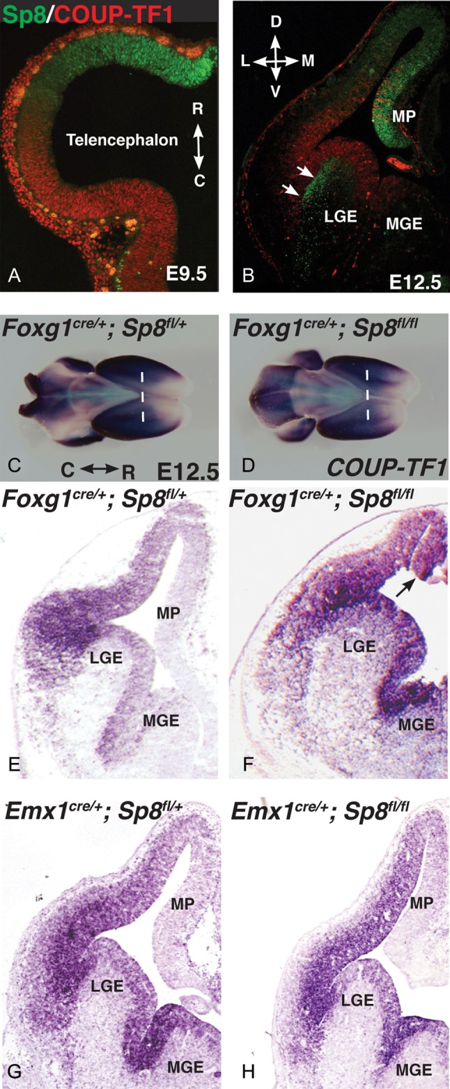
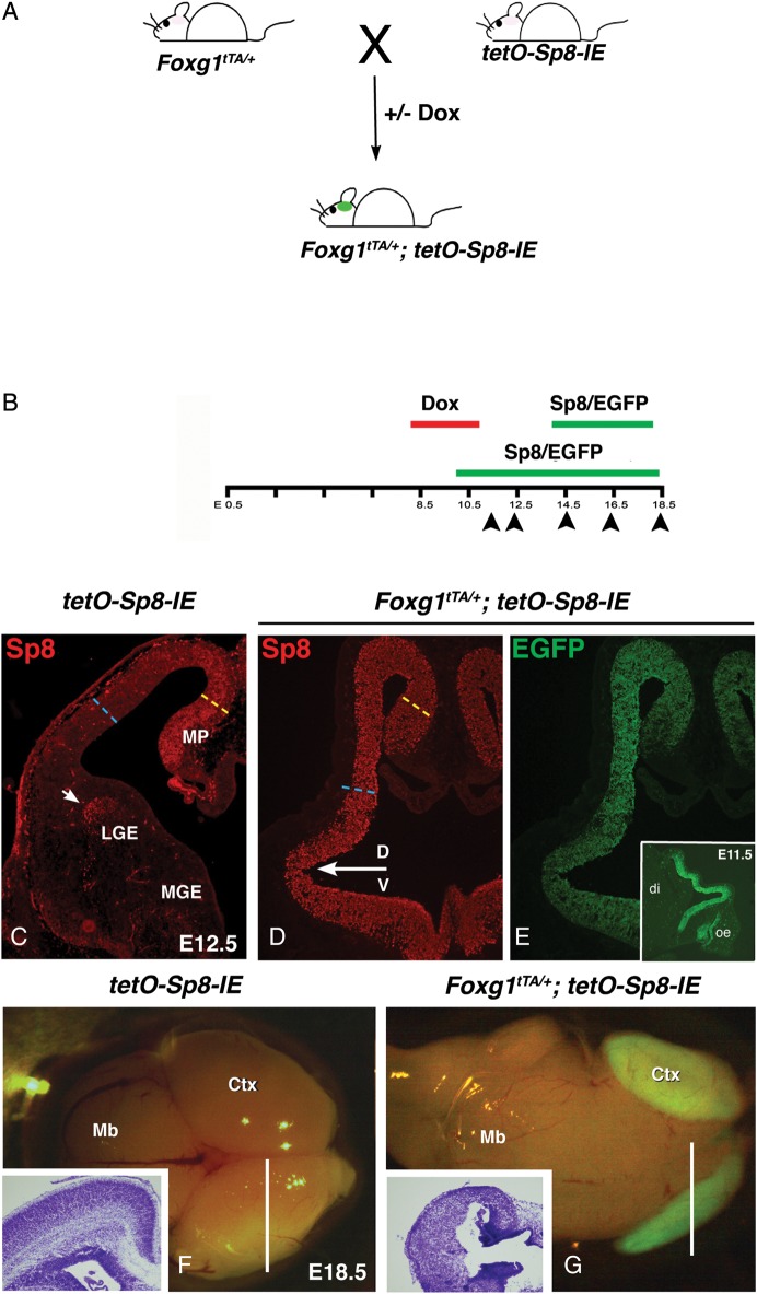
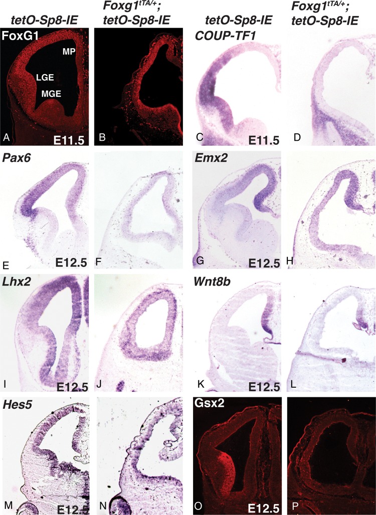
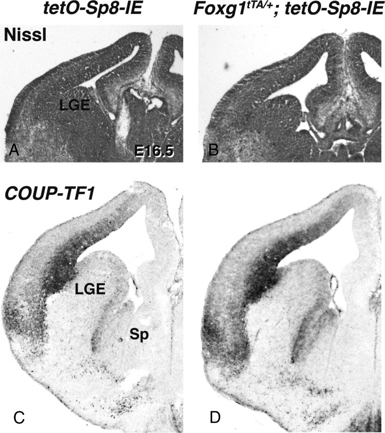
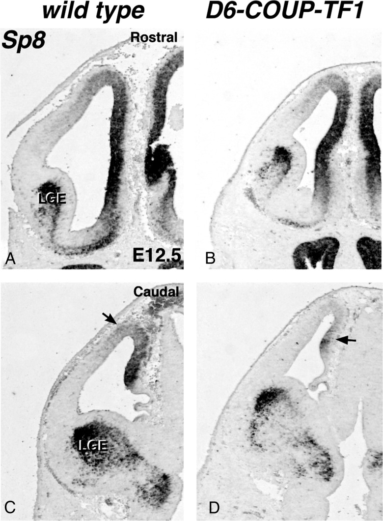
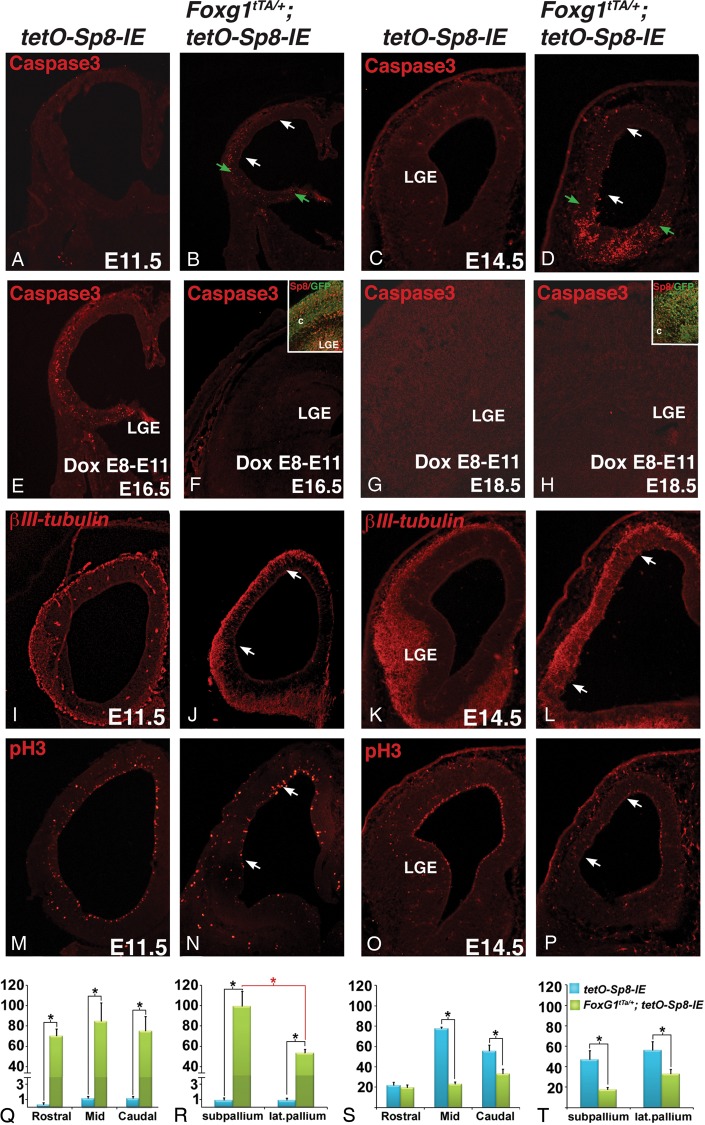
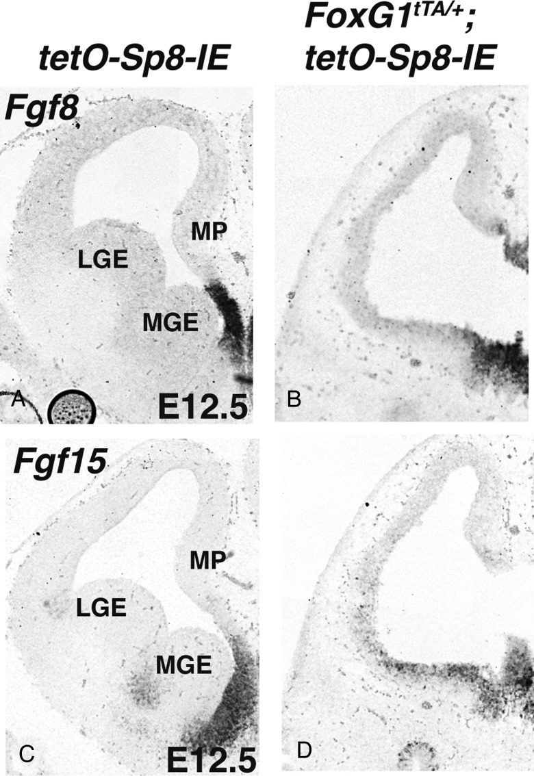
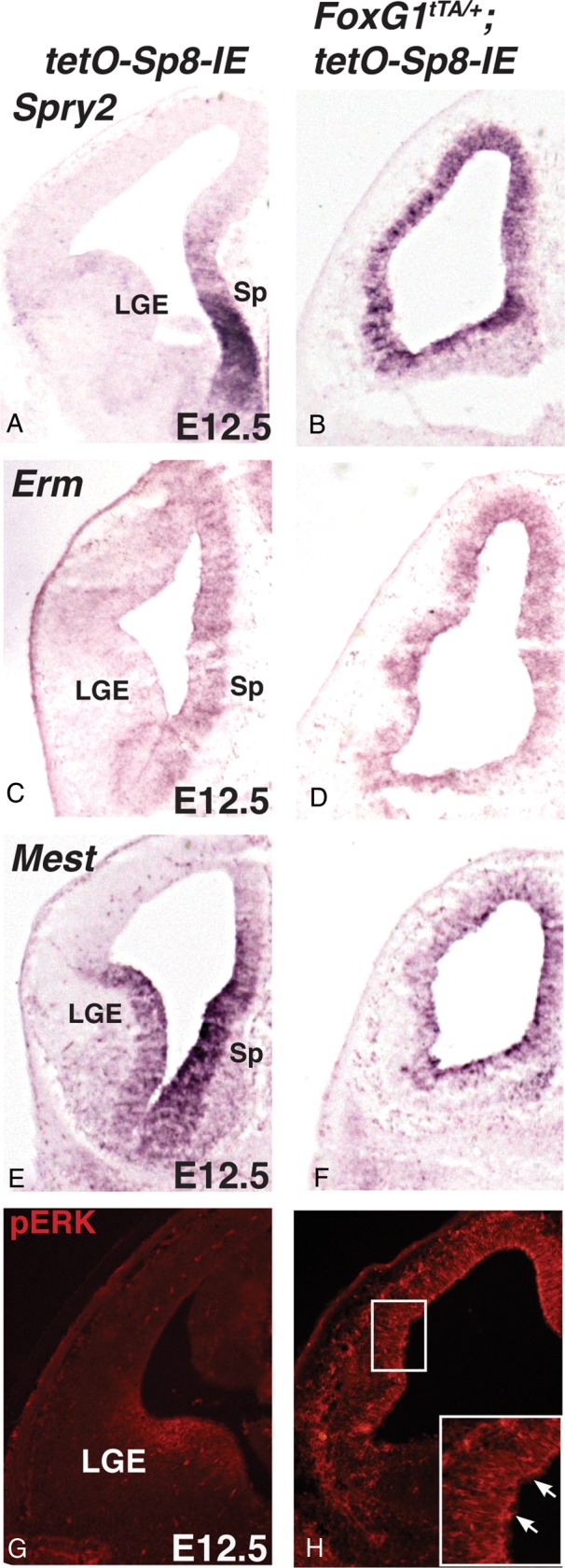
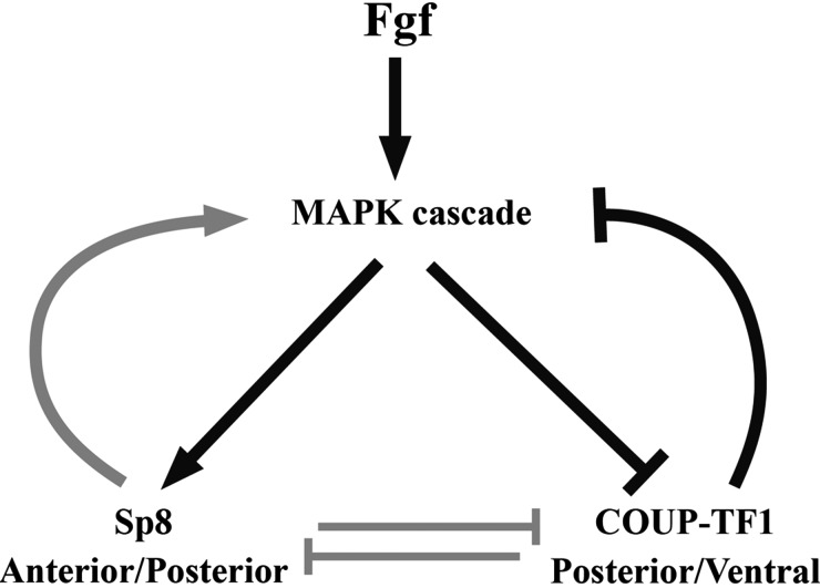
To gain new insights into the transcriptional regulation of cortical development, we examined the role of the transcription factor Sp8, which is downstream of Fgf8 signaling and known to promote rostral cortical development. We have used a binary transgenic system to express Sp8 throughout the mouse telencephalon in a temporally restricted manner. Our results show that misexpression of Sp8 throughout the telencephalon, at early but not late embryonic stages, results in cortical hypoplasia, which is accompanied by increased cell death, reduced proliferation, and precocious neuronal differentiation. Misexpression of Sp8 at early developmental stages represses COUP-TF1 expression, a negative effector of Fgf signaling and a key promoter of posterior cortical identity, while ablation of Sp8 has the opposite effect. In addition, transgenic misexpression of COUP-TF1 resulted in downregulation of Sp8, indicating a reciprocal cross-regulation between these 2 transcription factors. Although Sp8 has been suggested to induce and/or maintain Fgf8 expression in the embryonic telencephalon, neither Fgf8 nor Fgf15 was upregulated using our gain-of-function approach. However, misexpression of Sp8 greatly increased the expression of Fgf target molecules, suggesting enhanced Fgf signaling. Thus, we propose that Sp8 promotes rostral and dorsomedial cortical development by repressing COUP-TF1 and promoting Fgf signaling in pallial progenitors.

Keywords: Fgf signaling, neurogenesis; Sp8; corticogenesis; patterning; proliferation.


Figures

Figure 1.
Figure 1.
Sp8 and COUP-TF1 are expressed in complementary domains. As early as E9.5, Sp8 and COUP-TF1 form opposing gradients along the rostrocaudal (R-C) axis (A) and by E12.5 their opposing dorsoventral gradients are clearly established (B). Early deletion of Sp8, and not late deletion, results in the rostral and dorsal expansion of COUP-TF1. At E12.5 COUP-TF1 is expressed in a caudoventral high, rostral-dorsal low gradient, as seen in whole mount in situ hybridization in control Foxg1cre/+;Sp8fl/+ brains (C). In Foxg1cre/+;Sp8fl/fl animals, the expression of COUP-TF1 is expanded dorsally and rostrally (D). The dotted lines in C and D provide landmarks from which to compare the change in COUP-TF1 expression. Coronal section of E12.5 Foxg1cre/+;Sp8fl/fl mutant brains (F) show that COUP-TF1 is expanded dorsally compared with control Foxg1cre/+;Sp8fl/+ animals (E). Arrows in F indicate the dorsal expansion of COUP-TF1 expression. Expression of COUP-TF1 in E12.5 Emx1cre/+;Sp8fl/fl mutants (H) does not appear substantially changed from Emx1cre/+;Sp8fl/+ control animals (G). LGE, lateral ganglionic eminence; MGE, medial ganglionic eminence; MP, medial pallium.
Figure 2.
Figure 2.
To misexpress Sp8 throughout the telencephalon, Foxg1tTA/+ males were bred to tetO-Sp8-IE females to yield Foxg1tTA/+;tetO-Sp8-IE progeny (A). Foxg1tTA/+;tetO-Sp8-IE animals overexpress Sp8 and EGFP starting at E9.5, and this expression can be delayed by 5 days by administering doxycycline (Dox) to the mother between Days E8 and E11 (B). Foxg1tTA/+;tetO-Sp8-IE animals express the transgenes uniformly through the rostrocaudal extent of the telencephalon. Inset in E, shows EGFP expression in a sagittal section of an E11.5 animal, where the olfactory epithelium (oe) marks the rostral portion of the animals head and the diencephalon (di) indicates the caudal regions). Double transgenic animals also show uniform overexpression of Sp8 and EGFP along the dorsoventral (D/V) axis of the telencephalon (D and E, respectively, arrow marks the D/V border) as compared to tetO-Sp8-IE controls (C) that only express Sp8 in the medial pallium (MP) and the subventricular zone (arrow) of the LGE. Misexpression of Sp8 leads to reduction in ventrolateral pallial thickness at E12.5 (cyan line in D) compared with control animals (cyan line in C); the thickness of the dorsomedial pallium (yellow lines in C and D) shows no significant changes. This phenotype is amplified by E18.5 resulting in a severe hypoplasia of the cortex (Ctx) in Foxg1tTA/+;tetO-Sp8-IE animals (G) as compared to control tetO-Sp8-IE animals (F). Insets show Nissl-stained cross sections of the brains in Foxg1tTA/+;tetO-Sp8-IE (G) and control tetO-Sp8-IE animals (F) at the level indicated by the white line. The cortex of double transgenic animals (G) has large ventricles, which lack a clear laminar organization compared with the brains of control animals (F). LGE, lateral ganglionic eminence; MGE, medial ganglionic eminence; MP, medial pallium; Ctx, cortex; Mb, midbrain; oe, olfactory epithelium; di, diencephalon.
Figure 3.
Figure 3.
The brains of Foxg1tTA/+;tetO-Sp8-IE maintain their telencephalic identity as indicated by expression of Foxg1 (B). Foxg1 expression in tetO-Sp8-IE controls is shown in panel (A). However, early dorsoventral telencephalic patterning is altered after Sp8 misexpression; there is repression of ventrally expressed transcription factors COUP-TF1 at E11.5 (C and D), and Pax6 at E12.5 in Foxg1tTA/+;tetO-Sp8-IE animals (F) as compared to tetO-Sp8-IE controls (E). On the other hand, the domain of expression of the dorsally expressed gene Emx2 appears slightly increased in the lateral pallium in Foxg1tTA/+;tetO-Sp8-IE animals (H) as compared to tetO-Sp8-IE control (G). On the other hand, Lhx2 expression is downregulated in the embryos misexpressing Sp8 (I and J). Expression of the medial pallium and cortical hem marker, Wnt8b remains unchanged in Foxg1tTA/+;tetO-Sp8-IE animals (L) as compared to tetO-Sp8-IE controls (K). Hes5 expression, a marker for neuroepithelial progenitors and Notch signaling, is severely reduced in the Foxg1tTA/+;tetO-Sp8-IE animals (N) as compared to tetO-Sp8-IE controls (M). Additionally, the expression of the subpallial transcription factor Gsx2, is severely reduced in Foxg1tTA/+;tetO-Sp8-IE animals (P) as compared to tetO-Sp8-IE controls (O). LGE, lateral ganglionic eminence; MGE, medial ganglionic eminence; MP, medial pallium.
Figure 4.
Figure 4.
Misexpression of Sp8 in late-stage telencephalic progenitor cells has no obvious effects on cortical development. Sp8 misexpression was delayed until E14.5 by Dox administration. The effect of Sp8 misexpression in late-stage telencephalic progenitors was analyzed at E16.5. Nissl-stained tetO-Sp8-IE and Foxg1tTA/+;tetO-Sp8-IE cortices at E16.5 show similar morphology (A and B, respectively) and COUP-TF1 expression appears similar in tetO-Sp8-IE and Foxg1tTA/+;tetO-Sp8-IE cortices (C and D, respectively). LGE, lateral ganglionic eminence; Sp, septum.
Figure 5.
Figure 5.
COUP-TF1 overexpression in the medial pallium represses Sp8 expression. Weak overexpression of COUP-TF1 in the rostral pallium of D6-COUP-TF1 animals (B) causes a mild reduction in Sp8 expression as compared to wild-type controls at E12.5 (A). Strong (∼4-fold) overexpression of COUP-TF1 in the caudal pallium clearly reduces Sp8 expression in D6-COUP-TF1 animals (D) as compared to controls (C). Arrows in C and D point to reduced Sp8 expression in the dorsomedial cortex of the control and D6-COUP-TF1 embryos. LGE, lateral ganglionic eminence.
Figure 6.
Figure 6.
Misexpression of Sp8 alters cell death, proliferation, and differentiation of telencephalic progenitors. Cell death is strongly increased in the ventrolateral telencephalon of Foxg1tTA/+;tetO-Sp8-IE animals (B) compared with tetO-Sp8-IE controls (A) as early as E11.5. These apoptotic cells are concentrated ventrally in the telencephalon (in the subpallium and ventrolateral pallium) (R); on the other hand, they appear to be evenly distributed along the rostrocaudal axis (Q). Cell death in these regions is further increased by E14.5 (C and D). Green arrows in B and D indicate the approximate position of the pallial/subpallial boundary; the white arrows indicate the ventrolateral pallium. However, no increase in apoptosis is observed in E16.5 or E18.5 Dox-treated animals, in which Sp8 misexpression is delayed until E15 (cf. E with F and G with H). Insets in F and H show EGFP and Sp8 overexpression throughout the telencephalon at E16.5 and E18, after Dox treatment between E8 and E11. Another early effect of Sp8 overexpression is an increase in neuronal differentiation, as marked by βIII-tubulin. As early as E11.5, βIII-tubulin is increased in Foxg1tTA/+;tetO-Sp8-IE animals (J) compared with control tetO-Sp8-IE animals (I). βIII-tubulin immunoreactivity becomes even more pronounced by E14.5 (K and L) when the ventricular zone in lateral regions (arrows in L) of Foxg1tTA/+;tetO-Sp8-IE cortex shows precocious neuronal differentiation compared with tetO-Sp8-IE control (K). Cell proliferation, as indicated by phospho-Histone 3 (pH3), can be seen along the ventricular surface of control brains (M and N and O and P, E11.5 and E14.5, respectively) but is severely reduced in the lateral pallium (arrows in N and P) and subpallium of Foxg1tTA/+;tetO-Sp8-IE animals. The effects of Sp8 misexpression on cell proliferation are more pronounced at E14.5 (O and P) than at E11.5 (M and N). Proliferation is strongly reduced at mid and caudal levels of the telencephalon as shown in S; the reduction is not limited to either the lateral or ventral part of telencephalon (T). LGE, lateral ganglionic eminence, *P< 0.01.
Figure 7.
Figure 7.
Coronal sections of E12.5 brains show expression of Fgf8 (A and B) and Fgf15 (C and D). Fgf8 and Fgf15 expression persist in Foxg1tTA/+;tetO-Sp8-IE animals (B and D) compared with tetO-Sp8-IE controls (A and C). LGE, lateral ganglionic eminence; MGE, medial ganglionic eminence; MP, medial pallium.
Figure 8.
Figure 8.
Misexpression of Sp8 increases the expression of Fgf effector molecules within the telencephalon. Expression of Spry2 (A and B), Erm (C and D), and Mest (E and F) and immunoreactivity of pERK (G and H) are increased in Foxg1tTA/+;tetO-Sp8-IE (B, D, F, and H) brains as compared to tetO-Sp8-IE controls (A, C, E, and G). Inset in H is a higher magnification image showing increased pERK staining (arrows) in the ventrolateral pallium of the Foxg1tTA/+;tetO-Sp8-IE animals. LGE, lateral ganglionic eminence; Sp, septum.
Figure 9.
Figure 9.
Model of the Sp8 regulated gene network involved in cortical development. Fgfs produced at the cortical patterning center act on Sp8 and COUP-TF1 through the MAPK cascade. MAPK signaling upregulates Sp8 and downregulates COUP-TF1. COUP-TF1 in-turn downregulates Fgf signaling. Additionally, we show that COUP-TF1 and Sp8 cross-repress each other and that Sp8 can enhance MAPK signaling in cortical progenitor cells. A balance between the various branches of this gene network controls rostral-caudal and dorsoventral patterning of the cortex.

Similar articles

Cited by

References

    1. Armentano M, Chou SJ, Srubek Tomassy G, Leingartner A, O'Leary DD, Studer M. COUP-TFI regulates the balance of cortical patterning between frontal/motor and sensory areas. Nat Neurosci. 2007;10:1277–1286. - PubMed
    1. Bell SM, Schreiner CM, Waclaw RR, Campbell K, Potter SS, Scott WJ. Sp8 is crucial for limb outgrowth and neuropore closure. Proc Natl Acad Sci USA. 2003;100:12195–12200. - PMC - PubMed
    1. Bishop KM, Goudreau G, O'Leary DD. Regulation of area identity in the mammalian neocortex by Emx2 and Pax6. Science. 2000;288:344–349. - PubMed
    1. Bishop KM, Rubenstein JL, O'Leary DD. Distinct actions of Emx1, Emx2, and Pax6 in regulating the specification of areas in the developing neocortex. J Neurosci. 2002;22:7627–7638. - PMC - PubMed
    1. Borello U, Cobos I, Long JE, McWhirter JR, Murre C, Rubenstein JL. FGF15 promotes neurogenesis and opposes FGF8 function during neocortical development. Neural Dev. 2008;3:17. - PMC - PubMed

Publication types

MeSH terms