

Review
doi: 10.1126/science.1227901.
Microglia: scapegoat, saboteur, or something else?
Affiliations
- PMID: 23307732
- PMCID: PMC4431634
- DOI: 10.1126/science.1227901
Item in Clipboard
Review
Microglia: scapegoat, saboteur, or something else?
Science.
.
Abstract
Microglia are resident immune cells in the brain and spinal cord. These cells provide immune surveillance and are mobilized in response to disparate diseases and injuries. Although microglial activation is often considered neurotoxic, microglia are essential defenders against many neurodegenerative diseases. It also seems increasingly likely that microglial dysfunction can underlie certain neurological diseases without an obvious immune component.
Figures

Microglia derive from yolk-sac macrophages formed during primitive hematopoiesis beginning at embryonic day 7.5 (E7.5) (the embryo is depicted at E11.5 for illustrative purposes). Primitive macrophages migrate to the developing nervous system where they become microglia and reside throughout life. Local progenitors sustain the microglial population. In contrast, other CNS macrophages—found in the perivascular (Virchow-Robin) space, meninges, and choroid plexus—derive from precursor blood monocytes. Monocytes are formed in the BM from hematopoietic stem cells. Hematopoiesis in the BM is dependent on a transcription factor, Myb. However, this protein is dispensable for the formation of primitive macrophages.
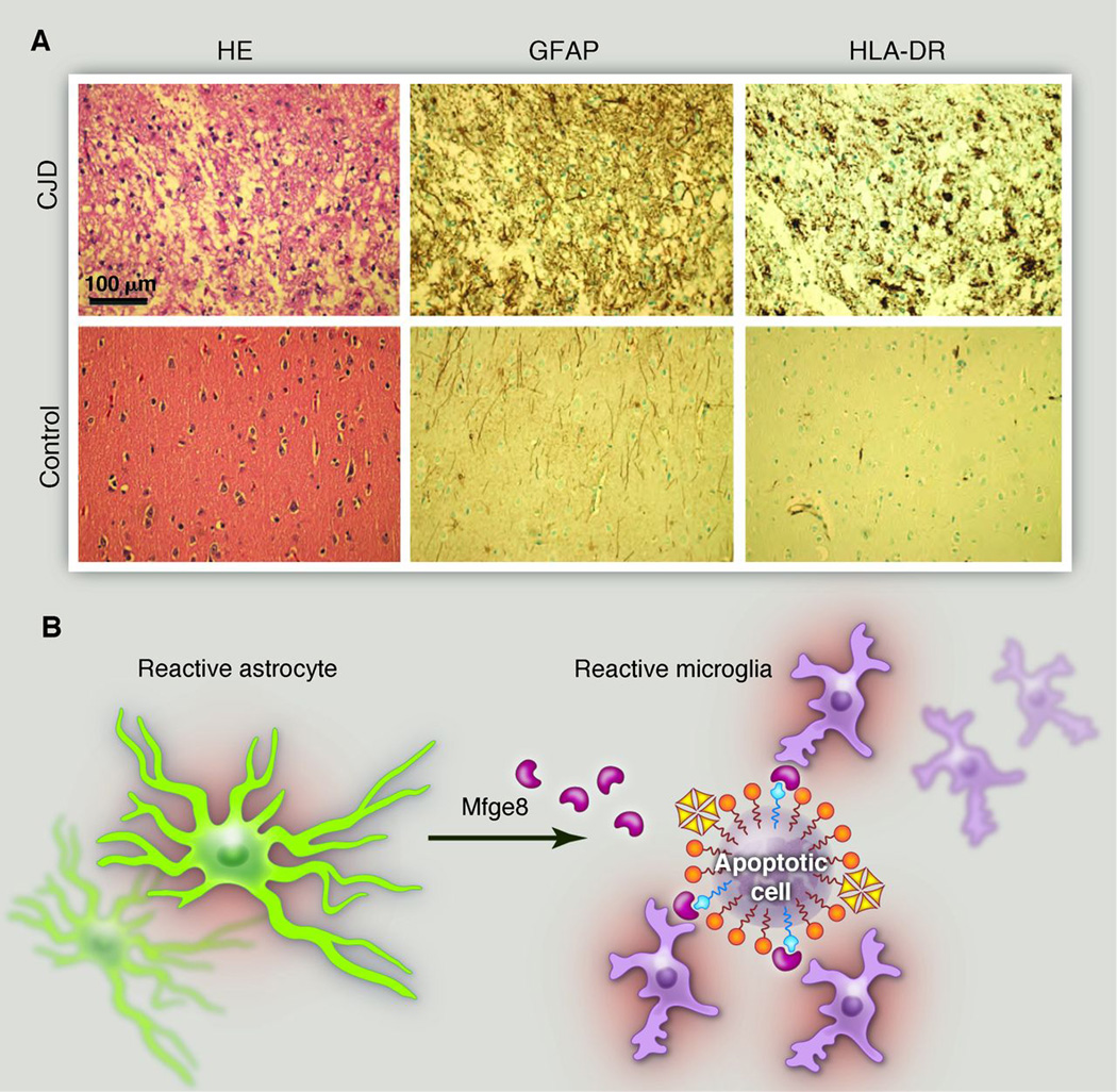
Microglia and prions. (A) The cerebral cortex of a patient who died of Creutzfeldt-Jakob disease (CJD). The pyramidal cells are almost entirely obliterated and replaced by a monoculture of microglia and, to a lesser extent, astrocytes. Note the pseudolaminar pattern of vacuolation (“status spongiosus”), which indicates a ribbon of pathological prion deposition. HE, hematoxylin and eosin staining; GFAP, glial fibrillary acidic protein (an astrocyte marker); HLA-DR, human leukocyte antigen class II. (B) By virtue of its dual binding domains for both phosphatidyl serine and integrins, the secreted factor Mfge8 can bridge apoptotic cells and phagocytes (56). The excessive accumulation of PrPSc in Mfge8-deficient mice infected with prions suggests that microglia use the Mfge8 pathway to clear aggregated and misfolded proteins (38). Mfge8 is produced by astrocytes in the brain and by follicular dendritic cells in lymphoid organs, demonstrating that stromal cells secrete a local “licensing factor” that arms hematopoietic cells and enables the disposal of toxic detritus (57, 58).

Microglia prune synapses during development in a complement-dependent manner. Segregation of eye-specific inputs in the LGN requires the expression of CR3 (A) and C3. KO, knockout. Scale bar, 100 µm. (B) Microglia in the developing LGN contain phagocytosed synaptic elements and cholera toxin B subunit–labeled axons from both the contralateral and ipsilateral eyes, except in mice with deficient C3 signaling. [(A) and (B) are reproduced with permission from Neuron (41)] (C) This developmental process, in which an unknown factor released by astrocytes induces expression of C1q by neurons and microglia to promote C3-dependent synapse elimination, may be reactivated in neurodegenerative disease. [Adapted from (43)]
References
-
- Celsus AC. De Medicina. Firenze, Italy: Nicolaus Laurentii; 1478.
-
- Cartier N, et al. Science. 2009;326:818. - PubMed
-
- Callaway EE. Nature. 2012;489:13. - PubMed
-
- Schweitzer PJ, Fallon BA, Mann JJ, Kumar JSD. Drug Discov. Today. 2010;15:933. - PubMed
-
- Alliot F, Godin I, Pessac B. Brain Res. Dev. Brain Res. 1999;117:145. - PubMed
Publication types
MeSH terms
Grants and funding
LinkOut - more resources
Full Text Sources
Other Literature Sources
Medical
