. 2013 Jan 11;339(6116):204-7.
doi: 10.1126/science.1229326.

Ezh2 orchestrates topographic migration and connectivity of mouse precerebellar neurons

Affiliations

Ezh2 orchestrates topographic migration and connectivity of mouse precerebellar neurons

Thomas Di Meglio et al. Science. .

Abstract

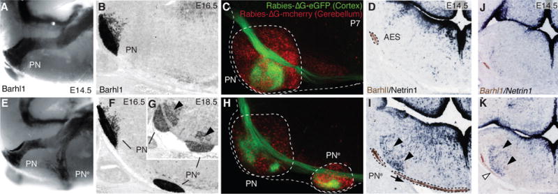
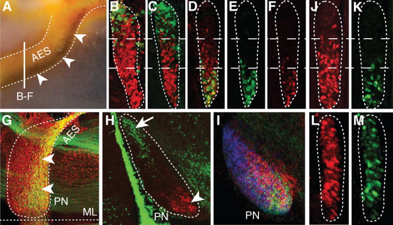
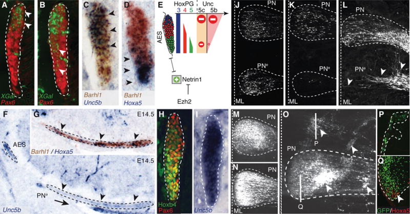
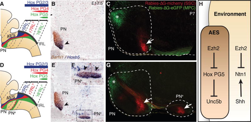
We investigated the role of histone methyltransferase Ezh2 in tangential migration of mouse precerebellar pontine nuclei, the main relay between neocortex and cerebellum. By counteracting the sonic hedgehog pathway, Ezh2 represses Netrin1 in dorsal hindbrain, which allows normal pontine neuron migration. In Ezh2 mutants, ectopic Netrin1 derepression results in abnormal migration and supernumerary nuclei integrating in brain circuitry. Moreover, intrinsic topographic organization of pontine nuclei according to rostrocaudal progenitor origin is maintained throughout migration and correlates with patterned cortical input. Ezh2 maintains spatially restricted Hox expression, which, in turn, regulates differential expression of the repulsive receptor Unc5b in migrating neurons; together, they generate subsets with distinct responsiveness to environmental Netrin1. Thus, Ezh2-dependent epigenetic regulation of intrinsic and extrinsic transcriptional programs controls topographic neuronal guidance and connectivity in the cortico-ponto-cerebellar pathway.


Figures

Fig. 1
Fig. 1. Ezh2 non-cell autonomous role in pontine neuron tangential migration
(A, B, E, F and G) Migratory phenotypes in control (A,B) and r5–6::Cre;Ezh2fl/fl mutants (E–G). Barhl1 in situ hybridization in E14.5 whole-mount (A,E), and E16.5 (B,F), E18.5 (G) sagittal sections. Pontine gray and reticulotegmental (arrowheads, G) nuclei (PN) are duplicated (PNe). (C and H) Tracings from P7 cortex (Rabies-ΔG-eGFP) and cerebellum (Rabies-ΔG-mCherry) in controls (C) and r5–6::Cre;Ezh2fl/fl mutants (H). Pontine (PN) and ectopic (PNe) nuclei are connected to cortex and cerebellum. (D, I, J and K) Barlh1/Netrin1 expression in E14.5 control (D), r5–6::Cre;Ezh2fl/fl (I), r5post::Cre;Ezh2fl/fl;Shhfl/+ (K), r5post::Cre;Ezh2fl/fl;Shhfl/fl (J) coronal sections. Ectopic Ntn1 (arrowheads,I,K) and PNe ectopic migration (arrows,I,K) are partially rescued (J).
Fig. 2
Fig. 2. Intrinsic topography of pontine migratory stream and nuclei and Ezh2 dependent Hox regulation
(A) Hoxb5/Barhl1 in situ hybridization on E15.5 whole-mount brain (lateral view). Arrowheads show Hoxb5+ neuron ventral restriction in anterior extramural stream (AES). (B to C) E15.5 r5–6::Cre;R26RZsGreen AES coronal sections co-stained with ZsGreen/Pax6 (red) (B) or ZsGreen/Hoxb4 (red) (C) showing complementary dorsal ZsGreen+/ventral Hoxb4+ cell distributions (bar in (A) shows section level). (D to F) E15.5 Hoxa5::Cre;R26RZsGreen AES sections showing partially overlapping ZsGreen/Hoxb4 (red) co-stainings with offset dorsal limits (D), while ZsGreen+ (E) and Hoxa5+ (F) neurons display similar ventral restriction. (G to H) E15.5 whole-mount Hoxa5::Cre;R26RZsGreen (G) or r5–6::Cre;R26RZsGreen (H) sagittal sections co-stained with ZsGreen/Pax6 (red) or ZsGreen/Hoxa5 (red), respectively, illustrating posterior pontine nuclei (PN) restriction of Hoxa5+ neurons (arrowheads, G,H), and anterior restriction of r6-derived neurons (arrow, H). (I) Hoxb3/Hoxb4/Hoxb5 nested in situ expression patterns on PN sagittal section (J to M) Hoxb4 (red) and Hoxa5 (green) immunostainings of E15.5 Wnt1::Cre;Ezh2fl/+ control (J,K) and Wnt1::Cre;Ezh2fl/fl mutant (L,M) AES. In (L,M), Hoxb4 and Hoxa5 loose their spatial restriction and are ectopically derepressed up to the dorsal edge of the AES. ML, ventral midline.
Fig. 3
Fig. 3. Ezh2 and Hox dependent regulation of Unc5b in pontine neuron migration
(A to B) X-gal (green)/Pax6 (red) co-stainings of E14.5 Unc5bbGal/+ heterozygotes (A) and Unc5bbGal/bGal homozygotes (B) showing X-gal-stained cell distribution in anterior extramural stream (AES) (arrowheads). (C to E) Unc5b/Barhl1 (C) and Hoxa5/Barhl1 (D) in situ hybridization in E14.5 AES showing complementary dorsoventral expression of Unc5b and Hoxa5 (arrowheads) and summary (E). (F to G) In r5–6::Cre;Ezh2fl/fl mutants, ectopic pontine nuclei (PNe) migrating neurons are Unc5b-negative (F) and Hoxa5+/Barhl1+ (G) (arrowheads). (H to I) In E14.5 Hoxa5−/−/Hoxb5−/−/Hoxc5−/− AES, Unc5b is up-regulated ventrally (I), while Hoxb4/Pax6 are normally expressed (H). (J to K) In utero electroporation (EP) in E14.5 r5–6::Cre;Ezh2fl/fl mutants of Unc5b/Unc5c/eGFP strongly reduces at E18.5 ectopically migrating PNe neurons (K), as compared to EP of eGFP (J), partially rescuing the phenotype. (L to Q) In E13.5 wild type, EP of Ntn1 results in posterior ectopic pontine neuron migration at E17.5, phenocopying r5–6::Cre;Ezh2fl/fl mutants (arrowheads, L). While EP at E13.5 of eGFP (M) or Unc5c/eGFP has no apparent effect on migration at E18.5 (N), EP of Unc5b/eGFP results in anterior ectopic migration and/or dorsal-lateral arrest (arrowheads, O). Immunostaining on sagittal sections shows that anterior ectopic GFP+/Unc5b+ electroporated cells are Hoxa5-negative (P); Hoxa5+ cells are normally restricted in posterior PN (Q).
Fig. 4
Fig. 4. Pontine nuclei regionalization and patterned cortical input
(A, B, D, E and F), Hox expression summary in migrating pontine neurons of control (A) and r5–6::Cre;Ezh2fl/fl mutants (D). Barhl1/Hoxb5 in situ hybridization on E17.5 sagittal sections (B,E,F). In r5–6::Cre;Ezh2fl/fl mutants (E,F), Hoxb5+ neurons spread throughout the rostrocaudal extent of the ectopic nucleus (PNe) (arrowheads, F), while in PN they are normally posteriorly restricted as in control (arrowhead, B,E). (C and G) Rabies-ΔG viruses injected in control visual/medioposterior (MPC) and somatosensory (SSC) cortex anterogradely trace fibers into anterior (green,*) and posterior (red, arrow) PN (C), respectively. In r5–6::Cre;Ezh2fl/fl mutants (G), PNe lacks innervation by MPC, while is innervated by SSC (arrow). H, Ezh2- and Hox-dependent genetic circuitry of intrinsic and extrinsic Unc5b/Ntn1 regulation.

References

    1. Lumsden A, Krumlauf R. Science. 1996;274:1109–1115. - PubMed
    1. Tümpel S, Wiedemann LM, Krumlauf R. Curr Top Dev Biol. 2009;88:103–137. - PubMed
    1. Farago AF, Awatramani RB, Dymecki SM. Neuron. 2006;50:205–218. - PubMed
    1. Altman J, Bayer SA. J Comp Neurol. 1987;257:529–552. - PubMed
    1. Rodriguez CI, Dymecki SM. Neuron. 2000;27:475–486. - PubMed

Publication types

MeSH terms

LinkOut - more resources