. 2013 Feb;4(1):111-8.
doi: 10.1007/s13244-012-0212-x. Epub 2013 Jan 10.

Desmoplastic small round cell tumour: the radiological, pathological and clinical features

Affiliations

Desmoplastic small round cell tumour: the radiological, pathological and clinical features

Robert Thomas et al. Insights Imaging. 2013 Feb.

Abstract

Objectives: Desmoplastic small round cell tumours (DSRCTs) are rare aggressive tumours of young adults that present late and have poor prognosis. This review discusses distinctive radiological features, histopathology and clinical course of this soft-tissue sarcoma.

Methods: From 1991 to 2012, the radiology of 20 patients with pathologically proven DSRCT was independently reviewed by two experienced radiologists. The clinical presentation, treatment and outcome were recorded.

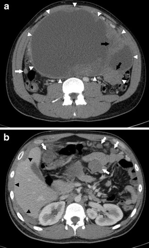
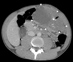
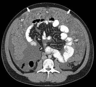
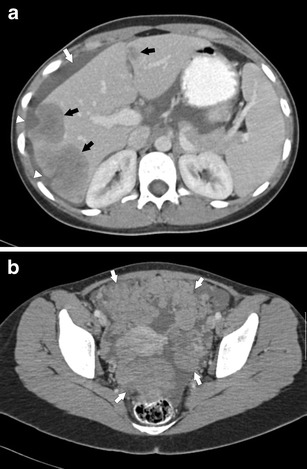
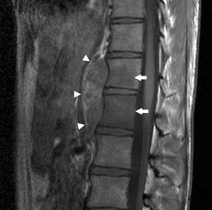
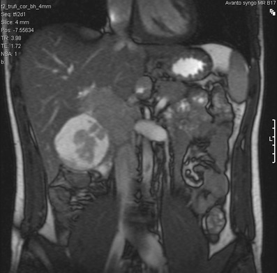
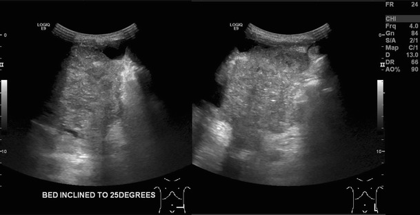
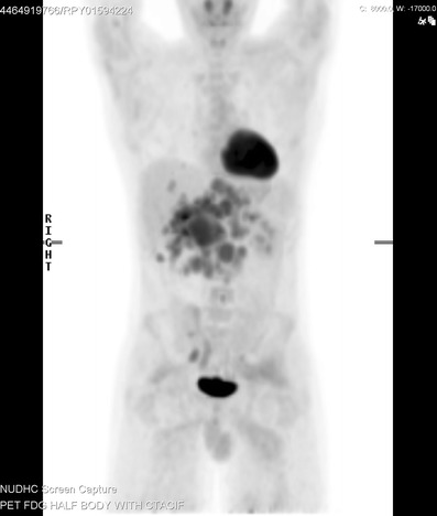
Patients: 16 men, four women; mean age 28.3 years. Computed tomography (CT) demonstrated peritoneal/omental masses without an organ of origin (94 %), with the majority of cases demonstrating large (>5 cm) dominant soft-tissue deposit (80 %) with multiple smaller foci. CT and magnetic resonance imaging (MRI) typically demonstrated heterogeneous soft-tissue enhancement with cystic degeneration. A minority (20 %) demonstrated calcification. Lymph node enlargement occurred in 50 % of cases. Distant metastatic disease occurred in 25 %. Painful abdominal masses were clinically predominant. Treatment strategies include combination chemotherapy with debulking surgery and/or radiotherapy. Median survival from diagnosis was 22.8 months.

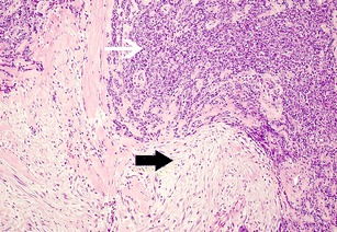
Conclusion: Features of multifocal peritoneal/omental masses, usually in combination with a dominant soft tissue deposit, are distinctive in this rare sarcoma. CT/MRI defines the extent of disease and characterises supporting imaging findings. Prolific desmoplastic reaction histologically separates DSRCT from similar subtypes. Combination treatment strategies can infer a survival benefit but prognosis remains poor. TEACHING POINTS : • DSRCTs are rare tumours of young adults (mean age 28.3 years) with a male predominance (4:1). • Painful abdominal masses clinically predominate. Non-specific features of malignancy can be present. • Multifocal peritoneal masses with a dominant soft tissue lesion is a distinctive imaging finding. • A large desmoplastic reaction differentiates DSRCTs from histologically similar round cell subtypes. • Despite debulking surgery with adjuvant chemotherapy, median survival from diagnosis is 22.3 months.


Figures

Fig. 1
Fig. 1
a Axial CECT of the abdomen in a 24-year-old man with DSRCT. There is a large, heterogeneous peritoneal mass in the abdominal cavity (white arrowheads). It is predominantly cystic but has solid enhancing tissue within it (black arrows). Peritoneal thickening and a peritoneal soft tissue nodule are seen separate to the mass (white arrow). b Axial CECT of the abdomen in a 24-year-old man with DSRCT. There are multiple mesenteric and peritoneal soft tissue nodules (white arrows). There is diffuse peritoneal thickening scalloping the edges of the liver (black arrowheads)
Fig. 2
Fig. 2
Axial CECT of the abdomen in a 25-year-old man with DSRCT. There is a large heterogeneous, mixed solid and cystic mass in the left upper quadrant (white arrowheads). The soft tissue component enhances. There is calcification within it (black arrow)
Fig. 3
Fig. 3
Axial CECT of the abdomen and pelvis in a 23-year-old male patient with DSRCT. There is diffuse omental thickening (white arrows) as well as moderate volume of ascites (black arrow)
Fig. 4
Fig. 4
a Axial CECT of the abdomen in a 22-year-old female patient with DSRCT. There are multiple hypo-attenuating, heterogeneous liver metastases (black arrows). There is diffuse peritoneal thickening scalloping the liver edges (white arrowheads). There is small volume ascites (white arrow). b Axial CECT of the pelvis in a 22-year-old woman with DSRCT. There are multiple peritoneal soft tissue nodules in the pelvis (white arrows)
Fig. 5
Fig. 5
Sagittal T1-weighted MRI of the lower thoracic spine in a 20-year-old man with DSRCT. There is a heterogeneous paravertebral mass lying anterior to the T11 and T12 vertebral bodies (white arrowheads). Whilst there is no direct invasion of the vertebral bodies, there is low signal intensity within the T11 and T12 vertebral bodies in keeping with bony metastatic disease (white arrows)
Fig. 6
Fig. 6
Coronal T2-weighted MRI of the abdomen in a 21-year-old man with DSRCT. There is a complex lesion in the gallbladder fossa, which demonstrates a measurable soft tissue component with a peripheral myxoid degeneration. There is no direct invasion into the adjacent right lobe of liver. The soft tissue returns signal that is marginally higher than the adjacent hepatic parenchyma and isointense to the spleen, with characteristic T2 fluid signal returned from the cystic component
Fig. 7
Fig. 7
Desmoplastic small round cell tumour. Histology shows sheets of tumour composed of small round cells (white arrow) which are surrounded by prominent sclerotic fibrous stroma (black arrow). (Haematoxylin and eosin, ×100)
Fig. 8
Fig. 8
Two trans abdominal ultrasound images acquired in the transverse and longitudinal plane in a 29-year-old man with DSRCT. They demonstrate a heterogeneously echogenic soft tissue mass within the left lower quadrant that is macroscopically inseparable from the adjacent peritoneum and bowel. There is associated ascites
Fig. 9
Fig. 9
Coronal 18F-FDG PET/CT imaging in a 21-year-old male patient with DSRCT. This image demonstrates the extent of disease with multiple areas of FDG uptake within the abdomen, which correspond to peritoneal deposits and retroperitoneal lymph node disease on the CT component. Imaging also confirms the presence of hepatic metastases which were covert on a previous contrast enhanced CT scan

References

    1. Gerald WL, Rosai J. Case 2. Desmoplastic small cell tumor with divergent differentiation Pediatr Pathol. 1989;9(2):177–183. - PubMed
    1. Ordonez NG, Zirkin R, Bloom RE. Malignant small-cell epithelial tumor of the peritoneum coexpressing mesenchymal-type intermediate filaments. Am J Surg Pathol. 1989;13(5):413–421. doi: 10.1097/00000478-198905000-00009. - DOI - PubMed
    1. Kushner BH, LaQuaglia MP, Wollner N, Meyers PA, Lindsley KL, Ghavimi F, et al. Desmoplastic small round-cell tumor: prolonged progression-free survival with aggressive multimodality therapy. J Clin Oncol. 1996;14(5):1526–1531. - PubMed
    1. Quaglia MP, Brennan MF. The clinical approach to desmoplastic small round cell tumor. Surg Oncol. 2000;9(2):77–81. doi: 10.1016/S0960-7404(00)00024-4. - DOI - PubMed
    1. Yachnis AT, Rorke LB, Biegel JA, Perilongo G, Zimmerman RA, Sutton LN. Desmoplastic primitive neuroectodermal tumor with divergent differentiation. Broadening the spectrum of desmoplastic infantile neuroepithelial tumors Am J Surg Pathol. 1992;16(10):998–1006. - PubMed

LinkOut - more resources