. 2013 Feb 12;127(6):710-9.
doi: 10.1161/CIRCULATIONAHA.112.116103. Epub 2013 Jan 10.

Molecular imaging of the paracrine proangiogenic effects of progenitor cell therapy in limb ischemia

Affiliations

Molecular imaging of the paracrine proangiogenic effects of progenitor cell therapy in limb ischemia

Jae Choon Ryu et al. Circulation. .

Abstract

Background: Stem cells are thought to enhance vascular remodeling in ischemic tissue in part through paracrine effects. Using molecular imaging, we tested the hypothesis that treatment of limb ischemia with multipotential adult progenitor cells (MAPCs) promotes recovery of blood flow through the recruitment of proangiogenic monocytes.

Methods and results: Hind-limb ischemia was produced in mice by iliac artery ligation, and MAPCs were administered intramuscularly on day 1. Optical imaging of luciferase-transfected MAPCs indicated that cells survived for 1 week. Contrast-enhanced ultrasound on days 3, 7, and 21 showed a more complete recovery of blood flow and greater expansion of microvascular blood volume in MAPC-treated mice than in controls. Fluorescent microangiography demonstrated more complete distribution of flow to microvascular units in MAPC-treated mice. On ultrasound molecular imaging, expression of endothelial P-selectin and intravascular recruitment of CX(3)CR-1-positive monocytes were significantly higher in MAPC-treated mice than in the control groups at days 3 and 7 after arterial ligation. Muscle immunohistology showed a >10-fold-greater infiltration of monocytes in MAPC-treated than control-treated ischemic limbs at all time points. Intravital microscopy of ischemic or tumor necrosis factor-α-treated cremaster muscle demonstrated that MAPCs migrate to perimicrovascular locations and potentiate selectin-dependent leukocyte rolling. In vitro migration of human CD14(+) monocytes was 10-fold greater in response to MAPC-conditioned than basal media.

Conclusions: In limb ischemia, MAPCs stimulate the recruitment of proangiogenic monocytes through endothelial activation and enhanced chemotaxis. These responses are sustained beyond the MAPC lifespan, suggesting that paracrine effects promote flow recovery by rebalancing the immune response toward a more regenerative phenotype.


Conflict of interest statement

CONFLICT OF INTEREST DISCLOSURES: Drs. Ting and Woda, and Mr. Lehman are employees of Athersys, Inc.

Figures

Figure 1
Figure 1
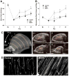
Microvascular perfusion imaging and fluorescent microangiography. (A) Mean (±SEM) microvascular blood flow in the hindllimb adductor muscles at baseline (BL) and after arterial ligation. *p<0.05 vs both control groups (B) Mean (±SEM) microvascular blood volume in the hindlimb adductor muscles. *p<0.05 vs both control groups. (C) Three-dimensional ultrasound composite image illustrating the distribution of MAPC injectate from the contrast-enhanced color-coded regions in the individual 1 mm elevational planes denoted 1 through 4. (D) Examples of fluorescent microangiograms from a PBS-treated control and MAPC-treated limb 7 days after ligation using similar optical settings. The number of subjects for each data point is listed in the Supplement Table.
Figure 2
Figure 2
Mean (±SEM) photon flux from luciferase activity (log scale) after injection of MAPC transfected with firefly luciferase in (A) immune competent, and (B) NK-deficient SCID mice (n=6 for each condition). The images illustrate examples of optical imaging of MAPC luciferase activity localized at the site of injection in the proximal ischemic limb over time.
Figure 3
Figure 3
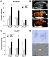
Molecular imaging for P-selectin and CX3CR-1. (A) Mean (±SEM) signal intensity from CEU molecular imaging with P-selectin-targeted microbubbles in control non-injected, sham PBS-injected, and MAPC-treated limbs. (B) Examples of P-selectin targeted imaging in the transvers-axis plane at day 7 illustrate the spatial distribution of signal in the proximal adductor muscle group. The B-mode 2-D image for the MAPC-treated mouse is provided at the top for anatomic reference (femoral acoustic shadowing at the right of the image). (C) Mean (±SEM) signal intensity from CEU molecular imaging with CX3CR-1-targeted microbubbles. (D) Microscopy showing attachment of CX3CR-1-targeted microbubbles (dark spheres) to murine homotypic monocyte aggregates (top); and lack of microbubble (fluorescently labeled green with DiO) attachment to a Ly-6chi (phycoerythrin-positive) monocyte. Scale bar = 10 μm. *p<0.05 vs. control and PBS; †p<0.05 vs. PBS. Number of animals for each group is provided in the Supplement Table.
Figure 4
Figure 4
Intravital microscopy data. (A) Illustration of a venule (delineated by dashed line) under transillumination (left) and fluorescent epi-illumination (right) illustrating perivascular localization of Di-I-labeled MAPC two days after intrascrotal injection of TNF-α and MAPC. (B) Mean (±SEM) leukocyte rolling flux fraction in cremasteric venules from animals treated with TNF-α alone (control, n=6) and TNF-α and MAPC (n=7) two days earlier. *p<0.05 vs control. (C) Histogram and median leukocyte rolling velocities in venules for control and MAPC-treated animals. Mann-Whitney p<0.05. (D) Image illustrating a perivenular Di-I-labeled MAPC (arrowhead) with extravasated or intravascular adhered leukocytes (arrows) in proximity. Scale bar = 20 μm.
Figure 5
Figure 5
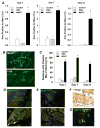
Monocyte immunohistochemistry. (A) Quantitative results of the area staining positive for monocyte Mac-2 from day 3, 7, and 21 (note different y-axis scales) (n=3 for each condition with >20 sections per subject analyzed). (B) Examples of Mac-2 staining at day 7 from MAPC-treated and sham PBS-treated ischemic muscle. (C) Percent of sections demonstrating any Mac-2-positive cells (≥80 fields for each condition). Histology from muscle at day 3 illustrating separate examples of arginase-positive (Arg1) cells (green) in MAPC-treated muscle (D) which were largely absent in untreated muscle (E). (F) Example of positive CX3CR-1 staining of monocytes from a MAPC-treated limb on day 3. (G) Example illustrating colocalization of Mac-2-positive monocytes with a Di-labeled MAPC. *p<0.05 vs. both control groups.

References

    1. Nahrendorf M, Swirski FK, Aikawa E, Stangenberg L, Wurdinger T, Figueiredo JL, Libby P, Weissleder R, Pittet MJ. The healing myocardium sequentially mobilizes two monocyte subsets with divergent and complementary functions. J Exp Med. 2007;204:3037–3047. - PMC - PubMed
    1. Ito WD, Arras M, Winkler B, Scholz D, Schaper J, Schaper W. Monocyte chemotactic protein-1 increases collateral and peripheral conductance after femoral artery occlusion. Circ Res. 1997;80:829–837. - PubMed
    1. Heil M, Ziegelhoeffer T, Wagner S, Fernandez B, Helisch A, Martin S, Tribulova S, Kuziel WA, Bachmann G, Schaper W. Collateral artery growth (arteriogenesis) after experimental arterial occlusion is impaired in mice lacking cc-chemokine receptor-2. Circ Res. 2004;94:671–677. - PubMed
    1. Milch HS, Schubert SY, Hammond S, Spiegel JH. Enhancement of ischemic wound healing by inducement of local angiogenesis. Laryngoscope. 2010;120:1744–1748. - PubMed
    1. Arras M, Ito WD, Scholz D, Winkler B, Schaper J, Schaper W. Monocyte activation in angiogenesis and collateral growth in the rabbit hindlimb. J Clin Invest. 1998;101:40–50. - PMC - PubMed

Publication types

MeSH terms