. 2013 Apr 1;19(7):1773-83.
doi: 10.1158/1078-0432.CCR-12-1281. Epub 2013 Jan 10.

Gene expression profile identifies tyrosine kinase c-Met as a targetable mediator of antiangiogenic therapy resistance

Affiliations

Gene expression profile identifies tyrosine kinase c-Met as a targetable mediator of antiangiogenic therapy resistance

Arman Jahangiri et al. Clin Cancer Res. .

Abstract

Purpose: To identify mediators of glioblastoma antiangiogenic therapy resistance and target these mediators in xenografts.

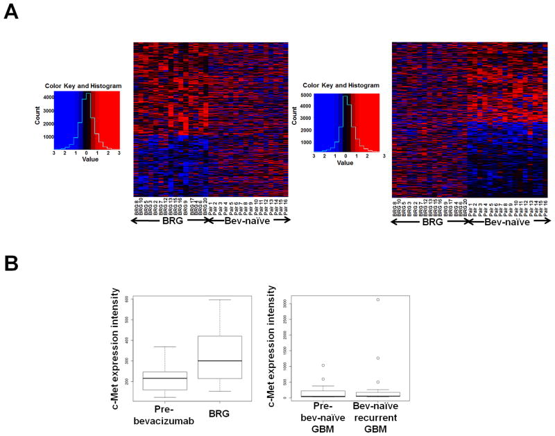
Experimental design: We conducted microarray analysis comparing bevacizumab-resistant glioblastomas (BRG) with pretreatment tumors from the same patients. We established novel xenograft models of antiangiogenic therapy resistance to target candidate resistance mediator(s).

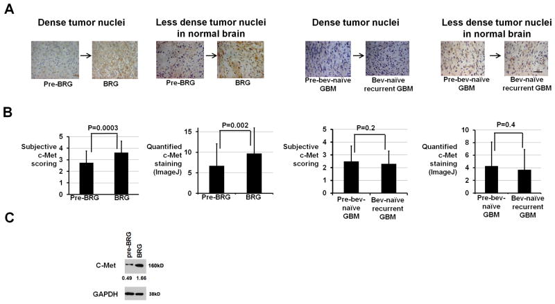
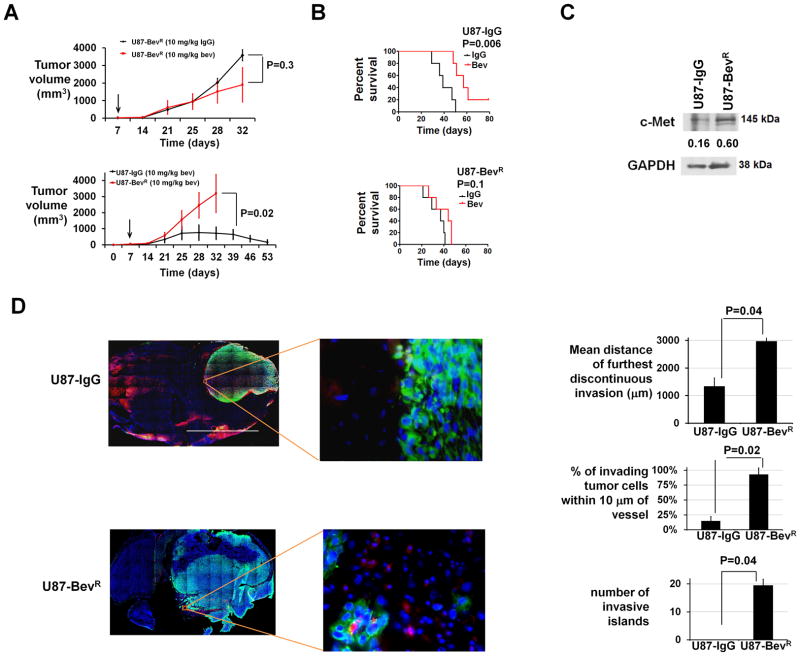
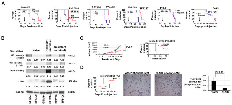
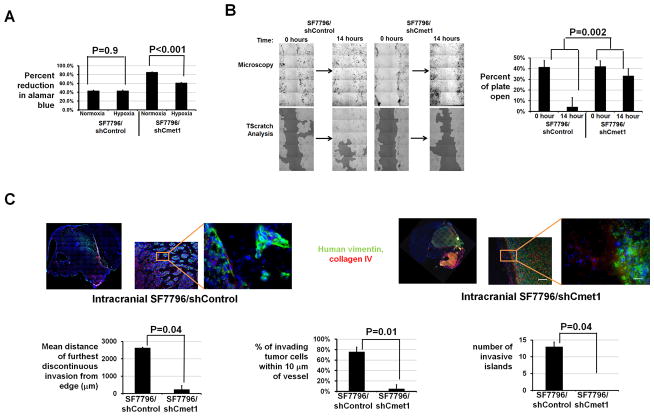
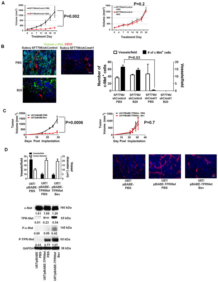
Results: BRG microarray analysis revealed upregulation versus pretreatment of receptor tyrosine kinase c-Met, which underwent further investigation because of its prior biologic plausibility as a bevacizumab resistance mediator. BRGs exhibited increased hypoxia versus pretreatment in a manner correlating with their c-Met upregulation, increased c-Met phosphorylation, and increased phosphorylation of c-Met-activated focal adhesion kinase and STAT3. We developed 2 novel xenograft models of antiangiogenic therapy resistance. In the first model, serial bevacizumab treatment of an initially responsive xenograft generated a xenograft with acquired bevacizumab resistance, which exhibited upregulated c-Met expression versus pretreatment. In the second model, a BRG-derived xenograft maintained refractoriness to the MRI tumor vasculature alterations and survival-promoting effects of bevacizumab. Growth of this BRG-derived xenograft was inhibited by a c-Met inhibitor. Transducing these xenograft cells with c-Met short hairpin RNA inhibited their invasion and survival in hypoxia, disrupted their mesenchymal morphology, and converted them from bevacizumab-resistant to bevacizumab-responsive. Engineering bevacizumab-responsive cells to express constitutively active c-Met caused these cells to form bevacizumab-resistant xenografts.

Conclusion: These findings support the role of c-Met in survival in hypoxia and invasion, features associated with antiangiogenic therapy resistance, and growth and therapeutic resistance of xenografts resistant to antiangiogenic therapy. Therapeutically targeting c-Met could prevent or overcome antiangiogenic therapy resistance.


Conflict of interest statement

Conflicts of interest: W.S.C. is co-Founder, CEO, and shareholder of OncoSynergy. M.K.A. is a Scientific Advisory Board Member and shareholder of OncoSynergy.

Figures

Figure 1
Figure 1. Transcriptional analysis of BRGs and bevacizumab-naïve recurrent GBMs versus paired earlier GBMs from the same patients
(A) Microarray analysis of 15 BRGs and their paired pre-treatment GBMs, along with 16 bevacizumab-naïve recurrent GBMs and their paired earlier GBMs produced heatmaps in which the 1000 most differentially expressed genes in BRGs (left) or bevacizumab-naïve recurrent GBMs (right) did not exhibit similar alteration patterns in the opposite group. Red/blue=increased/decreased gene expression. (B) Box and whisker plot summarizing microarray c-Met expression changes. Upon recurrence, BRGs exhibited nearly 40% c-Met upregulation, while bevacizumab-naïve recurrent GBMs exhibited unchanged c-Met. Plots show lower extreme (horizontal line below box), lower quartile (box base), median (box center), upper quartile (box roof), upper extreme (horizontal line above box), and outliers (dots outside extremes) for BRGs and their paired “pre-bevacizumab” GBMs (left), along with bevacizumab-naïve (“bev-naïve”) recurrent GBMs and their paired “pre-bev-naïve” GBMs (right).
Figure 2
Figure 2. C-Met upregulation in BRGs versus pre-treatment
(A) Shown are representative c-Met immunostainings from areas of dense tumor nuclei or less dense tumor nuclei in normal brain (630x; scale bar=10 μm). (B) Both subjective and automated ImageJ scoring revealed increased c-Met staining after bevacizumab resistance (P=0.0003–0.002) with unchanged c-Met staining after bevacizumab-naïve recurrence (P=0.2–0.4). (C) Western blot revealed 3.4-fold increased c-Met expression in a BRG versus the pre-treatment GBM from the same patient. Numbers represent band densities normalized to GAPDH from the same sample.
Figure 3
Figure 3. Xenograft model of acquired anti-angiogenic therapy resistance
(A) Subcutaneous U87-BevR was unresponsive to bevacizumab versus IgG (P=0.3; upper left), and subcutaneous U87-IgG tumors regressed during bevacizumab treatment while U87-BevR grew exponentially during bevacizumab treatment (P<0.05; lower left). Arrows indicate time bevacizumab treatment began. (B) U87-IgG and U87-BevR xenografts were responsive (P=0.006) and resistant (P=0.1), respectively, to bevacizumab in the orthotopic intracranial environment. (C) Western blot revealed 4-fold more c-Met in U87-BevR than U87-IgG cells. Numbers represent band densities normalized to GAPDH from the same sample. (D) Staining intracranial U87-IgG and U87-BevR xenografts for collagen IV and human vimentin revealed further discontinuous invasion, higher percentage of invasive cells 10 μm from a vessel, and more invasive islands in U87-BevR than U87-IgG (P<0.05). 2x/400x; scale bar=2000/20 μm
Figure 4
Figure 4. Xenograft model of intrinsic anti-angiogenic therapy resistance exhibits growth suppression with pharmacologic c-Met blockade
(A) While immunodeficient mice with intracranial U87, SF8557, SF7300, or SF7227 xenografts exhibited prolonged survival with B20-4.1.1 or bevacizumab versus ragweed control antibody or PBS treatment (n=10/group U87 and SF8557, n=5/group SF7300 and SF7227; P=0.0007 U87, P=0.0009 SF8557, P=0.002 SF7300, P=0.003 SF7227), B20-4.1.1 did not affect survival of mice with intracranial xenografts derived from a GBM with intrinsic (SF8244) or acquired (SF7796) bevacizumab resistance (n=10/group; P=0.4–0.9). Arrows indicate time of treatment initiation. (B) Western blot showing c-Met, human HGF, and mouse HGF expression of 3 xenografts from bevacizumab-naïve GBMs (SF7227, SF7300, and GBM14) and from GBMs with intrinsic (SF8244) or acquired (SF7796 and SF8106) bevacizumab resistance. Numbers represent band densities normalized to GAPDH from the same sample. (C) C-met inhibitor XL184 prevented volumetric growth of subcutaneous SF7796 xenografts after 4 treatment weeks (P=0.01; upper left; n=5/group) and prolonged survival (P<0.0001; upper right). (D) XL184 prolonged survival of mice with intracranial SF7796 xenografts (n=6/group; P=0.01) (lower left). Immunohistochemistry (lower right) revealed reduced phosphorylated c-Met in XL184-treated versus water-treated intracranial SF7796 xenografts (P=0.03). 630x; scale bar=10 μm.
Figure 5
Figure 5. Effects of c-Met shRNA on survival in hypoxia, in vitro migration, and in vivo invasion of cells from bevacizumab-resistant xenografts
(A) Percent Alomar blue reduction, indicating cell survival, was less in SF7796/shCmet1 in hypoxia versus normoxia (P<0.001), but unchanged in SF7796/shControl in hypoxia versus normoxia (P=0.9). (B) After 14 hours, scratches were 91% filled with SF7796/shControl cells, while only 21% scratch reduction occurred with SF7796/shCmet1 cells (P=0.002). (C) Intracranial SF7796/shCmet1-derived xenografts (n=3) exhibited reduced invasive distance from continuous tumor edge (P=0.04), percentage of invading tumor cells 10 μm from a vessel (P=0.01), and invasive islands (P=0.04) than intracranial SF7796/shControl tumors (n=3). Red=collagen IV (vessels); blue=Hoechst nuclear stain; green=human vimentin. 2x/100x/400x, scale bar=2000, 60/20μm.
Figure 6
Figure 6. Effects of genetic c-Met alteration on xenograft responsiveness to VEGF blockade
(A) SF7796/shCmet1-derived subcutaneous tumors exhibited B20-4.1.1 responsiveness (n=5/group; P=0.002), while SF7796/shControl-derived subcutaneous tumors exhibited B20-4.1.1 resistance (n=5/group; P=0.2). (B) Immunofluorescence revealed unaltered vessel density in PBS- versus B20-treated subcutaneous SF7796/shControl (P=0.8) and fewer c-Met-positive cells in PBS-treated subcutaneous SF7796/shCmet1 versus PBS-treated subcutaneous SF7796/shControl (P=0.03). B20-4.1.1-treated subcutaneous SF7796/shCmet1 xenografts regressed and could not be analyzed. 200x, scale bar=30 μm. (C) Treating U87/pBABE-Puro-Tpr-met subcutaneous xenografts with bevacizumab (Bev) caused no response versus PBS (n=5/group; P=0.7), while bevacizumab caused U87/pBABE-Puro subcutaneous xenograft regression (n=5/group; P=0.001). Error bars represent standard errors of the mean. (D) Immunohistochemistry revealed that U87/pBABE-Puro-Tpr-met xenografts exhibited fewer (P=0.04), larger (P=0.03) vessels than U87/pBABE-Puro xenografts, with bevacizumab not altering the U87/pBABE-Puro-Tpr-met vascular pattern (P=0.8–0.9). Bevacizumab-treated U87/pBABE-Puro xenografts regressed and could not be analyzed. 200x, scale bar=30 μm. Western blot identified Tpr-met and phosphorylated Tpr-met (P-Tpr-met) in U87/pBABE-Puro-Tpr-met xenografts, but not in U87/pBABE-Puro xenografts. Western blot numbers represent band densities normalized to GAPDH from the same sample.

Comment in

References

    1. Vredenburgh JJ, Desjardins A, Herndon JE, 2nd, Marcello J, Reardon DA, Quinn JA, et al. Bevacizumab plus irinotecan in recurrent glioblastoma multiforme. J Clin Oncol. 2007;25:4722–9. - PubMed
    1. Vredenburgh JJ, Desjardins A, Herndon JE, 2nd, Dowell JM, Reardon DA, Quinn JA, et al. Phase II trial of bevacizumab and irinotecan in recurrent malignant glioma. Clin Cancer Res. 2007;13:1253–9. - PubMed
    1. Jain RK, Duda DG, Clark JW, Loeffler JS. Lessons from phase III clinical trials on anti-VEGF therapy for cancer. Nat Clin Pract Oncol. 2006;3:24–40. - PubMed
    1. Clark AJ, Lamborn KR, Butowski NA, Chang SM, Prados MD, Clarke JL, et al. Neurosurgical management and prognosis of patients with glioblastoma that progress during bevacizumab treatment. Neurosurgery. 2011 - PubMed
    1. Bergers G, Hanahan D. Modes of resistance to anti-angiogenic therapy. Nat Rev Cancer. 2008;8:592–603. - PMC - PubMed

Publication types

MeSH terms

Substances