. 2013 Mar;193(3):785-801.
doi: 10.1534/genetics.112.146522. Epub 2013 Jan 10.

Gene copy-number variation in haploid and diploid strains of the yeast Saccharomyces cerevisiae

Affiliations

Gene copy-number variation in haploid and diploid strains of the yeast Saccharomyces cerevisiae

Hengshan Zhang et al. Genetics. 2013 Mar.

Abstract

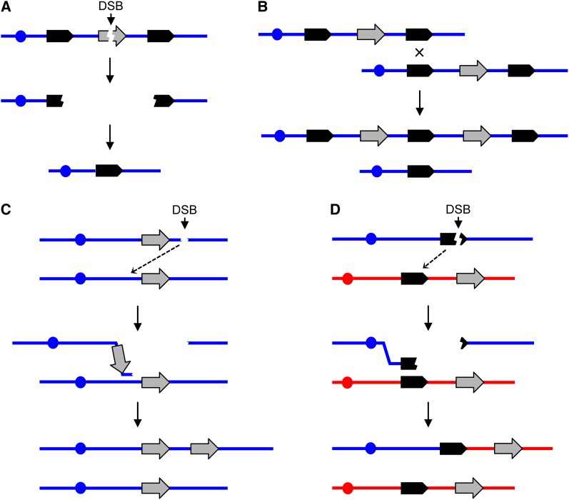
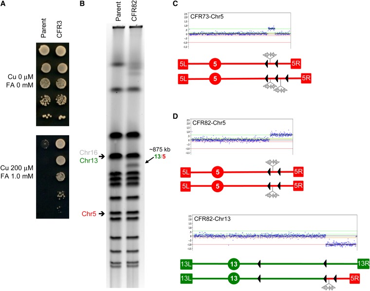
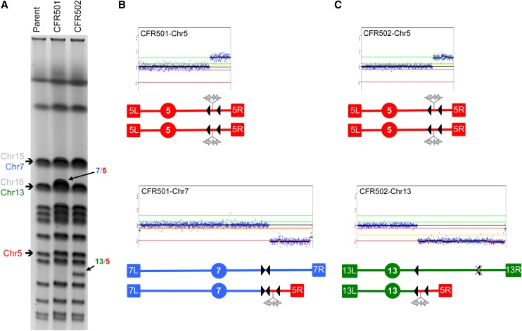
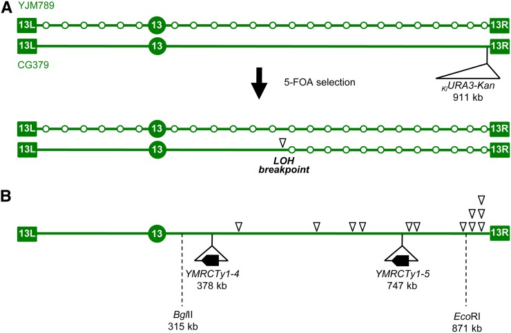
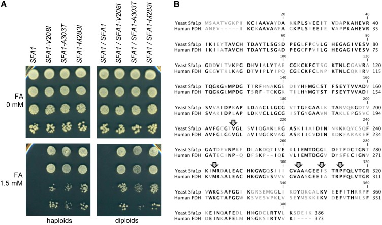
The increasing ability to sequence and compare multiple individual genomes within a species has highlighted the fact that copy-number variation (CNV) is a substantial and underappreciated source of genetic diversity. Chromosome-scale mutations occur at rates orders of magnitude higher than base substitutions, yet our understanding of the mechanisms leading to CNVs has been lagging. We examined CNV in a region of chromosome 5 (chr5) in haploid and diploid strains of Saccharomyces cerevisiae. We optimized a CNV detection assay based on a reporter cassette containing the SFA1 and CUP1 genes that confer gene dosage-dependent tolerance to formaldehyde and copper, respectively. This optimized reporter allowed the selection of low-order gene amplification events, going from one copy to two copies in haploids and from two to three copies in diploids. In haploid strains, most events involved tandem segmental duplications mediated by nonallelic homologous recombination between flanking direct repeats, primarily Ty1 elements. In diploids, most events involved the formation of a recurrent nonreciprocal translocation between a chr5 Ty1 element and another Ty1 repeat on chr13. In addition to amplification events, a subset of clones displaying elevated resistance to formaldehyde had point mutations within the SFA1 coding sequence. These mutations were all dominant and are proposed to result in hyperactive forms of the formaldehyde dehydrogenase enzyme.


Figures

Figure 1
Figure 1
General mechanisms for the generation of deletions and duplications. In this figure, chromosomes are shown as horizontal blue or red lines, DNA repeats as solid arrows, reporter genes as shaded arrows, and centromeres as circles. Single DNA strands are not represented. Variations of these models are also possible. (A) Deletion formation by the single-strand annealing (SSA) pathway. A double-strand DNA break (DSB) between the flanking repeats results in two broken ends that are resected 5′ to 3′. When complementary single-strand regions of the flanking repeats are exposed, reannealing occurs, resulting in loss of the sequences between the repeats (Paques and Haber 1999). (B) Unequal crossovers resulting in deletions and duplications. This homologous recombination event could involve either sister chromatids in haploids or diploids or homologous chromosomes in diploids. (C) Microhomology-mediated replication. A free 3′ end associated with centromere-containing DNA fragment invades a sister chromatid or a homologous chromosome using a nonallelic microhomology sequence. The subsequent break-induced replication (BIR) event generates a duplication in which the breakpoints share little sequence homology. It is assumed that the DNA fragment without a centromere is lost. (D) Coupled terminal duplication and deletion resulting in a nonreciprocal translocation. The different homologs are shown in blue and red. A DSB in or distal to a repeat in the blue chromosome is repaired by a BIR event using a repeat on a nonhomologous chromosome (red). This event results in a duplication of sequences from the red chromosome and a deletion of sequences from the blue chromosome.
Figure 2
Figure 2
Identification of chr5 amplifications in haploids: Segmental duplications. (A) Schematic representation of the region of chr5 where the SFA1–CUP1 CNV reporter was inserted (shaded arrows). The reporter also contained a drug-resistance marker, either Hph (hygromycin B), as shown, or Kan (G418–geneticin). Terminal boxes correspond to the left and right chr5 telomeres (5L and 5R, respectively), and the circle represents CEN5. Bottom, expanded view of the region involved in segmental amplifications (440–502 kb), showing full-length Ty1 elements as solid arrows and solo LTRs as arrowheads according to their orientation. (B) FA/Cu-resistance phenotypic differential between the parental haploid strain (JAY372) containing one copy of the reporter and CFR clones carrying two (CFR20) or three (CFR17) copies. Serial dilutions of these strains were spotted on plates containing Cu and FA at the concentrations indicated to the left. (C) PFGE showing the karyotypes of the parental haploid and three CFR clones carrying different segmental amplifications on chr5. The parental size chr5 is indicated to the left and the segmental amplifications are indicated to the right (5 SD). (D) Detailed view of chr5 array-CGH plots showing the Log2 Cy5/Cy3 signal intensity (copy number) and the rearrangement breakpoints for the three CFRs from C. The plots are aligned directly below to the expanded section of A such that the breakpoints correspond to the repeat sequences. Each blue dot corresponds to the signal of a specific probe in the region. The black horizontal lines correspond to the average signal for probes in a region of amplification. Neutral signal corresponds to no copy-number change (1× in haploids); positive signals corresponding to two copies (∼1.0) or three copies (∼1.5) are shown.
Figure 3
Figure 3
Identification of chr5 amplifications in diploids: chr13/chr5 translocation. Schematic representations are as in Figure 2. Chr5 is shown in red and chr13 is shown in green. (A) FA/Cu-resistance phenotype in the parental diploid strain JAY350 (two reporter copies) compared to a derivative resistant clone CFR3 (three copies). (B) PFGE showing the karyotype of the parental diploid strain JAY350 and of the derivative resistant clone CFR82 carrying the indicated recurrent 875 kb chr13/chr5 translocation. An identical translocation was also present in clone CFR3 shown in A. The parental size chr5 and chr13 are indicated to the left; chr16 comigrates with chr13 and is also indicated. (C) chr5 array-CGH plot (top) and schematic representation (bottom) of the karyotype in the CFR75 clone showing one parental size homolog and one homolog containing a segmental duplication between the YERCTy1-1 and YERCTy1-2 elements (solid arrowheads). (D) Array-CGH plots and schematic representation of chr5 and chr13 in the CFR82 clone. The breakpoints correspond to YERCTy1-1 in chr5 and YMRCTy1-5 in chr13. For position reference, the chr13 centromere-proximal (left) element YMRCTy1-4 is also represented (see also Figures 4C and 5B).
Figure 4
Figure 4
Alternative translocations in diploids lacking the YMRCTy1-5 repeat element. Schematic representations are as in Figure 2. Chr5 is shown in red, chr13 is shown in green, and chr7 is shown in blue. (A) PFGE showing the karyotypes of parent diploid strain JAY510 and of derivative clones CFR501 (containing a chr7/chr5 translocation) and CFR502 (containing a chr13/chr5 translocation). The parental bands for chr5, chr7, and chr13 involved in the rearrangements are indicated to the left; also indicated are chr16 and chr15, which comigrate with chr13 and chr7, respectively. (B) Array-CGH plots and schematic representation of chr5 and chr7 in the CFR501 clone. The breakpoints correspond to YERCTy1-1 in chr5 and YGRWTy2-2/YGRCTy1-3 in chr7. (C) Array-CGH plots and schematic representation of chr5 and chr13 in the CFR502 clone. The breakpoints correspond to YERCTy1-1 in chr5 and YMRCTy1-4 in chr13. Note the deletion of the YMRCTy1-5 element in this strain, represented by the gray X over the normal position of this element (distal/right arrowhead).
Figure 5
Figure 5
Initial mapping of a candidate fragile site on the right arm of chr13. (A) Experimental rationale. Schematic representation of chr13 in the hybrid diploid strain (JAY800 and JAY801), formed by mating haploids of the diverged strain backgrounds YJM789 and CG379. The open circles in the YJM789-derived chromosome represent SNP positions. The KlURA3-Kan marker was inserted in the CG379-derived chromosome, downstream of the ADH6 gene, near the right telomere. Mitotic crossovers initiated by DNA breaks in the CG379-derived chromosome and repaired using the YJM789 homolog as template result in clones that are resistant to 5-FOA and homozygous for the right end of the YJM789-derived chr13. Genotyping with SNP microarrays can determine the precise site of allelic mitotic recombination (LOH breakpoint, open arrowhead). This site should occur in close proximity to the precursor DNA lesion. (B) LOH breakpoint positions for 12 independent 5-FOA-resistant clones determined by SNP microarray genotyping showing a clustering of breakpoints near the right end of chr13. Also indicated are the relative positions of the two Ty1 elements involved in the chr13/chr5 translocations described in Figures 3 and 4 and the position of the SNP–RFLP markers BglII and EcoRI. The EcoRI marker is described in the Results. The genotype at BglII marker site was also determined and found to be heterozygous in most 5-FOAR clones (117/131; 89.3%).
Figure 6
Figure 6
Characterization of dominant mutant alleles of the SFA1 gene. (A) Formaldehyde (FA)-resistance phenotype in haploid (left) and heterozygous diploid (right) strains carrying three different mutant alleles of SFA1. The genotypes are indicated at the top. Serial dilutions were plated in media containing 0 or 1.5 mM FA as indicated to the left. No CuSO4 was added. (B) Amino acid residue sequence alignment between the S. cerevisiae Sfa1p and the human FDH enzymes. Conserved residues (60.9% identity) are shown in black letters, and diverged residues are shown in gray. Shaded arrows point to the five residues that were substituted in the dominant SFA1 mutations.

References

    1. Abrahams B. S., Geschwind D. H., 2008. Advances in autism genetics: on the threshold of a new neurobiology. Nat. Rev. Genet. 9: 341–355. - PMC - PubMed
    1. Argueso J. L., Westmoreland J., Mieczkowski P. A., Gawel M., Petes T. D., et al. , 2008. Double-strand breaks associated with repetitive DNA can reshape the genome. Proc. Natl. Acad. Sci. USA 105: 11845–11850. - PMC - PubMed
    1. Arlt M. F., Wilson T. E., Glover T. W., 2012. Replication stress and mechanisms of CNV formation. Curr. Opin. Genet. Dev. 22: 204–210. - PMC - PubMed
    1. Brewer B. J., Payen C., Raghuraman M. K., Dunham M. J., 2011. Origin-dependent inverted-repeat amplification: a replication-based model for generating palindromic amplicons. PLoS Genet. 7: e1002016. - PMC - PubMed
    1. Brown C. J., Todd K. M., Rosenzweig R. F., 1998. Multiple duplications of yeast hexose transport genes in response to selection in a glucose-limited environment. Mol. Biol. Evol. 15: 931–942. - PubMed

Publication types

Substances

LinkOut - more resources