. 2013 Apr 15;22(8):1525-38.
doi: 10.1093/hmg/ddt003. Epub 2013 Jan 9.

Enzyme replacement therapy rescues weakness and improves muscle pathology in mice with X-linked myotubular myopathy

Affiliations

Enzyme replacement therapy rescues weakness and improves muscle pathology in mice with X-linked myotubular myopathy

Michael W Lawlor et al. Hum Mol Genet. .

Abstract

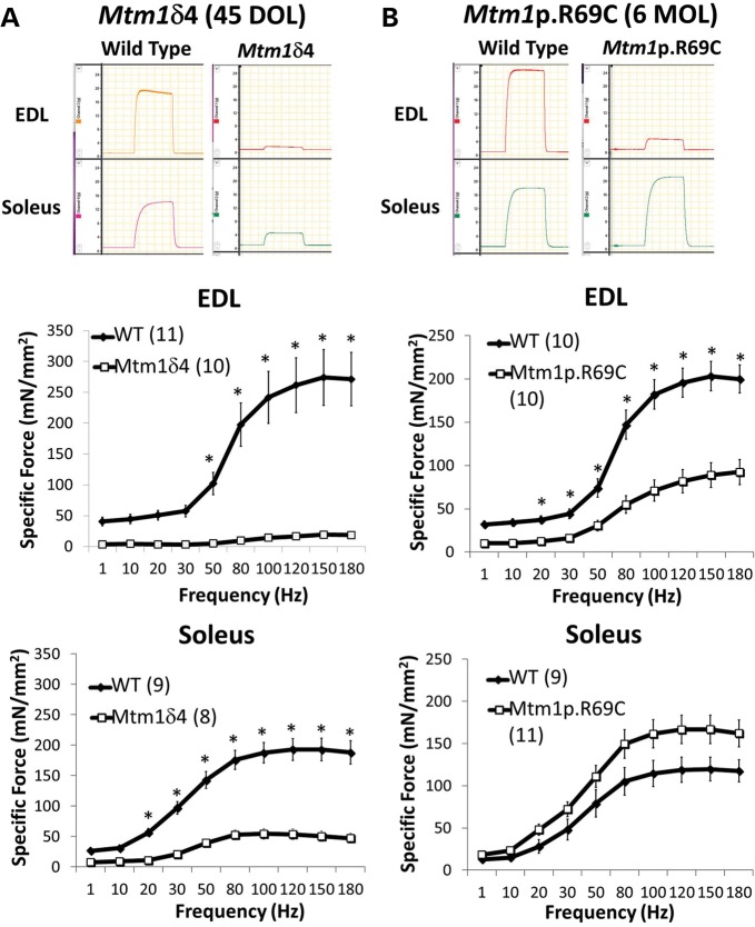
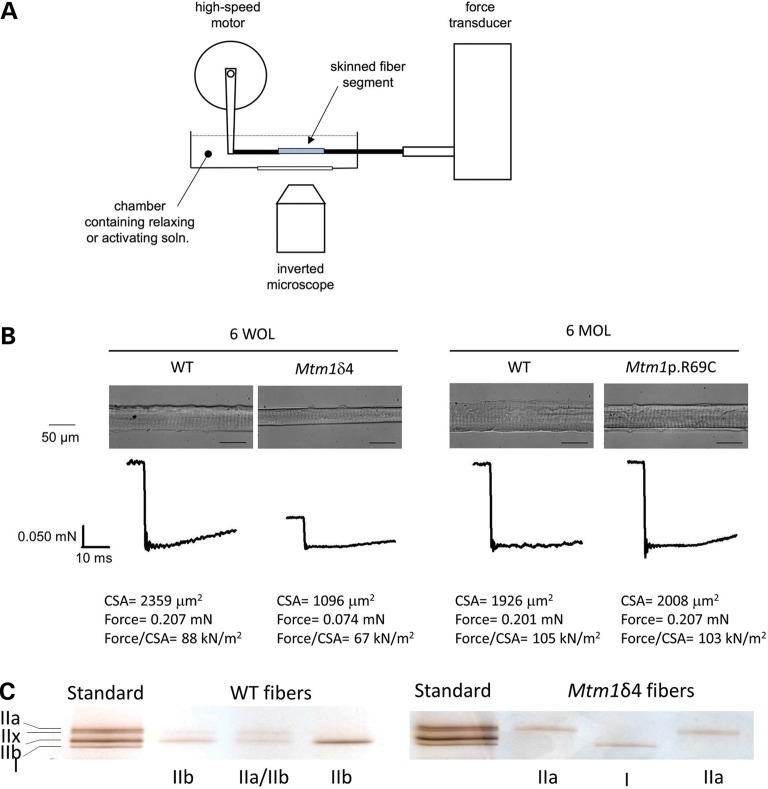
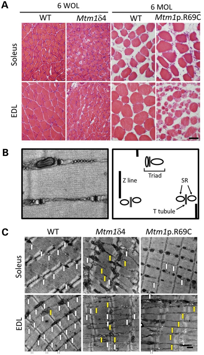
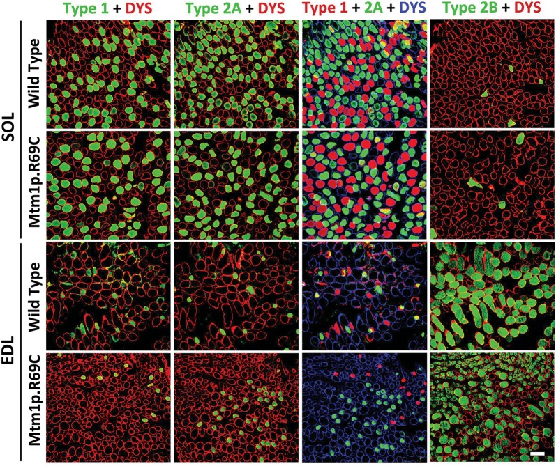
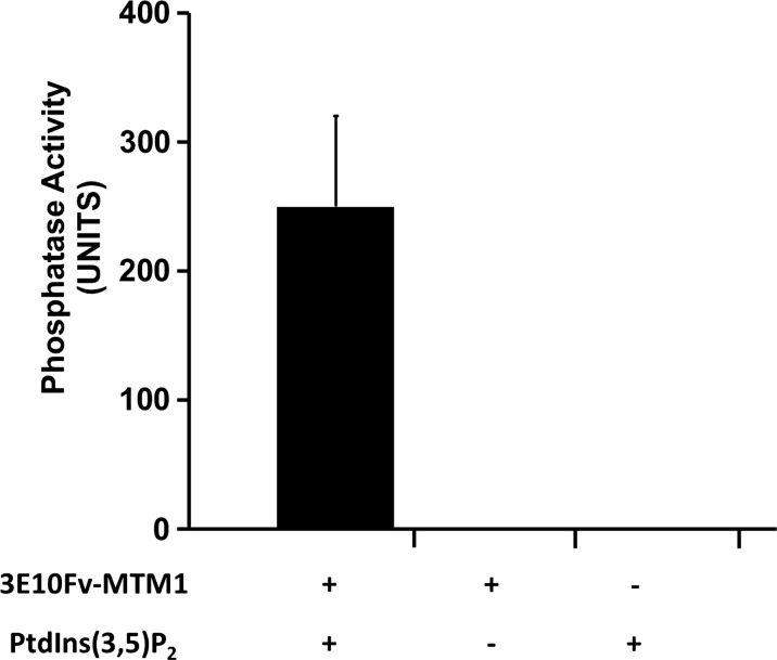
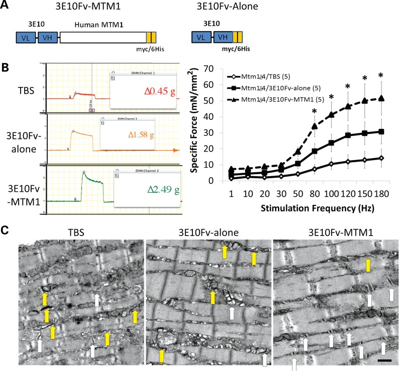
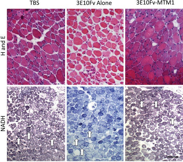
No effective treatment exists for patients with X-linked myotubular myopathy (XLMTM), a fatal congenital muscle disease caused by deficiency of the lipid phosphatase, myotubularin. The Mtm1δ4 and Mtm1 p.R69C mice model severely and moderately symptomatic XLMTM, respectively, due to differences in the degree of myotubularin deficiency. Contractile function of intact extensor digitorum longus (EDL) and soleus muscles from Mtm1δ4 mice, which produce no myotubularin, is markedly impaired. Contractile forces generated by chemically skinned single fiber preparations from Mtm1δ4 muscle were largely preserved, indicating that weakness was largely due to impaired excitation contraction coupling. Mtm1 p.R69C mice, which produce small amounts of myotubularin, showed impaired contractile function only in EDL muscles. Short-term replacement of myotubularin with a prototypical targeted protein replacement agent (3E10Fv-MTM1) in Mtm1δ4 mice improved contractile function and muscle pathology. These promising findings suggest that even low levels of myotubularin protein replacement can improve the muscle weakness and reverse the pathology that characterizes XLMTM.


Figures

Figure 1.
Figure 1.
Contractile performance of isolated EDL and soleus muscles in WT and myotubularin deficient mice. Ex vivo testing of isolated EDL and soleus muscles evaluates contractile performance over a range of stimulation frequencies, which provides a measurement of contractile force using intact muscle tissue and ECC (51). These studies also provide some insight into fiber-type-specific contractile function, since the soleus muscle is highly enriched for oxidative fibers and the EDL muscle is highly enriched for glycolytic fibers. Contractile performance was assessed by measuring the specific force-frequency relationships in the EDL and soleus muscles of (A) 6-week-old WT and Mtm1δ4 mice and (B) 6-month-old WT and Mtm1 p.R69C mice. Representative tracings of tetanic responses recorded at a frequency of 150 Hz depict representative maximum (Max) force in each group. Note that the vertical axis of all force tracings have the same scale, but that these measurements do not correct for differences in the cross-sectional area between muscles. Frequency/specific force (or stress) relationships, which account for both contractile force and normalize for the cross-sectional area, are shown for the EDL and soleus muscles of 6-week-old Mtm1δ4 and WT mice (left). Frequency/specific force relationships are shown for the EDL and soleus muscles of 6-month-old Mtm1 p.R69C and WT mice (right). Numbers of replicate muscles studies are in parentheses. *P < 0.05.
Figure 2.
Figure 2.
Skinned single fiber studies in WT and myotubularin-deficient mice. Chemically skinned single fiber preparations were performed to evaluate contractile performance independent of excitation contraction coupling. Chemically skinned single fiber preparations evaluate the contractile performance of single myofibers using exogenous calcium, which allows the measurement of contractile function independent of ECC. Force measurements can then be assessed with respect to the fiber cross-sectional area (CSA) to evaluate the impact of fiber size on contractile force. (A) Diagram of the equipment used in performing skinned single fiber studies. (B) Representative photographs and force measurements from fibers isolated from WT and myotubularin-deficient mice at 6 weeks of life (WOL) and 6 months of life (MOL). Data from these studies are summarized in Table 1. (C) Following force measurement, the myosin isoform (type I, IIa, IIb or IIx) expressed by the tested fiber was determined by gel electrophoresis and silver staining, and fiber type was determined by comparison with a standard.
Figure 3.
Figure 3.
Light microscopic pathology in myotubularin-deficient mice is dependent on mouse strain and the muscle examined. (A) Hematoxylin and eosin staining of EDL and soleus muscles from Mtm1δ4, Mtm1 p.R69C or age-matched WT littermate mice display variable levels of myofiber smallness and centrally nucleated fibers. Note that differences in fiber size when comparing the two WT populations is due to the normal growth between 6 weeks of life (WOL) and 6 months of life (MOL). (B) To evaluate sarcotubular organization at the ultrastructural level, the number of triads, T-tubules and L-tubules were quantified from longitudinal sections of EDL and soleus muscles from WT and Mtm1δ4 animals at six WOL and WT and Mtm1 p.R69C animals at six MOL. A picture and diagram of the normal sarcotubular architecture from a 6-week-old WT mouse is shown. (C) Representative images (×9300 magnification) of longitudinal sections of 6-week-old WT and Mtm1δ4 mice, which allow the quantification of T-tubules (white arrows) and L-tubules (yellow arrows). Representative images from a 6-month-old Mtm1 p.R69C mouse are also shown for comparison. Bar = 50 μm in (A) and 500 nm in (C).
Figure 4.
Figure 4.
Immunohistochemical evaluation of fiber types in Mtm1p.R69C EDL and soleus muscles. Immunohistochemistry for dystrophin (red or blue) and either type 1, type 2A or 2B (green) myosin reveals the fiber-type populations present within the EDL and soleus muscles of Mtm1p.R69C and age-matched WT mice. Fiber-type proportions were similar in 6-week-old Mtm1δ4 and WT mice. A pseudocolored overlay from adjacent muscle sections stained with type 1 and 2A myosin is provided to depict the total population of oxidative fibers in these muscles. Similar distributions of oxidative and glycolytic fibers were found in WT and Mtm1δ4 EDL and soleus muscles at 6 weeks of life, as described in Table 2. Bar = 100 μm.
Figure 5.
Figure 5.
3E10Fv-MTM1 possesses intact phosphatase activity. A malachite green phosphatase activity assay confirms the phosphatase activity of 100 nM 3E10Fv-MTM1expressed in, and purified from E. coli toward 50 µm PtdIns (3,5)P2 over a period of 20 min at 37°C as measured in n = 6 reactions over two experiments. An average derived from six reactions over two experiments measured in triplicate demonstrated a mean Pi generation of 249.83 ± 70.07 pmoles per 20 µl reaction over the course of 20 min, whereas reactions deficient in enzyme and/or substrate showed no detectable Pi release. Compared with previously published data on MTM1-alone phosphatase activity (52,53), the 3E10Fv-MTM1 protein showed comparable calculated activity, especially, when considering the proportional molecular weight of myotubularin as a percentage (∼70%) of the entire 3E10Fv-MTM1 fusion protein.
Figure 6.
Figure 6.
Improvement of functional deficits and ultrastructural abnormalities following intramuscular injection of 3E10Fv-MTM1 into Mtm1δ4 mice. Contractile performance was evaluated in EDL muscles of 6-week-old animals following 2 weeks of intramuscular injections into the tibialis anterior muscle with TBS, 3E10Fv-alone or 3E10Fv-MTM1. (A) Schematic diagrams of 3E10Fv-MTM1 and 3E10Fv-alone fusion proteins illustrating the linked VL and VH chains of mAB 3E10 (blue), full-length human myotubularin (white) and carboxy-terminal myc/6his tags utilized for affinity purification (orange). (B) Representative tracings of tetanic responses recorded at a frequency of 150 Hz depict representative maximum force in each group. Note that the vertical axes of all force tracings are on the same scale. Frequency/specific force relationships depict the force elicited in each group of animals while accounting for individual muscle cross-sectional area when expressing these data. Note that the specific force measurements for TBS-injected EDL muscles are similar to the measurements from uninjected Mtm1δ4 EDL muscles shown in Figure 1. *P < 0.01 when comparing 3E10Fv-MTM1 to TBS injection by two-way ANOVA. (C) Treated animals were evaluated ultrastructurally for the presence of triads, T-tubules and L-tubules, using the same methods described in Figure 3. White arrows identify the triads and yellow arrows identify the longitudinal (L-) tubules that are identifiable in each picture. Bar = 500 nm.
Figure 7.
Figure 7.
Pathology at the light microscopic level following 3E10Fv-MTM1 treatment of Mtm1δ4 mice. Hematoxylin and eosin (H and E) and NADH-TR staining of injected tibialis anterior muscles show pathological findings characteristic of myotubularin deficiency, including small fiber size, increased numbers of centrally nucleated fibers and fibers containing subsarcolemmal mitochondrial aggregates. No histological differences were noted at the light microscopic level when comparing vehicle and 3E10Fv-MTM1-injected muscles (Table 3). Arrows provide examples of fibers containing subsarcolemmal mitochondrial aggregates on NADH stain. Bar = 50μm for H and E images and 100 μm for NADH-TR images.

References

    1. Jungbluth H., Wallgren-Pettersson C., Laporte J. Centronuclear (myotubular) myopathy. Orphanet J. Rare Dis. 2008;3:26. doi:10.1186/1750-1172-3-26. - DOI - PMC - PubMed
    1. Heckmatt J.Z., Sewry C.A., Hodes D., Dubowitz V. Congenital centronuclear (myotubular) myopathy. A clinical, pathological and genetic study in eight children. Brain. 1985;108(Pt 4):941–964. doi:10.1093/brain/108.4.941. - DOI - PubMed
    1. Pierson C.R., Tomczak K., Agrawal P., Moghadaszadeh B., Beggs A.H. X-linked myotubular and centronuclear myopathies. J. Neuropathol. Exp. Neurol. 2005;64:555–564. - PubMed
    1. Tsujita K., Itoh T., Ijuin T., Yamamoto A., Shisheva A., Laporte J., Takenawa T. Myotubularin regulates the function of the late endosome through the gram domain-phosphatidylinositol 3,5-bisphosphate interaction. J. Biol. Chem. 2004;279:13817–13824. doi:10.1074/jbc.M312294200. - DOI - PubMed
    1. Buj-Bello A., Laugel V., Messaddeq N., Zahreddine H., Laporte J., Pellissier J.F., Mandel J.L. The lipid phosphatase myotubularin is essential for skeletal muscle maintenance but not for myogenesis in mice. Proc. Natl Acad. Sci. U S A. 2002;99:15060–15065. doi:10.1073/pnas.212498399. - DOI - PMC - PubMed

Publication types

MeSH terms

Substances