Comparative Study
. 2013 Feb 1;54(2):939-49.
doi: 10.1167/iovs.12-10536.

Alterations of retinal vasculature in cystathionine-Beta-synthase mutant mice, a model of hyperhomocysteinemia

Affiliations
Comparative Study

Alterations of retinal vasculature in cystathionine-Beta-synthase mutant mice, a model of hyperhomocysteinemia

Amany Tawfik et al. Invest Ophthalmol Vis Sci. .

Abstract

Purpose: Mice with moderate/severe hyperhomocysteinemia due to deficiency or absence of the cbs gene encoding cystathionine-beta-synthase (CBS) have marked retinal disruption, ganglion cell loss, optic nerve mitochondrial dysfunction, and ERG defects; those with mild hyperhomocysteinemia have delayed retinal morphological/functional phenotype. Excess homocysteine is a risk factor for cardiovascular diseases; however, it is not known whether excess homocysteine alters retinal vasculature.

Methods: Cbs(+/+), cbs(+/-), and cbs(-/-) mice (age ∼3 weeks) were subjected to angiography; retinas were harvested for cryosections, flat-mount preparations, or trypsin digestion and subjected to immunofluorescence microscopy to visualize vessels using isolectin-B4, to detect angiogenesis using anti-VEGF and anti-endoglin (anti-CD105) and activated glial cells (anti-glial fibrillary acidic protein [anti-GFAP]) and to investigate the blood-retinal barrier using the tight junction markers zonula occludens-1 (ZO-1) and occludin. Expression of vegf was determined by quantitative RT-PCR (qRT-PCR) and immunoblotting. Human retinal endothelial cells (HRECs) were treated with excess homocysteine to analyze permeability.

Results: Angiography revealed vascular leakage in cbs(-/-) mice; immunohistochemical analysis demonstrated vascular patterns consistent with ischemia; isolectin-B4 labeling revealed a capillary-free zone centrally and new vessels with capillary tufts midperipherally. This was associated with increased vegf mRNA and protein, CD105, and GFAP in cbs(-/-) retinas concomitant with a marked decrease in ZO-1 and occludin. Homocysteine-treated HRECs showed increased permeability.

Conclusions: Severe elevation of homocysteine in cbs(-/-) mutant mice is accompanied by alterations in retinal vasculature (ischemia, neovascularization, and incompetent blood-retinal barrier). The marked disruption of retinal structure and decreased visual function reported in cbs(-/-) mice may reflect vasculopathy as well as neuropathy.


Conflict of interest statement

Disclosure: A. Tawfik, None; M. Al-Shabrawey, None; P. Roon, None; S. Sonne, None; J.A. Covar, None; S. Matragoon, None; P.S. Ganapathy, None; S.S. Atherton, None; A. El-Remessy, None; V. Ganapathy, None; S.B. Smith, None

Figures

Figure 1.
Figure 1.
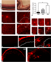
Altered retinal vasculature in cbs−/− mice. Histological assessment of retinal cryosections stained with hematoxylin and eosin (A, B), showing retinal hemorrhage (white arrow) and grossly dilated blood vessels in the cbs−/− retina (B) compared to the normal retina of wild-type (cbs+/+) mouse (A). Retinal flat mounts (CF) stained with isolectin-B4 (endothelial cell marker) labeling the retinal vasculature. Retina of cbs−/− mouse shows central capillary dropout (D) compared to healthy central vasculature of cbs+/+ mouse (C). Neovascular tufts (arrows) were observed in retinas of cbs−/− mice (F), which were not observed in the cbs+/+ mouse retina (E). Experiments were performed in five mice. Scale bar: 40 μm (B), 200 μm (D, F). Morphometric assessment of the central capillary-free areas showed significant increase in cbs−/− retinas compared to wild-type (cbs+/+) retinas ([G], *P < 0.05, n = 4 mice per group). Vasculature of the 3-week-old cbs+/− mice was mildly altered; retinal flat mounts were stained with isolectin-B4 (endothelial cell marker) to label retinal vasculature. (H, K) cbs+/+; (I, L) cbs+/−; (J, M) cbs−/−. There is marked central capillary dropout in the retina of the cbs−/− mouse (J) compared to mild dropout in the cbs+/− retina (I) and the healthy central vasculature of the cbs+/+ mouse (H). Neovascular tufts (large arrows) and capillary dilations (small arrows) were observed in retinas of cbs−/− mice (M), which were detected only occasionally in the cbs+/− retina (L) and were not observed in the cbs+/+ mouse retina (K). Experiments were performed in six retinas per group. Scale bar: 50 μm. Isolectin-B4–stained frozen sections confirmed the neovascularization in the cbs−/− mouse retinas (NR). Retinal cryosections from cbs+/+ (Q) and cbs−/− (R) mice were incubated with isolectin-B4, an endothelial cell–specific marker, to label blood vessels. There were new blood vessels growing into the vitreous in the cbs−/− mice retina, particularly in the midperipheral zone of the retina (P), while there were normal vascular patterns in the retina of the wild-type control cbs+/+ (O). Isolectin-B4 plus galactose (500 mM) was used as an isotype control (N). Scale bar: 500 μm. GCL, ganglion cell layer; IPL, inner plexiform layer; INL, inner nuclear layer; OPL, outer plexiform layer; ONL, outer nuclear layer; IS/OS, inner/outer segments of photoreceptor cells.
Figure 2.
Figure 2.
Increased endoglin (CD105) levels in cbs−/− mouse retinas. Fluorescent immunodetection of endoglin (red), a marker of neovascularization, was performed in retinal cryosections of 3-week-old mice; DAPI (blue) was used to label nuclei. Endoglin was minimally detected in cbs+/+ retinas (A, D), while endoglin levels were markedly increased in cbs−/− retinas (C, F), particularly in the ganglion cell layer and inner nuclear layer, where the blood vessels are predominant. Occasional endoglin-positive labeling was seen in cbs+/− retinas (B, E). The arrows represent neovascularization (new blood vessel formation). Scale bar: 50 μm. Experiments were performed in sections from six mice per group. Abbreviations for retinal layers are the same as for Figure 1.
Figure 3.
Figure 3.
Increased VEGF levels in cbs−/− mouse retinas. (A) Fluorescent immunodetection of VEGF (green), a marker of neovascularization, and isolectin-B4 (red), a marker of endothelial cells, was performed in retinal cryosections from 3-week cbs+/+, cbs+/−, and cbs−/− mice. The far left panel provides controls: an isotype (IgM) control for VEGF and a negative control (omission of the primary antibody), but with inclusion of the secondary antibody, for isolectin-B4. Scale bar: 50 μm. Abbreviations for retinal layers are the same as for Figure 1. (B) Quantification of the intensity levels of VEGF immunofluorescence, showing significantly greater VEGF levels in cbs−/− compared to cbs+/+ and cbs+/− mice (***P < 0.001, n = 6). (C) Expression of VEGF mRNA is increased in neural retinas of cbs−/− mice compared to cbs+/− and cbs+/+ mice. Fold change in expression of vegf120 and vegf164 was determined by quantitative RT-PCR (*P < 0.05). (D) Representative immunoblot to detect VEGF in retinas of cbs+/+ mice compared with cbs+/− and cbs−/− mice. Protein was extracted from retina and subjected to SDS-PAGE; immunoblotting was performed with an affinity-purified antibody against VEGF 164 and subsequently with an antibody against β-actin (internal loading control).
Figure 4.
Figure 4.
Increased GFAP expression in cbs−/− mouse retina. GFAP levels were detected by immunofluorescence in retinal cryosections and flat-mount preparations. (A) Retinal cryosections of 3-week cbs+/+, cbs+/−, and cbs−/− mice that were incubated with an antibody against GFAP followed by incubation with Alexa Fluor 488 (green)–labeled secondary antibody, showing increase in GFAP immune reactivity in both astrocytes and Müller cells in the cbs−/− retina compared to wild-type control cbs+/+, where GFAP is expressed in astrocytes only. (B) Retinal flat-mount preparations from cbs+/+, cbs+/−, and cbs−/− mice immunostained for isolectin-B4 (red) to visualize vasculature and GFAP (green), showing altered vasculature and ragged appearance of the astrocytes in cbs−/− mice compared to normal-shaped vasculature and astrocytes in the wild-type control cbs+/+ (n = 6). Scale bar: 50 μm. (C) Quantification of the data obtained from metamorphic analysis of color intensity of GFAP (significantly greater than wild-type, ***P < 0.001, n = 6).
Figure 5.
Figure 5.
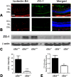
Decreased ZO-1 levels in cbs−/− mouse retina. Retinal cryosections from (A) cbs+/+ and (B) cbs−/− mice were incubated with an antibody against ZO-1 (green), a marker of tight junctions, and isolectin-B4 (red) to label blood vessels. Scale bar: 40 μm; abbreviations for retinal layers are the same as for Figure 1. (C) Retinal protein was extracted and subjected to SDS-PAGE, followed by immunoblotting to detect Z0-1 (molecular weight ([Mr] = 198 kD); β-actin was used as the loading control. (D) Quantification of immunohistochemical fluorescence intensity (***P < 0.001, n = 6). (E) Quantification of densitometric scans of protein bands shown in (C) (*P < 0.05).
Figure 6.
Figure 6.
Occludin expression in mouse retina. Retinal cryosections of eyes from cbs+/+ and cbs−/− mice, incubated with antibodies against occludin and isolectin-B4 followed by incubation with Alexa Fluor 488 (green)– and Texas Red avidin (red)–labeled secondary antibodies, show decreased occludin immune reactivity in the cbs−/− retina (B) compared to wild-type cbs+/+ (A). Trypsin-digested retina, immunostained for isolectin-B4 (red) and occludin (green), show decreased occludin expression in cbs−/− retina (D) compared to cbs+/+ (C). Scale bar: 50 μm (A, B), 20 μm (C, D).
Figure 7.
Figure 7.
In vivo and in vitro assessment of blood–retinal barrier function. (A) Data from fluorescein angiography, performed to evaluate the retinal vasculature and to determine whether permeability was altered in living mice (in vivo experiment). Left panel shows a representative angiogram of a cbs+/+ mouse; right panel shows an angiogram of a cbs−/− mouse. Data are representative of five mice studied in each group. (B) Data from an FITC–dextran flux permeability assay performed using HRECs in the absence (control) or presence of 50 μM homocysteine. Fluorescence intensity measured in the lower chamber versus the upper chamber of the transwell plate was determined photometrically, and the ratio of these values was significantly higher in homocysteine-treated HRECs versus PBS-incubated cells (**P < 0.01, n = 3).

Similar articles

Cited by

References

    1. Perła-Kaján J, Twardowski T, Jakubowski H. Mechanisms of homocysteine toxicity in humans. Amino Acids. 2007; 32: 561–572 - PubMed
    1. Mudd SH. Hypermethioninemias of genetic and non-genetic origin: a review. Am J Med Genet C Semin Med Genet. 2011; 157: 3–32 - PubMed
    1. Bleich S, Jünemann A, Von Ahsen N, et al. Homocysteine and risk of open-angle glaucoma. J Neural Transm. 2002; 109: 1499–1504 - PubMed
    1. Vessani RM, Ritch R, Liebmann JM, Jofe M. Plasma homocysteine is elevated in patients with exfoliation syndrome. Am J Ophthalmol. 2003; 136: 41–46 - PubMed
    1. Jaksic V, Markovic V, Milenkovic S, Stefanovic I, Jakovic N, Knezevic M. MTHFR. C677T homozygous mutation in a patient with pigmentary glaucoma and central retinal vein occlusion. Ophthalmic Res. 2010; 43: 193–196 - PubMed

Publication types

MeSH terms