

Nerves and neovessels inhibit each other in the cornea
- PMID: 23307967
- PMCID: PMC3562120
- DOI: 10.1167/iovs.11-8379
Nerves and neovessels inhibit each other in the cornea
Abstract
Purpose: To evaluate the regulatory cross-talk of the vascular and neural networks in the cornea.
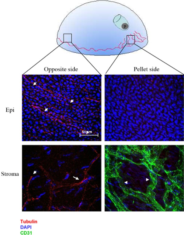
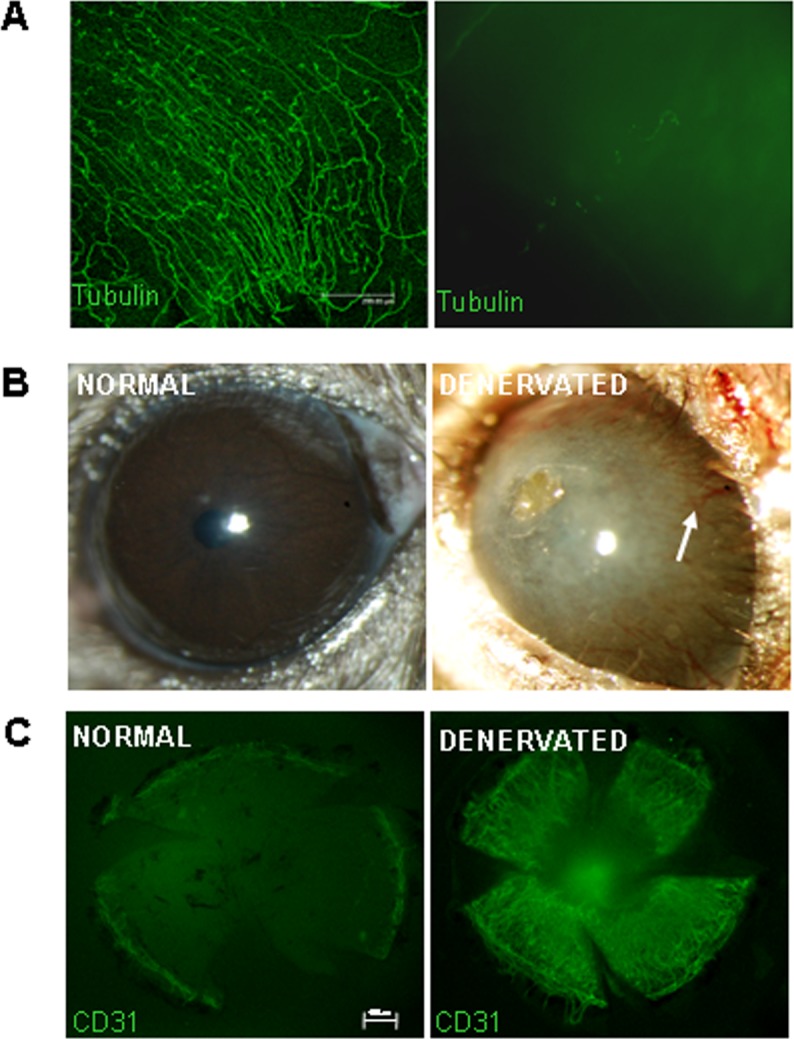
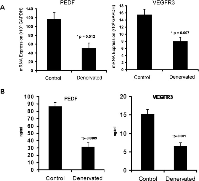
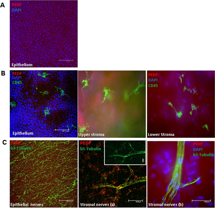
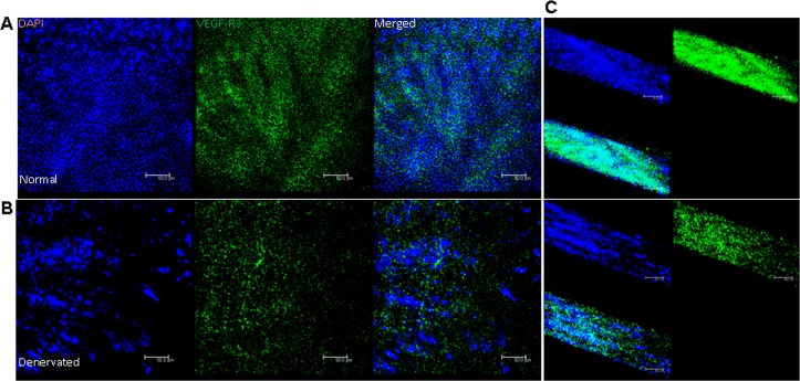
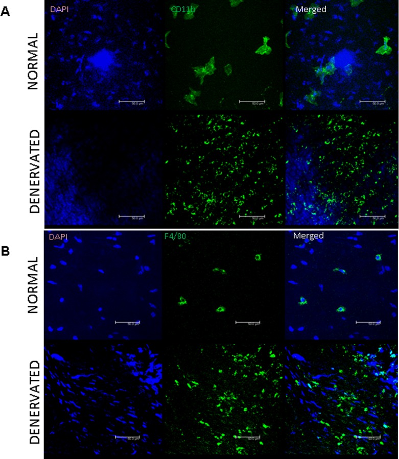
Methods: b-FGF micropellets (80 ng) were implanted in the temporal side of the cornea of healthy C57Bl/6 mice. On day 7, blood vessels (hemangiogenesis) and nerves were observed by immunofluorescence staining of corneal flat mounts. The next group of mice underwent either trigeminal stereotactic electrolysis (TSE), or sham operation, to ablate the ophthalmic branch of the trigeminal nerve. Blood vessel growth was detected by immunohistochemistry for PECAM-1 (CD31) following surgery. In another set of mice following TSE or sham operation, corneas were harvested for ELISA (VEGFR3 and pigment epithelium-derived factor [PEDF]) and for quantitative RT-PCR (VEGFR3, PEDF, and CD45). PEDF, VEGFR3, beta-3 tubulin, CD45, CD11b, and F4/80 expression in the cornea were evaluated using immunostaining.
Results: No nerves were detected in the areas subject to corneal neovascularization, whereas they persisted in the areas that were neovessel-free. Conversely, 7 days after denervation, significant angiogenesis was detected in the cornea, and this was associated with a significant decrease in VEGFR3 (57.5% reduction, P = 0.001) and PEDF protein expression (64% reduction, P < 0.001). Immunostaining also showed reduced expression of VEGFR3 in the corneal epithelial layer. Finally, an inflammatory cell infiltrate, including macrophages, was observed.
Conclusion: Our data suggest that sensory nerves and neovessels inhibit each other in the cornea. When vessel growth is stimulated, nerves disappear and, conversely, denervation induces angiogenesis. This phenomenon, here described in the eye, may have far-reaching implications in understanding angiogenesis.
Conflict of interest statement
Disclosure:
Figures
References
-
- Tessier-Lavigne M, Goodman CS. The molecular biology of axon guidance. Science. 1996; 274: 1123–1133 - PubMed
-
- Dickson BJ. Molecular mechanisms of axon guidance. Science. 2002; 298: 1959–1964 - PubMed
-
- Huber AB, Kolodkin AL, Ginty DD, Cloutier JF. Signaling at the growth cone: ligand-receptor complexes and the control of axon growth and guidance. Annu Rev Neurosci. 2003; 26: 509–563 - PubMed
-
- Carmeliet P, Smet FD, Loges S, Mazzone M. Branching morphogenesis and antiangiogenesis candidates: tip cells lead the way. Na. Rev Clin Oncol. 2009; 6: 315–326 - PubMed
Publication types
MeSH terms
Substances
Grants and funding
LinkOut - more resources
Full Text Sources
Other Literature Sources
Research Materials
Miscellaneous
